Intro Flashcards

(78 cards)

1
Q

Nearly 100% of human cancer has ____ pathway inactivation, >90% has ____ inactivation

A

Nearly 100% of human cancer has p16INK4a-Rb pathway inactivation, >90% has p53 inactivation

2
Q

What are 4 aspects of dysregulation present in the cellular processes of cancer?

A

I. Inappropriate proliferation

II. Resistance to differentiation and apoptosis

III. Genomic Instability

IV. Ability to grow where it ought not (i.e. “malignant growth”)

3
Q

Protooncogene:

A

Highly conserved eukaryotic genes, important in cellular growth and development, which can become oncogenes either by over/under expression or by mutation

4
Q

Cellular Oncogenes:

A

(c-onc); cellular genes involved in the development and/or maintenance of the malignant phenotype

5
Q

Viral oncogenes:

A

(v-onc); viral genes which are able to transform cells

6
Q

Oncogenic mechanisms (4):

A

•Growth factors

  • Mutated ras induces expression of normal growth factor genes (TGF-a)
  • The sis viral oncogene is highly homologous to PDGF, and can stimulate PDGF-R

•Signal transduction

  • Her-2/neu amplification in breast carcinoma

•Cell cycle control

•Regulation of gene expression

7
Q

Give 2 examples of growth factor involvement in oncogenic mechanisms:

A

–Mutated ras induces expression of normal growth factor genes (TGF-a)

–The sis viral oncogene is highly homologous to PDGF, and can stimulate PDGF-R

8
Q

Provide an example of oncogene development via signal transduction:

A

Her-2/neu amplification in breast carcinoma

9
Q

There are professional tumor suppressor genes (e.g. ____ and ____) that are dispensable for normal development, but serve only to prevent _______.

A

There are professional tumor suppressor genes (e.g. p16INK4a and p53) that are dispensable for normal development, but serve only to prevent transformation.

10
Q

Li-Fraumeni Syndrome:

A

Hereditary predisposition to cancer.

p53 protein normally

  • Arrests cell cycle when DNA damage occurs
  • Promotes apoptosis in damaged cells
  • Most commonly mutated gene in cancer

Persons with syndrome are born with one abnormal copy

  • Tumors have mutations at both alleles
  • Glioblastomas, leukemias, breast, lung, and pancreatic CA, Wilm, sarcomas
11
Q

Fill in the empty spaces

A
12
Q

FIll in the blanks (shows resistance mechanisms to apoptosis)

A
13
Q

Hyperplasia:

A

Increase in the cell number

Usually associated with an increase in tissue mass (hypertrophy)

Can be physiologic or pathologic

14
Q

Explain how hyperplasia can be physiologic or pathologic:

A

Physiologic:

  • Hormonal (glandular epithelium in breast during puberty)
  • Compensatory (contralateral kidney after nephrectomy)

Pathologic (breeding ground for cancer):

  • Endometrial hyperplasia
  • Benign prostatic hypertrophy (BPH)
15
Q

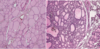
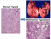
What types of cells are shown in these images?

A
16
Q

Hypertrophy:

A

•Increase in cell size

  • Production of new subcellular components (not swelling)

•Can be physiologic or pathologic

17
Q

Explain how hypertrophy can be physiologic or pathologic:

A

–Physiologic

  • Skeletal muscle

–Pathologic

  • Cardiomyocytes and increased afterload (HTN) (although limited proliferation may be possible!)
18
Q

What change is indicated in these images?

A

hypertrophy

19
Q

Define metaplasia:

A

•Change of one differentiated cell type into another differentiated cell type

–Usually an adaptive response

–Usually reversible

20
Q

List two common examples of metaplasia:

A

–Smoking and respiratory epithelium

–Barrett esophagus

21
Q

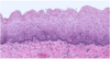
What change has occurred in these two images?

A

This is an image of the esophagus.

Left is NORMAL

Right is METAPLASTIC.
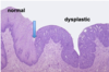
22
Q

Define dysplasia:

A

Atypical proliferation of cells with:

  • Abnormal appearance
    • Variation in size and shape (pleomorphism)
    • Nuclear enlargement (increased nuclear-to-cytoplasmic ratio; increased N:C)
    • Nuclear irregularity
    • Dark staining of nuclei (hyperchromasia)
  • Disorderly arrangement
    • Loss of polarity (disrupted order of expected growth)
    • Loss of maturation
    • Abnormal location of mitotic figures
23
Q

Describe this image:

A

Normal squamous epithelium

24
Q

What is shown in this image?

A

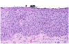
Dysplastic squamous epithelium

25
Explain this image:
On the left are normal cells; on the right are dysplastic
26
Define neoplasia:
•Abnormal growth of tissue * Well differentiated * Poorly differentiated * Anaplastic: lack of cellular differentiation * Excessive growth of tissue * “Uncoordinated” and autonomous * Benign or malignant…
27
Anaplastic:
Lack of cellular differentiation
28
How do benign neoplasms normally grow?
Without invasion or spread Usually grow slowly Remain localized
29
Morbidity associated with benign neoplasms:
Caused by: - size - anatomic location - production of hormone or other cell products (also in malignant neoplasm)
30
How are benign neoplasms generally named?
•Generally add an “-oma” * Osteoma (bone) * Chondroma (cartilage) * Adenoma (benign tumor of glandular tissue) •Suffix “oma” means “swelling” or “tumor”
31
Suffix "oma" means what?
swelling or tumor
32
Histological characteristics of benign neoplasms:
**•Usually resembles the normal counterpart** * Histologically well differentiated * Low mitotic rate * Generally well circumscribed * Do not metastasize
33
Adrenal adenoma:
•Benign tumor of adrenal cortex: –Usually nonfunctional –Cortisol: Cushings –Aldosterone: Conns –Androgens
34
Paraneoplastic syndrome:
Phenomemon where tumor produces something locally that then has a systemic effect. –Symptoms not due to the local presence of neoplastic cells –Secretion of humoral factors by tumor cells –Most commonly associated with malignant neoplasms
35
\_\_\_\_\_\_\_ have abnormal appearing cells and cellular architecture, but are REVERSIBLE. Can be mild, moderate, or severe depending on the extend of involvement.
Dysplasias
36
Carcinoma in situ:
Dysplastic changes involving the full thickness of the epithelium BUT cells do not extend beyond the basement membrane
37
A ____ is defined as a neoplasm that has invaded surrounding tissue - malignant cells with atypical morphology extend beyond the basement membrane.
Carcinoma
38
Mesenchymal malignant neoplasms are typically named:
Mesenchymal: **Sarcomas** * Osteogenic sarcoma (bone) * Chondrosarcoma (cartilage) * Rhabdosarcoma (skeletal muscle)
39
Epithelium-derived malignant neoplasms are typically named:
Epithelium: Carcinoma * Adenocarcinoma (glandular epithelium) * Squamous cell carcinoma
40
Histologic grade:
•Refers to level of differentiation: * Well differentiated * Moderately differentiated * Poorly differentiated •Presumed correlation between appearance and behavior * Imperfect (… renal cell carcinoma)
41
Anatomic stage:
•Refers to the **extent of spread** * Tumor size * Node involvement * Metastasis •Staging systems: * International (TNM) * Each scored individually * American Joint Committee on Cancer: 0-IV * Combined score
42
An adult patient is diagnosed with esophageal carcinoma. Histologically, the tumor is poorly differentiated. There is no evidence of metastatic disease. The tumor has a HIGH/LOW grade and HIGH/LOW stage:
High grade (poorly differentiated) Low stage (not spread)
43
Neoplasia:
The pathological process that results in the formation and growth of cells in a new location Can be benign or malignant (altered morphology or state of differentiation)
44
What is malignancy defined as?
A neoplasm that has acquired the ability to invade and damage/destroy host tissue
45
Neoplasm proliferation can result in:
A discrete mass Infiltration or replacement of normal structures (e.g. bone marrow in leukemia)
46
47
Carcinomas are cancers of ___ origin
Epithelial
48
Sarcomas are cancers of ____ origin
Connective and supportive tissue (bone, fat, muscle, etc.)
49
Leukemias are cancers of ___ origin
WBCs in the bone marrow
50
Lymphomas are cancers of ___ origin
lymphatic system
51
Myelomas are cancers of ___ origin
Plasma cells in the bone marrow
52
Fine needle aspiration:
Good for carcinomas, Bad to see tumor architecture (ie, bad if suspect lymphoma)
53
Core biopsy:
Adequate for most cancers Get an idea of architecture
54
Excisional Biopsy:
Surgical procedure that removes the entire suspected area. Common for melanoma treatment/diagnosis or lymphoma diagnosis.
55
What are 10 important hallmarks of cancer?
Sustaining proliferative signaling Evading growth suppressors Activating invasion and metastasis Enabling replicative immortality Inducing angiogenesis Resisting cell death Avoiding immune destruction Tumor-promoting inflammation Genome instability and mutation Deregulating cellular energetics
56
What are the cell cycle stages of normal, early growing tumors, and late growing tumors?
**Normal**: mostly non-cycling cells. Rate of cell birth = rate of death. Low growth fraction (~30%) **Early growing tumor**: mostly cycling cells. Rate of birth\>death. High growth fraction (~80%) **Late growing tumor:** necrotic center. Many non-cycling cells. Relatively low growt fraction. Poor drug penetration. Hypoxia --\> resistance to drugs, radiation.
57
Cell cycle nonspecific agents as treatment (chemo):
–Effects exerted at any phase of the cell cycle –Toxic effects occur during cell cycle and are expressed during attempt at cell division **–May be more effective given as a bolus dose** –Examples: * Alkylating agents * Anthracyclines * Platinum agents
58
Cell cycle specific agents as treatment for cancer (chemo):
–Effect exerted on **dividing cells only** –Not active in resting phase **–Administration as continuous infusion or multiple divided doses** –Examples: * Antimetabolites * Vincas * Taxanes * Topoisomerase Inhibitors
59
Why are combination regiments important for cancer therapy?
–Different mechanisms of action –Non-overlapping toxicities –Dose and time “intensity” –Eliminating minimal residual disease Single drugs are rarely curative (resistance)
60
Neoadjuvant therapy:
Therapy given prior to the primary therapy
61
Adjuvant therapy:
therapy given in addition to the primary therapy
62
Induction therapy:
Intensive initial treatment to get a handle on the cancer (followed by consolidation and maintenance)
63
Consolidation (chemo):
•Used to solidify a remission and further reduce tumor burden. –Less intensive therapy as a second step
64
Maintenance (chemo)
Used in some diseases to increase disease-free interval or catch the slow growing cells
65
Chemotherapy:
Inhibit the growth and division of cancer cells
66
Targeted cancer therapy:
Typically targets specific genes or proteins
67
Immunotherapy:
uses the body's own immune system to fight cancer
68
VEGF:
Normally, hypoxic conditions trigger VEGF Cancer cells can aberrantly secrete VEGF, leading to angiogenesis and thus tumor growth. VEGF inhibitors are good current targets for treatment.
69
Cancer **stage**:
* Define as precisely as possible the **extent** of disease * Rationale: Staging will determine * Treatment * Prognosis * Surveillance •A common currency for reporting treatment result
70
What is the TMN staging system?
**•T (tumor)** Stage * Denotes extent of primary tumor * Generally T1-T4 **•N (nodal)** Stage * Denotes extent of regional lymph node involvement * Generally N0-3 **•M (metastatic)** Stage * Denotes metastatic spread and site(s) * M0 to M1
71
What does the T stage reflect?
* T1 to T4 reflects **how deeply the tumor has been able to locally extend.** * For example, in colon cancer: –T1 is a superficial tumor –T2 is a muscle invasive tumor –T3 has grown through the muscularis propria –T4 has grown through the colon wall and serosa and is adherent to adjacent organs
72
Grade is a measure of \_\_\_\_\_
The microscopic appearance
73
Explain the resistance/evolution mechanisms of cancer:
Cancer has an evolution - can get subsets of the cancer that look different than other subsets due to variance in involvement of cell type, mutation, etc. So if you use a therapy that targets a certain gene, you might only be killing 85% of the tumor because the rest might not have a mutation in this gene. Can allow for this resistant area to grow after the rest has been treated.
74
What roles does surgery play in oncology?
–Initial diagnosis –Staging –Excision for cure or palliation –Prevention (genetic predisposition to breast or colon cancer) --Removal of bulk disease --Removal of metastatic lesion(s) * Small, few in number, slow growing * Brain, liver, lung --Oncologic emergencies * Spinal cord metastases, bowel perforation
75
"Gompertzian" Growth of tumors:
With slow dividing cells, might not be killing enough cells. Sometimes purpose is to prevent patient from hitting lethal limit and just keep the cancer under control (vs. totally wiping out the cancer)
76
Clinical Remission
Might hit a remission where you can't detect anymore, but you keep treating until immune system jumps in to kill the rest.
77
An ECOG Performance Score is used to measure WHAT
Comorbidities ## Footnote 0 = Fully active 1 = Some symptoms 2 = Needs some assistance 3 = Needs complete assistance 4 = Near death
78
How do people actually die from cancer?
•Local effects * Brain/spinal cord compression * End organ failure * Hemorrhage * Infection * VTE * Systemic effects * Anorexia * Cachexia * Sarcopenia