Intro to Derm Part 1 Flashcards
(65 cards)
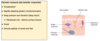
describe the embryological formation of the skin
Skin arises by juxtaposition of two major embryological elements:
- Epidermis - originates from ectoderm
- Dermis - arises from mesoderm that comes into contact with inner surface of epidermis
mesoderm is essential for inducing differentiation of epidermal structures (e.g. hair follicle)
describe the cellular changes that occur during the embryological development of skin
week 4 - Epidermis begins to form (from surface ectoderm) as single basal layer of cuboidal cells
week 5 - Secondary layer of squamous, non-keratinising cuboidal cells (periderm) develops on top of the basal layer
periderm generates vernix caseosa - white, waxy protective substance
week 11 - basal layer of cuboidal cells ( stratum germinativum) proliferates to form multilayered intermediate zone
weeks 9-13 - development of hair follicles in stratum germinativum and appearance of lanugo hair (Very fine hair)
weeks 10-17 - epidermal ridges protrude as troughs into developing dermis and neurovascular supply develops into dermal papillae
week 20 - 4 superifical strata have formed - spinosum, granulosum, lucidum, corneum
what is the funciton of the vernix caseosa
where is it produced
during gestation - protects fetus from amniotic fluid during gestation
during birth - protects baby from bacterial and environmental damage
produced by the periderm
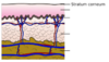
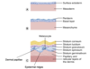
label the layers in the development of skin


where is the neruovascular supply in the skin
dermal papillae
(part of dermis and is beneath the epidermis)
what cells give skin, hair, eyes colour
give their sequence of embryological development
melanocytes - contain pigment
precursors are melanoblasts which are derived from the neural crest in the embryo
week 6-8 - melanoblasts migrate dorsally to the developing epidermis, dermis and hair follicles
by week 12-13 - most melanoblasts have reached their destination and differentiate into melanocytes
a subset of melanoblasts form melnaocyte stem cells in hair follicle bulge which are a reserve to replenish differentiated melanocytes
how are melanocytes regulated
with no exposure to external factors
Melanocortin 1 receptor (MC1R), a G protein-coupled receptor regulates quantity and quality of melanins produced:
MC1R is controlled by agonists α-melanocyte-stimulating hormone (αMSH) & adrenocorticotropic hormone (ACTH) and antagonist, Agouti signaling protein (ASP).
Activation of MC1R by agonist (αMSH or ACTH) → causes melanogenic cascade → synthesis of eumelanin (dark pigment)
ASP reverses those effects & elicits production of pheomelanin (pale pigment)
ACTH can also up-regulate expression of the MC1R gene
what effect does ACTH have on pigment produced
how does it do this
increases pigment production
by acting as a MC1R agonist and upregulating gene expression of MC1R on melanocytes
how are melanocytes regulated
using external factors
Exposure to UV
directly increases melanin:
results in Increased expression of MITF & downstream melanogenic proteins, including Pmel17, MART-1, TYR, TRP1, and DCT → increases melanin production
indirectly increases melanin:
also increases PAR2 in keratinocytes → increases uptake & distribution of melanosomes by keratinocytes
how can melanocytes be regulated
either by agonists (ACTH alphaMSH) and antagonist (ASP) of the MC1R receptor
or by UV light exposure
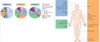
how is the structure of melanocytes related to their function
dendritic cells
processes allow to effectively distribute melanosomes to keratinocytes
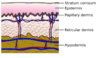
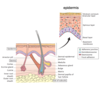
give an overview of the structure of the skin
superficial to deep:
epidermis - mostly made up of keratinocytes
basement membrane (dermal-epidermal junction)
dermis - mostly made up of connective tissue
subcutaneous fat

label this diagram of the skin

what is the extra layer of epidermis present in palms and soles of feet only
strutum lucidum
how are keratinocytes able to undergo progressive differentiation
cytoskeleton of keratinocytes is made up of keratin intermediate filaments
filaggrin - protein which regulates progressive differentiation/flattening of keratinocytes from cuboidal to flat
what is the structure of the epidermis
what process occurs in it and how does it happen
composed of keratinocytes
cells layers (superficial to deep):
stratum corneum
stratum lucidum (only present in palms and soles)
stratum granulosum
stratum spinosum
basal layer
progressive differentiation/flattening of keratinocytes occurs - cells progress from the basal layer to surface layer/stratum corneum in 30 days

what are the characteristics of cells in the different epidermal cell layers
basal layer - keratinocytes originate here and are simple cuboidal shaped
stratum spinosum
stratum lucidum - found only in palms and soles
stratum granulosum - cells contain granules of keratohyalin
stratum corneum - keratinocytes are flattened and have no nuclei or organelles, but have specific functions
what is the function of the keratinocytes in the stratum corneum
outer layer - absorb solutes
middle layer - absorbs water
lower/inner layer - mechanical defence barrier, contains lipids e.g. FFA, sterols to carry out its function
how does cellular progression of keratinocytes in erpidermis change in skin disease
it is accelerated
e.g. psoriasis
label this diagram of the epidermis


describe the intracellular structure of keratinocytes:
filamentous cytoskeleton: (from thickest to thinnest)
Tubulin‐containing microtubules (20-25nm)
Intermediate filaments (keratins) (7-10nm)
Actin‐containing microfilaments (7nm)
what is the function of keratins
Structural properties - part of cytoskeleton
Cell signalling
Stress response
Apoptosis
Wound healing
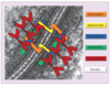
what are desmosomes
where are they found
what is their function
what is their structure
major adhesion complex
found in epidermis
anchors keratin intermediate filaments to cell membrane and bridges adjacent keratinocytes
allows cells to withstand trauma
composed of several smaller proteins - desmoglein, desmocollin, plakoglobin, plakophilin, desmoplakin, keratin
what are the cell to cell connections in the epidermis
desmosomes
gap junctions
tight junctions
adherens junction