Intro Virology and Replication Flashcards
(34 cards)
What is a Virus?
- Obligate intracellular parasite- needs host cell to survive (in order to replicate & complete its life cycle)
- Lacks organelles- no mitochondria/ energy source etc
- Extremely small - ‘filterable agents’- Need electron microscope to visualise
Virion
(an entire virus particle)
- In its most simple form comprises of:
- Nucleic acid- RNA/DNA (confers infectivitity)
- A capsid (confers specificity)- structure & symmetry of this is important.
- Additional features may include:
- An envelope- Host derived lipid bilayer contains virus encoded glycoproteins
-Viral enzymes- incorporated into the envelope/ within the capsid
DNA Viruses
- All monopartite (all viral genes on a single molecule)
- All ds (except parvoviridae and circoviridae)
RNA Viruses

- Mostly single stranded & can be segmented
- Segmentation- allows virus to ↑ its diversity very rapidly (reassortment)
- Need an RNA polymerase to copy their RNA genome (no equivalent enzyme in the host)
- RNA dependent RNA polymerase
- RNA polymerases are error prone- No proof reading capability, as a consequence of this:
- RNA viruses are more variable:
- Within a species of virus are more subtypes/serotypes
- Often zoonotic (jump from animals to humans and cause disease)
- Can evolve rapidly if needed
- If a virus jumps from one species to another, RNA viruses can more readily adapt
Capsids
(Structure)

Viral capsids: enclose the nucleic acid & have 3 forms of symmetry
1. Icosahedral-e.g.parvoviridae
- 12 vertices, 20 facets, 5:3:2 fold symmetry
- Composed of capsomers:
- *-Penton Capsomer**- 12 present, one at each vertex,has 5 capsid proteins
- *-Hexon Capsomer**- Composed of 6 capsid proteins
2. Helical Capsid-e.g.paramyxoviridae & rhabdoviridae
- Structural unit is one capsid protein- arranged as a helix
- All animal viruses with helical symmetry are enveloped
3. Complex Capsid-e.g.poxviruses
- Some of the large viruses have structures that are more complex
- >100 proteins -encode more proteins than other viruses
- Neither helical or icosahedral structure
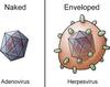
Enveloped VIrus

- Few viruses with icosahedral capsid
- All viruses with helical capsid
- Complex capsid
Naked Virus
- Icosahedral Capsid
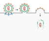
Viral Envelope

Many acquire an envelope as they bud through the host cell membranes
- Host membrane = lipid bilayer- ‘coating ‘is effectively inert
- BUT would not permit recognition of receptor molecules on the host cell
- SO viruses modify the envelope by synthesis of viral encoded proteins which are associated with the envelope
Biological Properties of Enveloped Viruses
- More pleomorphic (not a regular shape)
- More fragile than viruses with just a capsid
- More easily destroyed by: detergents, disinfectant & outside environment
Naked Capsids
- Components: Protein
- Properties: Environmentally stable to:
- Temperature, pH, Porteases, Detergents, Drying, Released by cell lysis
- Consequences:
- Can be spread easily
- Can dry out and retain infectivity
- Can survive adverse conditions in the gut
- Resisitant to detergents
- Lyses cell to release; usually cause acute infections
Enveloped Viruses
- Components: lipids, porteins, glyoproteins
- Prorperties: evironmentally labi
- destroyed by: acid, detergents, drying, heat, released by budding
- Consequences:
- Not easily spread (large droplets, secretions, transplants/transfusions)
- Must stay wet
- Cannot survive in the GIT
- Easilty destroyed by detergent
- Does not need to kill the cell to spread; can cause persistent infections
Baltimore Classification System
classified by the mechanism of generating positive strand mRNAs
- Seven fundamentally different groups
Hierarchical VIrus Classification System
classified according to characteristics:
- Presence or absence of viral envelope
- Capsid symmetry
- Size and shape
- Genome composition, polarity and structure
- Virus Taxonomy:
- Order (-virales)
- Example: family (-viridae)- Flaviridae, genus (- virus)- Pestivirus, species – Bovine viral diarrhoea virus 1
- Divided into genera containing species that further subdivide into serotypes and then subtypes
Antigenicity

- Viruses can be differentiated on basis of antigenic sites on their surface = “serotypes”
- Viruses can be divided into “serotypes” and further sub grouped into subtypes
- Classified on the basis of their reactivity with antibody (serological reactivity)
What viral proteins determine “serotype”
- Generally proteins on the virus surface that are involved in Virus entry & Antibody reactions
- In different serotypes, these proteins tend to vary in their precise amino acid composition–>immune system recognizes these proteins as slightly different
Virulence
- Viruses can be divided into pathogenic (virulent) and subclinical (avirulent- low pathogenic)
- isolates e.g. avian influenza virus and feline infectious peritonitis virus have both
Nucleotide Sequencing
- Viruses can be divided into genotypes depending on the nucleotide sequence of their genes
- e.g. BVDV1a ‐m, BVDV2a‐c
Capsid Proteins
(structural proteins)
- Structural components of the virus capsid (icosahedral, helical and complex)
- Protect the viral nucleic acid
Capsid Proteins of Naked Viruses
- Deliver the viral nucleic acid to the cell
- Receptors to attach to host cell
- Protein domains that fuse with the host membrane to allow entry
- Assembly‐capsid proteins self assembles into capsids during virus replication
- NB ‐contain sites that will induce an antibody response
Envelope Proteins
- Structural components of the virus, often glycosylated
- Embedded in a lipid bilayer, derived from host membrane (virus envelope)
- Contain receptors that allows the virus to attach and then enter the host cell
- Two step process
- Attachment/binding
- Fusion
- Targets of the host immune response
- Antibodies will recognise these surface exposed viral proteins (antigenic determinants)
- Antibodies often protective (neutralising)
- Virion Associated Enzymes: the virion contains an enzyme (RNA-dependent-DNA polymerase or reverse transcriptase) which uses this RNA as a template for the synthesis of double stranded DNA which integrates into the cell DNA
Non-structural proteins
- Are not structural components of the virus particle
- Often enzymes involved in viral replication Proteases; helicases; RdRpol(RNA dep RNA polymerase)
- Made in the virus infected cell following infection: Often proteins involved in transcription, replication & protein cleavage
Viral Attachment
- Viral surface proteins bind to receptor on cell surface
- Receptor is cellular protein that happens to fit viral protein
- Virus-receptor interaction determines specificity of viruses for cells and tissues (no receptor–> no entry)
Viral Entry

Two possible routes:
- Endocytosis: virus is released from endosome by pH change or fusion of viral envelope with endosomal membrane
- Some enveloped viruses fuse directly with the plasma Membrane
VIral Uncoating
Release of viral nucleic acid from viral capsid
- Process is variable: In some viruses, nucleic acids is still in a nucleoprotein complex,
- In other viruses the capsid is only partially disintegrated


