J de Zoysa: Renal Physiology; Potassium and Magnesium Flashcards
(37 cards)
Key roles of the the Kidneys
- ) Elimination of waste products
- ) Control of fluid balance
3.) Control of minerals: K+ and Mg2+
- ) Regulate acid-base balance
- ) Produce hormones
What is Magnesium and how much? distribution?
- Is an essential cation vital for numerous physiological functions.
- The total magnesium content of the human body is ~20 mmol/kg of fat-free tissue.
- ~1000mmol in average man
- Highly soluble
- 99% of Mg is found in bone, muscle and soft tissue.
- helps form the structure of the bone, hard to get at
- Decreases with age
- Intracellular Mg concentrations range from 5 to 20 mmol/L
- Extracellular Mg accounts for 1% (0.7-1mmol/L)
- some protein bound, some ionised, important for enzyme function
Physiological purpose of Magnesium?
- Bone formation
- Co-factor in > 300 enzymatic reactions.
- ATP metabolism, (key enzyme)
- Muscle contraction and relaxation,
- Normal neurological function
- Release of neurotransmitters
- Regulation of vascular tone
- Cardiac rhythm
- Platelet-activated thrombosis
Magnesium is highly soluble, what does that mean?
It binds a large amount of water molecules very easily, which increase its diameter and makes it hard to move through intracellular channels. Needs to be actively stripped from water molecules to move through IC

Describe magnesium homeostasis.
- Mostly sourced from diet
- 300mg from diet needed, some of this is from water (low amounts in NZ)
- cereals, nuts, green leafy veges great sources
- Absorbed in the GI tract
- Kidney has a key role in regulation responding to loads or depletion

How are the kidneys important to Magnesium?
- Serum Mg is controlled by its excretion in the urine.
- ~2400 mg of Mg is filtered by the glomeruli.
- ~95% is immediately reabsorbed
- 10-20% PCT
- 60-70% Thick ascending limb
- 10% DCT
- ~100 mg (5%) is excreted in the urine

Proximal convoluted tubule Magnesium reabsorbtion ?
- Mechanism unsure (10-20% reabsorbed)
- speculated that the Na/K ATPase could drag it through when pulling water across

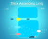
Thick ascending limb reabsorption of Magnesium?
The primary place of reabsorption
- Lots of water is reabsorbed in the descending limb, which concentrates the electrolyte concentration, so K+ and Mg2+ is high by the time it reaches the ascending limb
- Most important are the paracellular channels (formed by claudine 16 and Claudin 19) which are vital for magnesium reabsorb
- those with mutations in this paracellular channel develop hypomagnesia with a low conc of magnesium in the blood, and can develop issues with this
- Other channels: Na/K transport channels, K channels that impact Mg reabsorption
- arters syndrome; hypermagnesemia
Reabsorption of Mg in the DCT?
- transient receptor potential channels important
- Mutations can occur here → hyper magnesic → secondary hyper calcaemic → chance of developing kidney stones
- abnormalities of Mg and Ca in children can present in these cases and lead top kidney failure

How do we assess Mg?
- Serum Mg: most common, tightly regulated (0.7-1), sometimes not a good measure of bone stores
- Red cell Mg: if low they may need Mg
- 24 hour excretion: will tell us if there’s renal loss occuring (happens more over night)
- Mg retention test : give them an oral load, if it doesn’t rise then there’s an absorbtion issue
- Isotope analysis: to see distribution of Mg - NOT typically done
Causes of Hypomagnesamia
- Decreased dietary intake (water, meats, nuts)
- GI malabsorption and loss
- Endocrine – hyperaldosteronism, DM, SIADH, “hungry bone” syndrome
- Renal loss
- Congenital
- Acquired: eg; Drug-induced
Drugs that can cause acquired Hypomagnesia?
• Aminoglycosides
• Amphotericin B
- CNI
- Cisplatin
- Cetuximab
• Omeprazole: Protein pump inhibitor, now more commonly used
- Pentamidine
- Foscarnet
Symptoms of Hypomagnesaemia are?
- Weakness and fatigue
In severe cases:
- Fasciculations /cramps
- Tetany/carpopedal spasm
- Numbness paresthesiae
- Seizures
- Arrhythmias
although it’s uncommon in the community this is common in the hospital setting due to diet, drugs
How do you treat hypomagnesemia
- IV magnesium (MgS) - acute cases in hospital
- Oral is used in most cases
When does hypermagnesaemia occur?
Usually very rare!
• In advanced CKD the compensatory mechanisms start to become inadequate and hypermagnesaemia may develop. (but usually these patient have problems with anorexia so this doesn’t occur often)
• Excessive oral administration of magnesium salts or magnesium-containing drugs: lots of reflux medication.
• Typically = Iatrogenic: resp depression, seizures, arrhythmias
What is Potassium? our levels? Homeostasis?
- Potassium is the most abundant cation in the intracellular fluid.
- Maintaining the proper distribution of potassium across the cell membrane is critical for normal cell function.
- Adult contains ~3500mmol K+
- 300mmol in skeleton,
- 80mmol in ECF,
- Majority 90% in cells (IC) (2700mmol in muscle cells)
- Daily oral intake 1560-5850mg/day (pregnant/lactating women need more)
- At healthy steady state: 90 – 95% excreted in urine
- 5 – 10% excreted in faeces
Internal balance of Potassium is maintained by? How?
- The kidney is primarily responsible for maintaining total body K.
- Initial changes in extracellular K are initially buffered by movement of K into or out of skeletal muscle regulated by insulin and catecholamines.
Short term: buffered by K+ moving in/out of cells
Long term: buffered by the kidneys
Tonicity and pH can affect K+ how?
Tonicity: Hyperglycaemia will lead to K efflux from the cell. Patients who present with TKA will often have a high initial K
pH:
- Acidosis can also drive K efflux. (to balance unbound H+)
- Alkalosis will leave to K influx. (to balance bound H+)
Whats the overview of K+ reabsorption
- Freely filtered at the glomerulus
- 60-70% at PCT
- 30% at Thick ascending limb
- DCT and collecting ducts have a huge amount of control
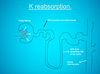
K+ reabsorption in the PCT
- paracellular mechanism driving reabsorbtion
- Na/K+ ATP ase drives K+ intracellularly, which causes paracellular movement of K+ into the blood from the lumen

K+ reabsorption in the Thick ascending limb
- Active reabsorption of K+ via a Na/K/ClCl channel and the ROMK channel, and the Na/K ATPase pump

But lots of regulation occurs at the DCT via hormones such as : Aldosterone (and angiotensin)
How

- If there’s a reduction in the EC vol or a reduction in salt, this will drive renin → angiotensin and AT II → aldosterone production

What affects Serum K+ levels?
- K+ intake (diet)
- K+ losses
- K redistributed from ECF in/out of cells
Hypokaelaemia is defined as? What are the symptoms
- Serum K < 3.5mmol/L -5mmol/L
- Symptoms:
- Muscle Weakness
- Paralysis (severe cases)
- Cardiac Conduction Abnormalities - dizzy, light-headed
- Cramps
- Constipation
