L 47-50 Glomerulus 1 & 2 Flashcards

(55 cards)

1
Q

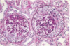
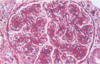
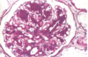
Describe the anatomy of the glomerulus

A

Note location of the afferent and efferent arterioles, mesangial cells, podocytes, bowman’s capsule, etc.

2
Q

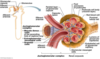
Where do the tubular capillary bed vessels branch off from?

A

Tubular capillary beds are derived from efferent arterioles which are the vessels leaving the glomerulus. This means that damage to the glomerulus will have an effect on the blood flow to the cortex and medulla of the kidney.

The medulla is the least perfused and therefore the first to be damaged by alterations in blood flow.

3
Q

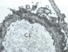
What are the layers of epithelium that line the glomerular capillaries?

A

The first layer of epithelium is the fenestrated capillary wall called the Visceral epithelium, this is often described specifically as the podocytes

Second layer is the Parietal which lines Bowman’s space, this is the outer layer of the capsule and the filtered contents should not cross this layer

4
Q

What are the layers that filter the contents of the blood into Bowman’s space?

A

1) Fenestrated capillary wall
2) Basement membrane
3) Podocytes

5
Q

Juxtaglomerular apparatus

A

Juxtaglomerular cells contain the renin and are also called granular cells

Macula densa

Nongranular cells = Lacis cells = extraglomerular mesangial cells

6
Q

What are the most common causes of glomerular, tubule, interstitial pathology?

A

Glomerular–immunologic

Tubule/Interstitial–toxic/infectious

7
Q

Azotemia vs Uremia

A

Azotemia: elevation of BUN and creatinine, can be prerenal and postrenal

Uremia: azotemia with clinical SSx and biochemical abnormalities

8
Q

What are the three major renal syndromes?

A

Acute nephritic syndrome: glomerular, acute; has visible hematuria, HTN, and mild-moderate proteinuria

Nephrotic syndrome: heavy prteinuria (greater than 3.5 gm/day), lipiduria, hypoalbuminemia, edema

Asymptomatic hematuria or proteinuria: mild glomerular abnormalities

9
Q

What kind of renal failure is described by Oliguria or Anuria with recent onset of azotemia?

A

This describes acute renal failure

10
Q

What is polyuria?

A

Producing abnormally large volumes of dilute urine

11
Q

Name the 5 Glomerular syndromes

A

Nephritic Syndrome

Rapdily progressive glomerulonephritis

Nephrotic Syndrome

Chronic renal failure

Isolated urinary abnormalities

12
Q

What are crescents?

A

Accumulation of cells composed of proliferating epithelial cells and infiltrating leukocytes

13
Q

Explain the terminology used in the histology of glomeruli such as:

Diffuse, Global, Focal, Segmental, Mesangial

A

Diffuse: invlolves all glomeruli

Global: involves the entire glomerulus

Focal: involves only some of the glomeruli

Segmental: involves only part of each glomerulus

Mesangial: primarily the mesangial region

14
Q

What are the two forms of antibody associated injury to the glomerulus?

A

1) In-situ immune complex deposition
2) Deposition of circulating immune complex

15
Q

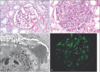
What type of antibody associated injury could be pictured?

A

The image has a Granular appearance indicating the antibody associated damage could be a number of things including deposition of circulating complexes or in-situ complexes anywhere except in the BM.

16
Q

What antibody associated injury is pictured?

A

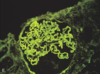
The image displays a linear pattern consistent with Anti-GBM Nephritis

17
Q

Describe anti-GBM Nephritis

A

Fixed intrinsic normal antigens in the GBM are targeted by antibodies inducing a diffuse, linear, immunofluorescent pattern

The antibodies also cross react with other BM’s in the body, namely those in the alveoli. This is called Good Pasture’s Syndrome

18
Q

Describe Circulating Immune Complex Nephritis

A

Ag-Ab complexes from elsewhere in the body lodge in the glomerulus and cause damage when complement gets activated. Antigens may be endogenous like in SLE, or exogenous like bacteria.

19
Q

Describe Nephritic Syndrom

A

Hematuria, azotemia, variable proteinurua, oliguria, edema, HTN

20
Q

Describe rapidly progressive glomerulonephritis

A

Acute nephritis, proteinuria, acute renal failure

21
Q

Describe nephrotic syndrome

A

Greater than 3.5 mg/day proteinuria, hypoalbuminemia, hyperlipidemia, lipiduria

22
Q

Describe Chronic renal failure syndrome

A

Azotemia leading to uremia and progressing for months to years

23
Q

Describe the glomerular syndrome related to isolated urinary abnormalities

A

Glomerular hematuria and/or subnephrotic proteinuria

24
Q

Describe the syndrome of acute nephritis

A

Inflammatory alterations to the glomeruli

Hematuria, red cell casts, azotemia, oliguria, mild-mod HTN

Proteinuria not as pronounced as Nephrotic syndromes

Typically charcteristic of acute proliferative glomerulonephritis and crescent GN

25
Acute Glomerulonephritis
Also called Acute Proliferative GN (Poststreptococcal, Postinfectious) 1-4 wk after strep infection of skin or pharynx Morphology: enlarged, hypercellular glomeruli, infiltration of PMNs and monocytes, diffuse changes, tubules have red cell casts Immunofluorescence: granular deposits of IgG, IgM, C3 in mesangium and along BM EM: discrete amorphous deposits on epithelial side of BM "humps" Clinical: child, abrupt onset of malaise, fever, nausea, oliguria, hematuria 1-2 weeks after recovery from sore throat; find red casts in urine, mild proteinuria, periorbital edema, mild to mod HTN Labs: elevated anti-streptococcal ab titers, low serum C3 (being used up) Recovery: 95% kids recover completely, adults: 60% recover completely
26
If they say "Crescenteric," we say...
Rapidly Progressive GN
27
Explain type I RPGN
Type I RPGN is an anti-GBM disease May cross react with pulmonary alveolar BM causing hemoptysis and hemorrhage = Good Pasture's Syndrome Immunofluorescence shows linear deposits IgG and C3 Crescents can be seen on light microscopy which are a proliferation of cells from the parietal epithelium that fill the bowman's space, may also include macrophages and monocytes Tends to go chronic more than other forms Clinical: hematuria with red cell casts, proteinuria, HTN, edema, hemoptysis and pulmonary hemorrhage in goodpasture Death within weeks to months if not treated with plasmapharesis, cytotoxic agents, steroids
28
On light microscopy you see a crescent shape and on immuno you see a linear pattern. Patient also has hemoptysis and periorbital edema. What disease are you thinkning this is?
The linear immuno is indicative of anti gbm Crescent shape always = Type I Rapidly Progressing GN The above accompanied with hemoptysis = goodpasture syndrome
29
If you see on EM humps on the epithelial side of the BM, and granular deposits on immuno, and red cell casts, and hypercellular glomeruli with PMN and monocyte infiltrates, What disease are you thinking?
Poststreptococcal GN–a form of acute GN
30
What are the four major features of nephrotic syndrome?
Massive proteinuria (more than 3.5 gm/day) Hypoalbuminemia Generalized edema Hyerlipidemia and lipiduria
31
Name the 3 main Nephrotic Syndromes and the ages of people who get them
Membranous nephropathy–adults Minimal-change disease–kids Focal segmental glomerulosclerosis–adults
32
Membranous GN morphology
Light Microscopy: diffuse, uniform thickening of capillary wall, silver stain shows spikes of BM between deposits Immuno: granular deposits of IgG and C3 EM: irregular dense subepithelial deposits that crawl into the BM and make it appear thicker, epithelial foot processes are lost
33
What disease is the image showing? And what is the major feature being shown?
Image shows uniform thickening of the capillary walls This is indicative of Membranous GN which is a type of Nephrotic Syndrome
34
Clinical picture for Membranous GN
Insidious onset, mild HTN and hematuria Course irregular but indolent Does not respond well to steroids Proteinuria persists in 60%, but only 10% renal failure in 10 years Overall good outlook, not excellent or bad
35
45 y/o Patient presents with mild HTN and hematuria, proteinuria found in the nephrotic range Immuno shows granular deposits of IgG and C3 EM shows dense subepitheliam deposits Light microscopy is as shown What is the condition?
Image shows uniformly thickend membranes of the glomerulus That combined with EM showing subepithelial deposits that invade the BM and make it appear thicker, and this being a nephrotic syndrome, and an adult indicate this is Membranous GN Has good outcome thoug many patients will have long-term proteinuria
36
Young boy presents with edema, lethargy, albuminuria, lipiduria, 4g protein/day Glomeruli appear normal on light microscopy EM is as shown below
This is a nephrotic syndrome in a child. The most common cause of this presentation is Minimal Change Disease (Lipoid Nephrosis) Light microscopy appears normal Only finding is that of effeced foot processes on EM
37
Characteristics of Minimal Change Disease
Nephrotic syndrome of kids Glomeruli appear normal on light microscopy EM shows effacement of foot processes Massive proteinura, lipiduria Responds well to steroids Adults respond slower to treatment but long term prognosis is good
38
What is the disease represented by the image?
Image shows a focal/segmental pattern of sclerosis in the glomeruli Focal because it is not all the glomeruli, and segmental because it only affects a portion of the glomerulus Therefore, the disease is Focal Segmental Glomerulosclerosis
39
What are the features of Focal Segmental Glomerulosclerosis?
Focal–affects some but not all glomeruli Segmental–affects portion of capillary tuft Idiopathic, may represent evolution from minimal change disease Differs from MCD in that it has higher incidence of hematuria, reduced GFR, and HTN, does not respond well to steroids, proteinuria more often nonselective, many progress to chronic EM might show pronounced focal detachment of epthelial cells–an advanced form of MCD
40
What is membranoproliferative GN?
Alterations in BM and proliferation of glomerular cells with leukocyte infiltration Seen in older children and young adults Divided into two types, look similar on light micro (large glomeruli, hypercelllar, lobular appearance, GBM thickened, capillary walls appear split (tram track) on silver stain) but different on EM, immuno and pathogenesis of each Type I: subendothelial deposits, granular on immuno with C3, IgG deposits Type II: intramembranous deposit, granular with C3, but also linear in BM's and mesangium, IgG is absent
41
Patient presents with recurrent hematuria, IgA staining shows up as seen in the image. What is it?
Classic presentation for IgA nephropathy
42
Characteristics for IgA nephropathy
Prominent IgA in the mesangium Frequent cause of recurrent hematuria Respiratory or GI exposure to environmental antigens leads to disregulation of mucosal IgA tha activate alternative complement and cause injury to the glomerulus Best way to ID is on Immuno showing mesangial deposits of IgA Affects older children and young adults, may ovvur several days after a mucosal infection or respiratory, GI or UTI Hematuria for several days then subsides for weeks to months Initially benign, but slowly progressive, failure in 20 years
43
Young male patient presents with hematuria, recent onset deafness and eye disorders, EM shows thinned membranes in the glomerulus, labs show microscopic hematuria in parents as well. What is the cause?
Hereditary Nephritis ALso known as Alport syndrome
44
Describe features of Hereditary Nephritis or Alport syndrome
Hereditary renal disease that affects males more. Defined as a think membrane disease caused by defective GBM synthesis. Accompanied by nerve deafness and various eye disorders. Clinical: gross or microscopic hematuria, SSx at age 5-20, renal failure in 20-50 years Renal function is usually normal and prognosis is good
45
Summarize the prognosis for the glomerular diseases discussed in terms of becoming chronic and leading to potential death
Best outcome: MCD, Poststreptococcal Intermediate outcome: IgA nephropathy, membranous nephropathy, little worse is membranoproliferative GN Worst outcome: Crescenteric (RPGN), Focal Segmental glomerulosclerosis
46
Describe Chronic Glomerulonephritis
This is the end stage of glomerular disease Morphology: kidneys are symmetrically contracted, hyaline obliteration of the glomeruli, acellular, eosinophilic, PAS positive masses, atrophy of the tubules, interstitial fibrosis, lymphocytic infiltrate Outside Kidney: uremic state, uremic pericarditis, gastroenteritis, secondary hypoparathyroidism with nephrocalcinosis and renal osteodystrophy, LVH 2nd to HTN Clinical: Most patients progress insidiously to death, most have HTN with cerebral or CV manifestations, must be on dialysis or recieve transplant
47
What is the pathogenesis of SLE damage in the kidney?
Similar to other glomerular diseases that involve immune complexes. SLE cause deposition of DNA-anti-DNA complexes Immuno will show granular Ig's and complement
48
What form of glomerular disease shows up with SLE?
SLE can present with almost any form of glomerular disease EM can show deposits in all areas of the glomerulus Chracteristic wire loop lesions
49
Basic clinical presentation of Henoch-Schonlein Purpura
Generally presents in kids between 3-8 yrs Onset often after a respiratory infection SSx: Purpuric skin lesions, abd pain, vomiting, GI bleeding, nonmigratory arthralgias, renal abnormalities: proteinuria, hematuria, nephrotic syndrome Renal lesions: mesangial proliferation, crescent GN, immuno shows deposition of IgA in mesangium Course: variable from recurrent hematuria to renal failure if crescents are present
50
How is bacterial endocarditis related to kidney disease?
Bacterial-Ab complexes can deposit in the kidney and cause a varying amount of histological lesions
51
Amyloidosis in the kidney
Congo Red positive fibrillary amyloid deposits eventually obliterate the glomerulus causing nephrotic syndrome and death from uremia
52
What is the pathogenesis of diabetic glomerulosclerosis?
Advanced glycosylation end products casue thick GBM, and increased mesangium Glomerular hypertrophy causes glomerulosclerosis Non-enzymatic glycosylation of proteins
53
Morphology of diabetic glomerulosclerosis
Capillary BM thickening Diffuse glomerulosclerosis–PAS positive mesangial matrix deposition Nodular glomerulosclerosis–Kimmelstiel Wilson disease: spherical, laminated hyaline masses
54
What is the clinical progression of diabetic glomerulosclerosis?
Increased GFR with microalbuminuria Proteinuria develops becoming nephrotic Progressive loss of GFR and end stage renal failure Dialysis needed Blood sugar control could prevent the disease ACE inhibitors helpful
55