Lab 2: Blood Circulation Flashcards

(168 cards)

1
Q

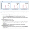
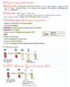
Blood Pressure: Definition

A
2
Q

Normal Blood Pressure Range

A

“will you be so kind to specify under which conditions and where?”

question:

“What is the blood pressure in the temporal artery?”

Answer:

“I can not say specifaclly because i do not know your systemic pressure. i can make an estimaion based on the fact that you look healthy and by using the approximite distance of the temporal artery from your heart.”

3
Q

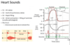
Systolic BP definition

A

Systolic: The highest pressure anywhere (where there is a pulsation) during the cardiac cycle

4
Q

Siastolic BP Definition

A

Diastolic: The lowest arterial pressure during the relaxation of the left ventricle

5
Q

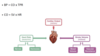
Cardiac Output Definition

A

Volume of blood pumped from each ventricle per minute

6
Q

CO equations

A
7
Q

Stroke Volume: definition, equations, factor that affect it

A
8
Q

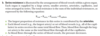
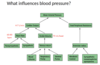
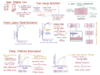
What factors influence blood pressure (scheme)?

A
9
Q

Preload definition:

A
  • The extent to which heart muscle fibers are stretched before the onset of systole
  • This depends greatly on the end-diastolic ventricular volume (EDV): volume load created by blood entering ventricles at end of diastole before contraction
  • It is not possible to measure preload in vivo, but we can use the EDV as an indicator
10
Q

Factors affecting Preload

A

**add

11
Q

Afterload Definition

A

•The extent to which heart muscle fibers are stretched during systole/ejections

…The ressistance ventricles must overcome during systole

12
Q

Factors affecting Afterload

A
13
Q

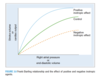
Frank-Starling Law: definition

A

Definition: a law that describes the relationship between end-diastolic volume and cardiac stroke volume

  • Cardiac contractility is directly related to the wall tension of the myocardium.

An increase in end-diastolic volume (preload) will cause the myocardium to stretch (↑ end-diastolic length of cardiac muscle fibers), which increases contractility (↑ force of contraction) and results in increased stroke volume in order to maintain cardiac output.

This relationship between end-diastolic volume and stroke volume is shown in the Frank-Starling curve.

Aim: maintain CO by modulating contractility and SV

↑EDV= ↑STRECH= ↑CONTRACTILITY= ↑SV= ↑CO

14
Q

Diffrence in systemic and pulmonary circulation: pressure, resistance, compliance

A
15
Q

Compliance Deffinintion:

A

?

16
Q

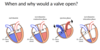
Geeral stages of the cardiac cycle

A
17
Q

When and why do the hears valves open?

A
18
Q

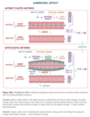
Relative proportion of blood volume throughout the cardiovascular system:

A
19
Q

What is Pressure Pulse:

A

is the difference between the systolic and diastolic pressures.

Pulse pressure = P(systolic) - P(diastolic)

■   The most important determinant of pulse pressure is stroke volume. As blood is ejected

from the left ventricle into the arterial system, arterial pressure increases because

of the relatively low capacitance of the arteries. Because diastolic pressure remains

unchanged during ventricular systole, the pulse pressure increases to the same extent

as the systolic pressure.

■■   Decreases in capacitance, such as those that occur with the aging process, cause

increases in pulse pressure

20
Q

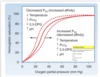
Mean Arterial Pressure

A

■■   is the average arterial pressure with respect to time.

■■   is not the simple average of diastolic and systolic pressures (because a greater fraction

of the cardiac cycle is spent in diastole).

■■   can be calculated approximately as diastolic pressure plus one-third of pulse pressure.

* to know the exact vaule we must do an integral of the graph (area under the graph)

21
Q

Ejection Fraction deffinition:

A
22
Q

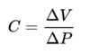
What is arterial compliance?

A

Compliance is the ability of a hollow organ (vessel) to distend and increase volume with increasing transmural pressure

* In compliance, an increase in volume occurs in a vessel when the pressure in that vessel is increased. The tendency of the arteries and veins to stretch in response to pressure has a large effect on perfusion and blood pressure. This physically means that blood vessels with a higher compliance deform easier than lower compliance blood vessels under the same pressure and volume conditions.[1] Venous compliance is approximately 30 times larger than arterial compliance.[2] Compliance is calculated using the following equation, where ΔV is the change in volume (mL), and ΔP is the change in pressure (mmHg):
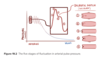
23
Q

Windkessel effect

A

Windkessel effect is a term used in medicine to account for the shape of the arterial blood pressure waveform in terms of the interaction between the stroke volume and the compliance of the aorta and large elastic arteries (Windkessel vessels) and the resistance of the smaller arteries and arterioles.

24
Q

Distribution of blood flow ( as a % of CO)

A

..

25
Blooc pressure values inside the heart chambers
26
Blood pressure regulation: classifications
* Myogenic —\> stretch reflex of the smooth muscles in the vessel walls * Neural (Short Term) * Humoral (Long Term - RAAS)
27
Heart Sounds
28
Pulse wave velocity (PWV)
* Pulse: alternate expansion and recoil of elastic arteries due to a travelling pressure wave after each ventricular systole * PWV: The velocity at which bp pulse propagates through the an artery or combined length of arteries * Clinical: Measure of arterial stiffness (due to biological ageing and arteriosclerosis) —\> arteries become less compliant * Gold Standard: Palpation of carotid artery and femoral artery at the same time —\> femoral will cause a pulse pressure wave later that the carotid artery
29
Measurments of blood pressure: Classifications
a) Indirect = Non-invasive • Auscultation • Palpation • Oscillatory b) Direct = Invasive • Catheterisation
30
BP measurment: Auscultatory Method
31
BP measurment: Palpation and Oscillatory meathod
32
Influence of gravity on blood content in blood vessels
?
33
34
What is the effect of body position on heart rate?
?
35
central venous pressure
Central venous pressure (CVP) is the blood pressure in the venae cavae, near the right atrium of the heart. CVP reflects the amount of blood returning to the heart and the ability of the heart to pump the blood back into the arterial system. CVP is often a good approximation of right atrial pressure (RAP),[1] although the two terms are not identical, as a pressure differential can sometimes exist between the venae cavae and the right atrium. CVP and RAP can differ when arterial tone is altered.
36
Determination of central venous pressure
37
How does HR and BP change after exercise:
you need to ask: - What type of excersize? what happens to the bp: when? during the beggining? middle? end? - where are we measuring the BP? During what part? The diastolic pressure can decrease a little bit (due to vasodilationin the muscle- increased metabolic request)
38
Important equations: Blood flow Resistance CO SV Ejection fractions PP MAP
39
Important Pressure Values
40
Pressure Value: Pulmonary Vein
Pulmonary Vein = 2-15mmHg
41
Pressure value: Left Atria
Left Atria = 0-10mmHg
42
Pressure Value: Left Ventricles
Left Ventricles = 0-120mmHg | (250/unchanged during excersize)
43
Pressure Values: Aorta
Aorta = 80-120mmHg
44
Pressure Values: Systemic Capillaries
Systemic Capillaries = 15-35mmHg (μ = 17mHg) (Note: exception is Renal capillary) * Venule side = 10-15mmHg * Arteriole Side = 35mmHg
45
Pressure Values: Vena Cava
Vena Cava = 0-5mmHg
46
Pressure Values: Right Atria
Right Atria = 0-5mmHg
47
Pressure Values: Right Ventricle
Right Ventricle = 0-25mmHg
48
Pressure Values: Pulmonary Artery
Pulmonary Artery = 8-25mmHg
49
Pressure Values: Pulmonary Capillaries
Pulmonary Capillaries = 7mmHg
50
Pressure Values: Renal Capillaries
Renal Capillaries = 55mmHg
51
Pressure Values: Renal Vein
Renal Vein = 10-15mmHg
52
Pressure Values: Hepatic Artery
Hepatic Artery = 90mmHg
53
Pressure Values: Portal Vein
Portal Vein = 5-10mmHg (Note this is close to 0mmHg as venous system)
54
Length–tension relationship in the ventricles
■   describes the effect of ventricular muscle cell length on the force of contraction. ■   is analogous to the relationship in skeletal muscle. 1. Preload ■■   is end-diastolic volume, which is related to right atrial pressure. ■■   When venous return increases, end-diastolic volume increases and stretches or lengthens the ventricular muscle fibers (see Frank-Starling relationship, IV D 5). 2. Afterload ■■   for the left ventricle is aortic pressure. Increases in aortic pressure cause an increase in afterload on the left ventricle. ■■   for the right ventricle is pulmonary artery pressure. Increases in pulmonary artery pressure cause an increase in afterload on the right ventricle.
55
Changes in the Ventricular-volume loop during increased: preload/afterload/contractility
56
Velocity of Blood flow
• The velocity of blood flow is the rate of displacement of blood per unit time. (Linear velocity)
57
Blood flow
58
Vascular resistance
Definition: resistance offered by the circulatory system that must be overcome to create blood flow (R = ΔP / Q)
59
Poiseuille equation
60
Resistance of vessels in parallel
61
Resistance of vessels in series
62
Laminar flow versus turbulent flow
63
Shear
64
Compliance
65
Windkessel Effect
66
67
What does blood pressure of value 0 mean?
0= pressure that is equal to the atmospheric pressure
68
Where can we find blood pressure that is equal to 0?
RA= LA= LV= diastol Majority of the venous system
69
Why arent we measuring the flow of blood instead of blood pressure?
* Its hard and expensive to measure (example: dopler system) * need a qualidied technishion BP: cheap, non invasive, easy
70
What is the BP of the glomerulus?
=55mmHg The glomerular filtration rate The rate at which kidneys filter blood is called the glomerular filtration rate. The main driving force for the filtering process, or outward pressure is the blood pressure as it enters the glomerulus. This is counteracted to some extent by inward pressure due to the hydrostatic pressure of the fluid within the urinary space, and the pressure generated by the proteins left in the capillaries that tend to pull water back into the circulatory system (colloidal osmotic pressure). The net filtration pressure is the outward pressure minus the inward pressure.
71
BP Vasa recta?
72
How to regard BP with relation to gravity at the level blow and above the heart?
* hydrostatic pressure and gravity * murcury is 13 time more dense than blood.... * What are the claculations? * for 1 cm below the heart there is a 0.77nnHg rise in BP
73
Blood groups: defintion
* Definition: Categorisation of blood types by presence or absence of specific antigens * Antigens found = Agglutinogens * Antibody to agglutinogens = Agglutinins
74
Importance of knowing blood types:
; transfusion, organ transplantation and certain pregnancies
75
ABO Blood groups:
76
Rh blood group + incompability
* There are six common types of Rh antigens, each of which is called an Rh factor: * C D E, c d e * A person with C antigen won't have c antigen, and person without C antigen will surely have c antigen (same for D-d and E-e). * The type D antigen is widely prevalent in the population and considerable more antigenic than the other Rh antigens. * Anyone who has this type of antigen is said to be Rh positive, whereas a person who doesn't have type D antigen is said to be Rh negative. * 85% of white people are Rh positive (D), 15% Rh negative (d). * Antibodies do not occur naturally in the Rh system (unlike the AB0 system). * This means that there are no antibodies to the D antigen in the body of an Rh-negative individual. * They can form in the body in the event of prolonged contact with Rh-positive blood. Rh incompatibility blood transfusion * When RBCs containing Rh factor (D) are injected into a person whose blood doesn't contain the Rh factor (into a Rh negative person) anti-Rh agglutinins develop slowly * If a Rh-negative person has never before been exposed to Rh-positive blood, transfusion of Rh+ blood into this person likely cause no immediate reaction, but anti-Rh antibodies can develop in sufficient quantities during the next 2-4 weeks to cause agglutination of the transfused cells which will then hemolyzed by the macrophage system. * Thus, a delayed transfusion reaction occurs, usually mild. * Upon subsequent transfusion of Rh-positive blood into the same person, the transfusion reaction is greatly enhanced and can be immediate and severe.
77
Rh incompatibility of mother and fetus
78
79
80
81
82
83
84
85
86
87
88
89
90
91
92
93
94
95
96
97
98
99
100
101
102
103
104
105
106
107
108
109
110
Bleeding time: Clotting time: Prothrombin time: Activated partial thrombin time:
111
Rh vs ABO
112
aggulation test:
113
114
115
116
117
118
119
120
121
122
123
124
125
126
127
128
129
130
131
132
133
134
135
136
137
138
139
140
141
142
143
144
145
146
147
148
149
150
151
152
153
154
I think it is yellow, but not suree
155
156
157
158
159
160
Formed Blood Elements Number per μL
161
Hemoglobin general characteristics
1% of oxygen is dissolved in the blood. Each hemoglobin molecule is not strictly bound to 4 molecules of oxygen. Concentration of hemoglobin is 150g/L in women and 160g/L in men. It is less in females due to monthly blood loss through menstruation Always give a range for Hb 130-150g/L and 160-170g/L – BUT CHECK THESE VALUES every book is different Hb storage of Oxygen (1L) Talk about 75% deoxygenated blood Variable in the case of fetal hemoglobin, embryonic hemoglobin and some pathological states such as Sickle cell anemia, HbE etc. FETAL Hb à (37 AAs residues different from those of Beta chains.) CO2 binds to the amine group in the chain Whereas O2 binds to the iron along with CO, compete
162
Types of Hemoglobin
163
\> Oxygen- Hemoglobin dissociation curve
* Saturation in arterial blood is around 100% (97.5% Guyton) * Saturation in venous blood is around 75% * Not a straight line as initial oxygenation of one of the hemoglobin subunits will cause an increased affinity for oxygen in the other hemoglobin subunits (T configuration à R configuration) * Sigmoidal shape: COOPERATIVE BINDING * Cooperative bind is the binding of the first molecule of oxygen increases the chances of binding/ affinity for the next molecules of oxygen due to a conformational change in the Hb molecule. * Hence Hb affinity for oxygen increases as more molecules of oxygen bind. More molecules of oxygen bind as the partial pressure of oxygen increases until a maximum value is obtained (i.e. when Hb is fully saturated). * When this value is obtained the curve flattens out (plateaus) as very little additional oxygen is able to bind. * The plateau portion of the curve exists in the pulmonary capillaries. * At higher partial pressures (towards the right side of the curve) the curve is not as steep which means the oxygen content (Hb saturation) does not change drastically despite large changes in oxygen partial pressure. * In the middle part of the curve (steepest area) this is where gaseous exchange takes place in the systemic capillaries. At this point on the graph the oxygen is being unloaded to respiring tissues and as a result hemoglobins affinity to oxygen decreases. A small change in oxygen partial pressure leads to a large difference in the hemoglobin saturation hence a release in large amounts of oxygen for respiring cells. * P50: the conventional measure of heamoglobin affinity for oxygen. It is calculated using the graph as you simply just record what the oxygen partial pressure is at 50% heamoglobin saturation. * Left shift? Increased affinity for oxygen à more loading of oxygen à Fetal Hb * Right shift? Decreased affinity for oxygen à more unloading of oxygen à Exercise * CADET = CO2, acidity, 2,3 DPG, exercise & temperature * FETAL HB: They don’t have beta-chains therefore pockets wide therefore o2 retained by Hb/ can be caught more easily, which is important so it can take up o2 from placental circulation. * Temperature= increased temperature is able to weaken/denature the bond between the Ferrous iron and the O2 molecule (only really evident with extreme changes in temperature) * pH= Bohr effect, it is observed that deoxygenated hemoglobin is better at buffering (binding H+ ) therefore there is a shift towards this form * 2,3 biphosphoglycerate= glycolysis by-product of erythrocytes (anaerobic respiration), it promotes the release of oxygen to the tissue, in hypoxic conditions there is an overall increase in 2,3 BPG production. It holds the 2 beta globin chains and cross links them therefore pockets become narrower and o2 jumps out of Hb. * CO2 = Haldane Effect, deoxygenated hemoglobin is better at binding CO2 than oxygenated hemoglobin, this normally occurs at the same time as the Bohr effect as increased CO2 will lead to a decrease in pH too
164
Bohr Effect
165
Hemoglobinometry
166
Colorimetry
167
168
The principle of Drabkin's solution:
Drabkin's Reagent is used for the quantitative, colorimetric determination of hemoglobin concentration in whole blood at 540 nm. Drabkin's Solution reacts with all forms of hemoglobin except sulfhemoglobin, a pigment that normally occurs in only minute concentrations in blood. * Drabkin′s reagent is used in measuring haemoglobin from blood samples. * It comprises potassium ferricyanide, potassium cyanide and potassium dihydrogen phosphate as components. * Potassium ferricyanide **oxidizes** haemoglobin to methemoglobin and then to cyanmethemoglobin.