Lab 5: Histology Flashcards
(39 cards)
What is the level of the duodenum in relation to the spinal column?
L1 to L4
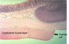
Describe taeniae coli
- 3 longitudinal ribbon of smooth muscle
- Thickened area of smooth muscle
- Located only on the large intestine
- One on the ascending colon
- Another on the descending colon
- Finally on the sigmoid colon

Name these different parts of the small and large intestine?


Describe mesocolon?
The peritoneal process attaching the colon to the posterior abdominal wall.
Ascending, descending, or transverse, according to the portion of the colon to which it attaches.
Fans out from their root regions

What is the difference between Mesocolon and mesentery?
Mesentery is the membrane that attaches the intestines to the wall of the abdomen, maintaining their position in the abdominal cavity, and supplying them with blood vessels, nerves, and lymphatics.
Mesocolon is the part of the mesentery that attaches the large intestine to the abdominal wall
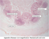
Describe appendices epiploicae?
They are small pouches of the peritoneum filled with fat and situated along the large intestine, but are absent in the rectum

Describe haustrations?
Small pouches caused by sacculation (sac formation) giving the large intestine its segmentated appearance.

What is the colon?
The large intestine
What does the “bowel” refer to?
Intestines
Could be either large or small
Describe Meckel’s diverticulum?
- Outpouches in the lower small intestine
- Congenital abnormalities (i.e. present at birth)
- Descended from left over yolk stalk
- Usually asymptomatic but problems may arise e.g. inflammation.
- Each has an opening to the colonic lumen through a narrow neck

Describe Plicae circulares?
The numerous permanent crescentic folds of mucous membrane found in the small intestine especially in the lower part of the duodenum and the jejunum

What regions of the GI tract are descended from the midgut?
Extends from the major duodenal papilla to the proximal 2/3 of the transverse colon.
What regions of the GI tract are descended from the hindgut?
Extends from the distal 1/3 of the transverse colon to the upper anal canal.
Which blood supplies the midgut and hindgut?
Midgut: supplied by the superior mesenteric artery (structures extending distal from the major duodenal papilla to the proximal 2/3 of the tranverse colon).
Hindgut: supplied by the inferior mesenteric artery (structures extending from the distal 1/3 of the transverse colon to the upper anal canal)
What is the major anatomical distinction between the structures that are descended from the midgut and hindgut?
Supplied by different blood supplies.
Midgut is supplied by the superior mesenteric artery.
Hindgut is supplied by the inferior mesenteric artery.
What are the major differences in mucosa between the large and small intestine?
- The muscularis externa of the large intestine is different from that of the small intestine in that the outer longitudinal layer of smooth muscle varies in thickness and forms three thick longitudinal bands, the taeniae coli.

Describe the histology of the taeniae coli?
The taeniae coli is due to the outer longitudinal layer of smooth muscle varying in thickness. Forming three thick longitudinal bands, the taeniae coli.
Name these different parts of the intestines on a radiology?


Is this a section for a large or small intestine and why?

Small intestine
Why? villi and plicae circulares present
Is this a section for a large or small intestine and why?

Large intestine
Why? no villi present, invaginations present down from the “true surface” of the mucosa
Indicate the true surface of the mucosa in this image?

Image of the small intestine- as the “true surface” appears lower because the villi are projections from the true surface

Indicate the true surface of the mucosa in this image?

Appears invaginations: these are not villi but intestinal glands (colon crypts)

Name these parts of the small intestine on the histology


Where are brunner’s glands located?
In the duodenum only