Lecture Review Flashcards

(161 cards)

1
Q

What parasite is this image associated with?

A

Babesia

(presence of tetrad)

2
Q

What specific parasite is this associated with?

A

Plasmodium Falciparum

(Banana gametocyte)

3
Q

ID the parasite this image is associated with

A

Acid fast of Cryptosporidium oocysts

4
Q

ID the parasite this image is associated with

A

Fluoresence of Cryptosporidium Oocysts

5
Q

What parasite is this?

A

Giardia

6
Q

What organism is this associated with?

A

This is a cyst for Giardia

7
Q
A

Trichomonas Vaginalis

8
Q

What is this and what is it associated with?

A

Amastigote

Intracellular form of Leishmania/Trypanosomiasis found in human

9
Q

This is the result of what?

A

Cutaneous Leishmaniasis

10
Q

What is this from?

A

DFA (fluoresence) of Pneumocystis Jiroveci (Carinii)

11
Q

What is this?

A

Silver Stain of Pneumocystis Jiroveci (Carinii). Shows empty cysts which have expelled the protozoa.

12
Q

Interpret this Serology:

HBsAg: negative

anti-HBc: negative

anti-HBs: negative

A

Susceptible

13
Q

Interpret this Serology:

HBsAg: negative

anti-HBc: positive

anti-HBs: positive

A

Immune due to natural infection

14
Q

Interpret this Serology:

HBsAg: negative

anti-HBc: negative

anti-HBs: positive

A

Immune due to hepatitis B vaccination

15
Q

Interpret this Serology:

HBsAg: positive

anti-HBc: positve

IgM anti-HBc: positive

anti-HBs: negative

A

Acutely infected

16
Q

Interpret this Serology:

HBsAg: positive

anti-HBc: positve

IgM anti-HBc: negative

anti-HBs: negative

A

Chronically infected

17
Q

Interpret this Serology:

HBsAg: negative

anti-HBc: positve

anti-HBs: negative

A

Interpretation unclear! (4 possibilities)

  1. Resolved infection (most common)
  2. False positive anti-HBc (thus susceptible)
  3. “Low level” chronic infection
  4. Resolving acute infection
18
Q

What liver disease is this associated with?

(hint: these are plasma cells)

What else might you see?

A

Autoimmune hepatitis

This image shows plasma cells creeping into the lobule. You would also expect to see interface hepatitis.

19
Q

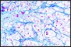
Positive blue staining for iron in liver as in the image, is indicative of what disease process?

A

Hemochromatosis

A disease of iron overload

20
Q

What is the phenomenom in the bottom left photo called and what is it associated with?

A

“Scalloped edges”

Celiac Disease

21
Q

What is pyloric stenosis and what are its symptoms (3)?

A

Congenital hypertrophy of the smooth muscle of the pylorus

Sx: projectile vomiting in first 2-6 weeks of life, visible peristalsis, olive-like mass in abdomen.

22
Q

What is the most likely cause for acute gastritis? chronic?

A

acute: impairment of protective system (via NSAIDs, direct injury, ingestion, etc.)

chronic: H. Pylori

23
Q

What stains are useful for highlighting H. pylori (2)?

A
  • methylene blue stain
  • Warthrin-starry stain
24
Q

Describe the pathogenesis of Autoimmune chronic gastritis and what occurs as a result.

Is the risk of adenocarcinoma affected?

A
  • There are antibodies to parietal cells and IF
  • Antrum is spared
  • Results in B12 deficiency (IF loss) and resulting anemia
  • Also decreased pepsinogen (acid) levels (chief cells loss)
  • Increased risk of gastric adenocarcinoma
25
What are the symptoms and _acute_ PUD? _Chronic_? Describe the ulcers in each case.
_Acute_: * N/V w/ coffee-ground hematemesis * Ulcers: \<1cm and sharply demarcated _Chronic_: * Epigastric burning or aching that is worse at night * Relieved w/ akali or food * Nausea, bloating, belching * Could be caused by ZE * Ulcers: Usually solitary and sharply punched out
26
What is the relationship between location and prognosis for a carcinoid tumor? What kind of tissue do these tumors arise from?
These tumors arise from neuroendocrine organs (G-cells of stomach) Location determines prognosis: 1. Foregut is resectable 2. Midgut is most aggresive 3. Can also be located in the hindgut
27
Gastrointestinal Stromal Tumor (GIST) Pathogenesis? What does it look like? Treatment?
* Mesenchymal neoplasm of interstitial cells of cajal * 75-80% w/ GOF mutation in gene encoding tyrosine kinase c-KIT * Solitary, well circumscribed, fleshy, submucosal mass * Tx: Surgical resection (if possible); Imatinib (there is often resistance)
28
What are the (3) general causes for Gastric Adenocarcinoma?
1. H. Pylori 2. EBV 3. Mutation
29
Describe the (2) types of mutation which can lead to Gastric Adenocarcinoma. What are the mutations? What type of adenocarcinoma do they cause? What are the differences in the tumor types and sexes affected? Other key points w/ treatment and clinical findings?
_Diffuse Type_ * Caused by mutation of CHD1 causing **E-cadherin** fxn loss * Infiltrative growth and discohesive cells w/ large mucin vacuoles (signet ring cells) * Equal incidence between men and women * Linitis plastica (thick stomach wall) _Intestinal type_ * Familial **APC** mutations * Bulky, glandular, exophytic mass or ulcerated tumor * Men twice as common * Tx: Surgical resection (**chemo is _not_ effective**) *
30
Intestinal type _M__etastatic_ Gastric Adenocarcinoma is called what and is located where? Diffuse type?
_Intestinal_: Sistery Mary Joseph nodule- Mets to Periumbilical region _Diffues_: Krukenberg tumor- Mets to Bilateral ovaries
31
What type of muscle makes up the upper 2/3rds of the esophagus? lower 1/3rd?
Striated = upper smooth= lower
32
What is _achlasia_ and what is one of the key secondary etiologies? Pathogenesis? How does it look on barium swallow?
* Incomplete relaxation of LES and disordered motility of the esophagus (anti-peristalsis); Esophageal dilatation * Pathogenesis: destruction of myenteric plexus * Birds beak deformity on barium swallow Associated w/ **_Chagas disease_**
33
Boerhaave syndrome What is it and what is a key finding associated?
**Transmural** esophageal rupture from severe vomiting Very high morbidity/mortality, so always a _medical emergency_ Associated with Subcutaneous emphysema of skin (rice krispy feeling when you press on patient's skin)
34
What are the (3) M's of Herpes Simplex virus associated with one of the forms of esophagitis?
Multinucleation, Molding and Margination
35
All forms of Esophagitis show what (3) histologic changes?
1. Basal layer hyperplasia (\>20% of mucosal thickness) 2. Lamina Propria papillae which reach top 1/3rd of mucosa 3. Intra-epithelial eosinophils
36
Name the corresponding LNs that SCC in each of the following parts of the esophagus spreads to: Upper 1/3rd Middle 1/3rd Lower 1/3rd
Upper: Cervical LNs Middle: Mediastinal/ tracheobronchial LNs Lower: Celiac/gastric LNs
37
Name the Vibrio Cholerae exotoxin responsible for causing disease.
AB Type ADP-ribosylating toxin
38
What level of inoculum is needed for a campylobacter infection?
Low! As low as 500 organisms
39
Clinical manifestation of Campylobacter Jejuni What disease is it affliated with?
Fever (1 day) followed by sever abdominal pain and diarrhea w/ blood, pus and campy in feces Associated w/ Guillain-Barre Syndrome (ascending paralysis)
40
What are the roles of Bab A, Vac A and Cag A, in h.pylori infection?
_Bab A:_ Used for adherence. Binds to lewis blood group antigens _Vac A_: Vacualiting cytotoxin that inserts into epithelial cell membranes forming nutrient releasing poor. Also inserts into mitochondria to release cytochrome C and induce apoptosis. _Cag A_: Functions to promote cytokine production
41
Volvulus Where does it tend to happen for children? adults?
Twisting of the bowel around its mesentery. Leads to obstruction and infarction. Tends to be midgut for children and sigmoid for adults.
42
Meckel's diverticulum
Persistence of the omphalomesenteric duct. Known as the disease of "2's" (2% of pop.; 2x likely for men; 2 main complications- pain w/ inflammation and hemorrhage w/ ulcer)
43
Hirschsprung Disease
Form of megacolon in which the absence of ganglion cells (enteric neurons) leads to premature death of neural crest cells migrating from cecum to rectum. Mostly in males.
44
What effect does pancreatic insufficiency have on neutral fat? D-xylose absorption test?
* Increased neutral fat * Normal D-xylose absorption test (urinary excretion)
45
Abetalipoproteinemia
Decreased synthesis of apo B which results in decreased ability to generate chylomicrons.
46
What is the skin issue associated with Celiac disease? How do we diagnose this disease (be specific)?
* Dermatitis herpetimormis (skin blistering disease) * Diagnosis via: 1. _Biopsy_: Villous atrophy w/ inc. intraepithelial lymphocytes (CD8) 2. _Serotype_: anti-TTG, anti-deaminated gliadin, anti-endomysial antibodies (If pts are IgA deficienct, IgG tests are needed)
47
Both collagenous and lymphocytic colitis cause what clinical presentation?
Chronic watery diarrhea (3-20 non-bloody stools per day)
48
What is the pathogenesis and clinical presentation in whipple disease?
_Pathogenesis_: Tropheryma whippleli bug is engulfed by macrophages _Sx_: Malabsorption, lymphadenopathy (due to congestion of lymph vessels w/ macrophages), and arthritis
49
What are the key differences between _Crohn's Disease_ and _Ulcerative Colitis_, in terms of... Pt. Demographic? Location? Gross mrophology? Microscopic changes? Complications? Intestinal Manifestation? Key Extraintestinal manifestations?
_Crohn's_: White Jewish folks, with _non-bloody_ diarrhea, cobblestoning, skip lesions throughout GI tract, and strictures _UC_: Wealthy western folks, with _bloody_ diarrhea, pseudopolyps limited to the colon, and megacolon
50
Juvenile polyps and malignancy
Mostly occur sporadically in rectum of children \< 5y/o If _single_, no malignant potential. If _multiple_, increased risk of adenocarcinoma.
51
Hamatomatous (Peutz-Jeghers syndrome) Pathophysiology? Presentation? Malignant potential?
* Multiple non-malignant hamartomas throughout the GI tract * Sx: Hyperpigmented melanotic macules of mouth, lips, genatalia and hands * Increased CRC risk (although the polyps themselves have _NO_ malignant potential)
52
What are adenomatous polyps and what are they precursors to? What are the different types (2) and which is more likely to be dangerous?
These are benign polyps that are precursors to the majority of colorectal adenocarcinomas. There are peduculated types and sessile types. Sessile is more likely to grow to cancer.
53
Where do sessile serrated adenoma occur? What is the main mechanism for their pathogenesis?
Right colon DNA mismatch repair
54
Familial Adenomatous Polyposis What is it and what does it lead to? What causes it?
It is a condition in which patients develop 100-1000 polyps, due to an autosomal dominant defect in the _APC_ gene (Ch5q21) 100% development into colorectal adenocarcinoma
55
What are the (2) variants of FAP? Describe them.
**Gardner syndrome**- same as FAP + osteomas (mandible, skull, and longbones), epidermal cysts, desmoid and thyroid tumors, and dental abnormalities **Turcots syndrome**- Intestinal adenomas + tumors of CNS which depend on the the type of mutation (APC: medulloblastoma/ repair mutation: glioblastoma)
56
Hereditary nonpolyposis colorectal cancer (Lynch syndrome) What is it and what causes it?
Associated w/ FAP but fewer number of polyps. Cancer occurs at a younger age than sporodic cancers. Caused by mutation in DNA mismatch repair genes
57
What are the (2) main molecular pathways to CRC and what specific conditions are they each associated with?
1. _APC/WNT pathway:_ APC is a neg. regulator of **Beta-Catenin**. Double knockout of APC increases B-catenin, which translocates to the nucleus and activates transcription of **MYC, Cyclin D1,** etc. Associated w/ **FAP and sporodic colon cancer.** 2. _DNA Mismatch Repair_: spelling error in coding region. Affect on MLH, MSH, and PMS genes. Associated w/ **HNPCC**
58
Right sided vs Left sided CRC
**Right**: asymptomatic for longer because colon diameter is larger. First symptom for right side is anemia due to blood loss/ulceration **Left**: smaller diameter so obstruction occurs much quicker (changes in BM habits). Tend to see napkin ring lesion.
59
What cancer type is most commonly found in the rectum? The anus?
**Rectum**: adenocarcinoma **Anus**: SCC
60
Name/describe the (3) Neoplasm types associated w/ the appendix
1. _Mucocele_: benign dilatation o lumen by mucinous secretion 2. _Mucinous cystadenoma_: proliferation of benign neoplastic cells and dilation which may rupture 3. _Mucinous cystadenocarcinoma_- invasion of neoplastic cells
61
Pseudomyxoma peritonei
Distention of peritoneal cavity by the presence of semisolid mucin and epithelial mucin producing implants and/or malignant cells. _Possible complication of mucinous tumor of the appendix_.
62
What separates the upper GI system from the lower GI system?
Ligament of Triez
63
Hematochezia
Bright Red Blood Per Rectum (BRBPR) Usually denotes LGI bleed but could also be UGI bleed
64
What are the top (4) differentials for an UGI bleed?
1. PUD 2. Esophageal varices 3. Erosive esophagitis 4. Mallory-Weiss tears
65
Nasogastric Aspiration What counts as a (+) result? What counts as a (-) result? WHat result types are indeterminant?
(+): blood, +/- bile (-): bile, no blood Indeterminate (fake negative): no blood _and_ no bile EGD usually ends up being done anyway.
66
When dealing w/ a UGI bleed, what is the usefullness of a pre-endoscopic IV PPI? post-endoscopic IV PPI?
_Pre-endoscopic IV PPI:_ **No impact** on rebleeding, mortality, or need for surgery. **Does** accelerate resolution of bleeding and decreased need for endoscopic therapy. _Post-endoscopic IV PPI_: **Very effective** for decreasing likelihood of rebleeding, need for surgery and mortality
67
What are the top (4) differentials for LGI bleeding?
1. Diverticulosis 2. Colitis (Ischemic, IBD, Radiation) 3. Hemorrhoids 4. Postpolypectomy
68
What are the (3) different ways to define diarrhea?
1. High stool frequency (\>3/day) 2. High quantity of stool (\>200g of stool/day) 3. Abnormally loose stool
69
What are the special populations to keep an eye out on if they hve diarrhea?
1. Elderly 2. Immunocompromised 3. IBD pts 4. Pregnant pts
70
How common are infectious causes of chronic diarrhea?
Uncommon for immunocompetent people. Consider giardia if patient is exposed to young children or in contact w/ lakes/streams
71
What are the (5) classifications for chronic diarrhea?
1. Secretory 2. Osmotic 3. Steatorrhea 4. Impaired Mobility (less important) 5. Inflammatory
72
What is the most common cause for Steatorrhea?
Small intestine bacterial overgrowth! (Can also occur due to celiac, whipples, etc.)
73
What are the (5) ways to define constipation?
1. Straining during defecation 2. Passage of lumpy or hard stool 3. Sensation of incomplete defecation 4. Use of manual maneuvers to facilitate BM 5. Freq. of less than (3) BMs per week
74
What are the general alarm signs associated with constipation? (5)
1. Age \> 50 2. New onset in the elderly 3. Weight loss 4. History of cancer (personal and family) or radiation or resection 5. Palpable mass on ecam
75
What are the criteria to qualify for IBS?
1. Recurrent abdominal pain 3x/mo 2. 2 or more of the following: * Improvement with defecation * Change in freq. of stool * Change in appearance of stool (No further testing unless alarm symptoms present)
76
What is the stool pattern for IBS? How does this impact treatment?
Could be either constipation or diarrhea. Treatment is based on the stool pattern. _Constipation_- psyllium/miralax _Diarrhea_- loperamide
77
80% of acute pancreatitis cases are associated with... (2 things)
Biliary tract disease of alcoholism
78
What is the clinical presentation for acute pancreatitis? (6)
1. Abdominal pain 2. **Elevated plasma amylase/lipase** 3. Hypocalcemia 4. ARDS 5. DIC 6. Fluid sequestration
79
Predisposing factors for chronic pancreatitis?
1. Longterm alcohol abuse 2. Long standing obstruction 3. CF 4. Idiopathic
80
5 key symptoms/presentations associated with Chronic Pancreatitis
1. Recurrent attacks of abdominal pain 2. Recurrent attacks of jaundice or vague indigestion 3. Exocrine pancreatic insufficiency (Steatorrhea) 4. Endocrine pancreatic insufficiency (DM) 5. Pancreatic calcifications of imaging
81
What is the precursor lesion for Pancreatic Carcinoma (aka infiltrating ductal adenocarcinoma of the pancreas)
Pancreatic Intraepithelial Neoplasia (PanIN) (caused by mutations in K-RAS, p16 and p53)
82
Most common location for pancreatic carcinoma (head, body, tail, or diffuse?) What basic symptom often separates the (3) types?
Head of the pancreas is most common Leads to obstructive jaundice (unlike the others which tend to be clinically silent)
83
Desmoplastic response
Proliferation of dense stromal fibrosis in response to a pancreatic tumor
84
What is one of the most common symptoms associated w/ pancreatic carcinoma, and why?
Pain because it often invades into adjacent nerves
85
Key tumor markers for pancreatic carcinoma
Elevated CA19-9 and CEA
86
Trousseau Sign
Migratory thrombophlebitis (Spontaneously appearing and disappearing venous thromboses which occur in 10% of Pts with pancreatic carcinoma.)
87
Key points about serous cystadenoma (Behavior, gross morphology, histopath, key clinical points)
Benign, small cysts, cuboidal cells, non-specific symptoms
88
Key points about Mucinous Cystic Neoplasms (Behavior, gross morphology, histopath, key clinical points)
Almost exclusively **women**, could be benign or malignant, not connected to main pancreatic duct, lined by columnar cells, **Body/Tail** pancreas
89
Key points about Intraductal papillary mucinous neoplasms (IPMN) (Behavior, gross morphology, histopath, key clinical points)
Benign to malignant, arise in main pancreatic duct/major branch, lined by columnar cells, Men \> women, **Head of Pancreas**
90
The Sand fly serves as a vector for what? Tsetse fly? Triatomid bug?
_Sand fly_- Leishmania _Tsetse fly_- African trypanosomiasis (T. brucei gambiense and rhodesiense) _Triatomid bug_- American trypanosomiasis (T. Cruzi)
91
What kind of bilirubin is detected in bilirubinuria? What are the causes (common and rare)?
Conjugated bilirubin (unconjugated is water insoluble). _Hepatocellular disease_ is the most common causes, but Dubin-Johnson and Rotor Syndrome can also cause it (due to inability of hepatocytes to secrete conjugated bilirubin into the bile).
92
What is the etiology of primary Biliary Cirrhosis? What tests would we run and what would we expect?
It is autoimmune + antimitochondrial antibody, inc. alkaline phosphatase, and inc. total bilirubin (advanced disease)
93
What is the key histological finding for Primary Biliary Cirrhosis? Describe it.
Florrid duct lesion Periductal inflammation w/ granuloma formation and duct destruction. Indicitive of lymphocyte infiltrate.
94
What is Primary Sclerosing Cholangitis and what disease is it associated with? What does it progress to?
_Onion skinned_ pattern of obliterative fibrosis of intrahepatic and extrahepatic bile ducts w/ inflammation and dilation of preserved segments (**beading**). Progreses to end stage liver disease.
95
GIlbert's Syndrome
Isolated elevation of total and direct bilirubin after physical activity in particular, due to genetic defect in UGT (so can't conjugate). Mostly in men. No follow-up needed.
96
Courvoiser sign
States that in the presence of an enlarged gallbladder which is nontender and accompanied with mild jaundice, the cause is unlikely to be gallstones.
97
What signs/ tests make you think a patients hyperbilirubinemia is intrahepatic? Extrahepatic?
**Hepatocellular**: * Transaminases elevated \> 700 * Dec. serum albumin * Alkaline phospatase only 1.5-2x normal * Thrombocytopenia **Obstructive:** * Transaminases ( * Alkaline phosphatase 5-10x normal
98
What key molecules are absorbed in the duodenum? In the Ileum?
**Duodenum**: Calcium and Iron **Ileum**: Vitamin B12 and Bile salts Everything else is the jejunum (90% of absorption)
99
Colonic Salvage
Bacteria act on carb/fiber to produce short-chain fatty acids and the colon can absorb up to 500 kcal/d as SCFAs/lactate. This is particularly useful in short gut syndrome.
100
What are the (2) Helminths that have the ability to multiply w/in the human host?
1. Strongyloides 2. Hymenolepsis nana (dwarf tapeworm) Can autoinfect and are therefore chronic
101
List the (3) clinical phases of trichinosis spiralis
**Phase I**: * 24-72 hours * Diarrhea, N/V, and abdominal pain **Phase II**: * 2-3 weeks * Fever, myalgia, periorbital edema * CNS/cardiac (major cause of death) **Phase III:** Recovery
102
Clinical presentation of strongyloides stercoralis. Who does it effect?
* Severe diarrhea * Pneumonia * Eosinophilia * Septicemia Effects the immunocompromised
103
What nematodes have microfilariae which circulate in the bloodstream? (2)
1. Wuchereria Bancrofti (Lymphatic Filariasis) 2. Loa loa
104
What is the optimal method for diagnosis for Onchocerciasis (river blindness)?
Skin snips
105
What are the (4) tapeowrms w/ adult stages w/in the human intestine? What foods are they associated w/?
1. Taenia saginata (Beef) 2. Taenia solium (Pork) 3. Diphyllobothrium latum (Fish) 4. Hymenolepsis nana (n/a)
106
How are the shistome species acquired? Where do they develop into adults? Where do they lay eggs?
Schistosome species are acquired from skin exposed to infested water. They develop into adults while in the liver, and then mate. Ova are then produced in the large intestine and passed into the stool.
107
What organism is critical for the schistosoma life cycle?
The snail!
108
Mechanism for human acquisition of infection by Opisthorchis species? Paragonimus westermani? What is another name for each of these species?
**Opisthorchis** (liver fluke)- Undercooked fish w/ cysts **Paragonimus** (lung fluke)**-** Crayfish/crab
109
What is the pathogenesis of swimmer's itch?
Also known as cercarial dermatitis. Accidental infection of human swimmers w/ duck schistosome, which can't penetrate beyond the skin.
110
Wucheria Bancrofti symptoms/presentation (4)
1. Cough from microfilariae in lungs 2. Lymphadenopathy 3. Elephantiasis (long standing lymphedema) 4. Eosinophilia
111
What type of transmits loa loa? wuchereria bancrofti?
loa loa: Deer fly wuchereria bancrofti: mosquito
112
What chromosome is CFTR located on?
Chromosome 7
113
Name the (5) mutation classes for CF. Which is most common and what is the genetic change associated with it?
1. No synthesis 2. No maturation (destroyed before proper packaging) \* most common - F508 mutation 3. Blocked regulation 4. Dec. Cl- conductance 5. Dec. abudance of CFTRs
114
Name the (2) theories of ASL defect associated with CFTR
Airway Surface Liquid 1. ASL low volume (leads to mucus stasis) 2. ASL too salty (salty sweat)
115
What are the GI symptoms of CF?
1. Meconium Ileus (black, tarry meconium) 2. Hypoplastic gall bladder 3. Focal biliary cirrhosis 4. Fatty liver
116
Pancreatic effects of CF
Duct obstruction due to inspissated (thickened) secretions, which leads to dilation, destruction and fibrosis.
117
98% of men with CF are...
Infertile
118
Quantitative pilocarpine iontophoresis is also known as...
A sweat test (for CF)
119
What is the antibiotic that is nebulized for CF patients? What are the mucolytics used by CF patients?
antibiotic: tobramycin mucolytics: rhDNAse
120
What role do potentiators play in CF? What is its name? What are the chaperones names?
Used to deal with class 3 CF by shifting the CFTR to proper position for use. Ex. Ivacaftor Chaperone ex. is Lumacaflor
121
Eggshell calcifications in cysts on liver CT is associated with what?
Echinococcus granulosus
122
Describe the pain associated with pancreatitis (acute/chronic)
Epigastric abdominal pain that radiates to the back
123
What is the presentation of acute cholecystitis
Right upper quadrant pain, often radiating to right scapula.
124
Choledocholithias? Cholangitis?
Choledocholithias- presence of stones w/in biliary tree Cholangitis- Acute inflammation of the bile ducts. Includes ascending infection (usually bacterial)
125
Murphy Sign
An arrest in inspiration during direct palpation of the right upper quandrant. Associated with acute cholecystitis.
126
Bile stone ileus
Impacttion of a gallstone in the intestine (usually a large stone which enters through a cholecystoeteric fistula).
127
Acalculous Cholecystitis What is it? What does it result from? Who does it affect and how?
Acute cholecystitis w/o gallstones. Results from bladder ischemia. Usually seen in patients hospitalized w/ other serious conditions and has a high mortality rate.
128
Porcelain gallbladders are particularly prone to developing...
Gallbladder cancer (adenocarcinoma)
129
Cholangiocarcinoma? What % is extahepatic? Intrahepatic?
Cancer of the biliary tree. 90% is extrahepatic; 10% is intrahepatic
130
What is _ballooning degeneration_ and what diseases processes is it associated with?
Swelling of hepatocytes Associated with acute hepatitis, acute drug injury, steatohepatitis (EtOH), and NASH
131
What is feathery degeneration and and what diseases is it associated with? (1)
Retains biliary material in swollen hepatocytes Disease: chronic cholestasis disorders
132
Macrovesicular vs Microvesicular Steatosis
Both are accumulation of fat droplets in hepatocytes Macro (displaced nucleus): associated w/ EtOH, NASH, HCV and diabetes Micro (no displaced nucleus): acute fatty liver of pregnancy, reyes, valporic acid
133
Key marker(s) of liver cancer? pancreatic cancer? CRC?
Liver: AFP (alpha-fetoprotein) Pancreatic: CA19-9 and CEA CRC: CEA only
134
Name the liver diseases in which AST is greater than ALT (3)
AWA-lem Alcohol Wilson's Advanced Fibrosis
135
Causes of extremely elevated aminotransferase levels (\> 1000u/l) (5)
**Dark CASA** 1. Drug/toxin induced injury [usually acetaminophen] (not alcohol alone) 2. Common bile duct obstruction 3. Acute viral hepatitis 4. Shock liver 5. Autoimmune
136
Key markers for cholestasis (4) Which is most specific to the liver?
1. Alkaline phosphatase 2. GGT 3. 5' nucleotidase (most specific to liver) 4. Bilirubin
137
What (3) things define acute liver failure?
1. Sudden loss of hepatic fxn in person w/o evidence of preexisting liver disease 2. Coagulopathy (INR \> 1.5) **AND** 3. Hepatic encephalopathy
138
When would see this image? What is it called?
It's Chicken wire fibrosis aka pericellular fibrosis. Associated with alcohol/ non-alcohol liver disease
139
How do the splanchics respond to cirrhosis?
Autoregulatory vasodilation and angiogenesis
140
What is the end result for most patients with Hep B? Hep C?
**Hep B**: Full recovery (as healthy state carrier) **Hep C**: Transition to chronic
141
How is Hep E spread? Where is it endemic? Likely to become cancer?
Fecal-oral Tropical/subtropical countries Unlikely risk of cancer
142
What are the non-viral etiologies for chronic hepatitis? (4)
1. Autoimmune (most common) 2. Drugs 3. Wilson's disease 4. a-1 AT deficiency
143
Staging of hepatitis tells us what? Grading? List the stages.
Staging = degree of fibrosis Grading= degree of inflammation _Stages_: 1. Portal 2. Periportal 3. Bridging 4. Cirrhosis
144
What do you give to counteract APAP in APAP-related liver injury?
NAC
145
Describe the pathogenesis for Alcohol-related liver disease. What are (3) key findings? What is the key complication
Acetaldehyde (alcohol metabolite) causes damage to cells. Characterized by: 1. Swelling of hepatocytes w/ formation mallory bodies 2. Necrosis 3. Acute inflammation Can lead to cirrhosis
146
What are the key risk factors for Non-alcoholic Fatty Liver Disease (NAFLD)? What is the general pathology and what can it lead to?
Associated w/ obesity (dyslipidemia/hyperinsulinemia/insulin resistance) and metabolic syndrome. These patients have Non-alcoholic steatohepatitis (NASH) which can progress to _cirrhosis_
147
What is the cause of primary hemochromatosis? Secondary? How does it present? What association w/ cancer?
**Primary**: mutation in the HFE gene (usually C282Y) **Secondary**: Transfusion/hemolysis Presents w/ classic triad of cirrhosis, secondary DM and bronze skin 200x inc. HCC risk in setting of cirrhosis.
148
What is the key defect associated with Wilsons disease? Treatment?
Autosomal recessive defect (ATP7B gene) in ATP-mediated hepatocyte copper transport. Results in loack of copper transport into bile and lack of copper incorporation into ceruloplasmin, allowing it to build up into hepatocytes. Treat w/ D-penicillamine (copper chelation)
149
Sinusoidal Obstructive Syndrome (Veno-Occlusive Disease) Pathophysiology? Etiology? Sx (triad)? Dx?
* **Toxic injury** to the sinusoidal endothelium w/ resulting _fibrotic occlusions_ of small hepatic veins * Caused by 1. BM transplant, 2. chemo, 3. drugs (ex. azathioprine for immunosuppresion) * Sx.: hepatomegaly, ascites, jaundice * **NO BIOPSY!** _T__oo dangerous_
150
Defining features of focal nodular hyperplasia (FNH)
**Central stellate scar**! No malignant potential, surgery not recommended, product of long term BC or steroid use
151
What is Nodular Regenerative Hyperplasia and what causes it? (4)
Widespread transformation of the hepatic parenchyma into small regenerative nodules **w/o fibrosis.** Can lead to Non-cirrhotic portal HTN. Associated w/... 1. Solid organ transplant 2. BM transplant 3. Vasculitis 4. HIV
152
Describe a Hepatic Adenoma Who is it common in?
A mass (difficult to distinguish from cancer), where the cords of normal hepatocytes while portal tracts (triads) _are absent_. Common in **young women** on **BC**
153
What are the Sx w/ Hepatic adenoma? Tx?
**Sx**.: Solitary lesion w/ very rare risk of rupture and rar transformation to HCC. **Dx**. Discontinue BC and resect, _only_, if greater than 5cm (inc. transformation risk)
154
Hemangioma
Most common benign tumor
155
Angiosarcoma
Most common primary sarcoma of the liver-- highly aggressive.
156
Hepatoblastoma What is it? Sx? Types?
Most common primary malignant liver tumor of _childhood_. **Sx**: Abdominal enlargement w/ RUQ mass; elevated AFP **Types**: Epithelial type and mixed epithelial/mesenchymal
157
What are the (5) overall causes for Hepatocellular Carcinoma (HCC)?
1. Cirrhosis of any cause 2. Chronic Hep B (w/ or w/o cirrhosis) 3. NASH 4. Aflatoxin (food contaminents) 5. Hereditary Tyrosinemia (highest risk-- 40%)
158
Describe the gross presentation of HCC
Single lesion, multifocal, and diffusely infiltrative path: Accumulation of mutations due to repeated cycles of cell death and regeneration. Or integration of viral DNA (HBV)
159
Describe the Fibrolamellar variant of HCC
Found in younger people, this form of HCC is not associated w/ cirrhosis and has a moderately better prognosis.
160
What is a Cholangiocarcinoma? What is the largest risk factor? What are the two general categories?
Malignancy of the bile ducts Main factor is primary sclerosing cholangitis (PSC). Can be intrahepatic (10%) or extrahepatic (90%)
161
Klatskin tumor
A particular variant of extrahepatic cholangiocarcinoma in which the perhilar/ hilar region of the biliary tree is blocked. Causes severe jaundice.