LRC 2 Flashcards

(66 cards)

1
Q

Which 4 muscles flex the hip?

A
  1. Quadriceps femoris - rectus femoris
  2. Sartorius
  3. Tensor fascia latae
  4. Iliopsoas
2
Q

Which 4 muscles rotate the pelvic limb laterally?

A
  1. Internal obturator
  2. Gemelli
  3. Quadratus femoris
  4. External obturator
3
Q

What muscles make up the four small pelvic association muscles?

A
  1. Internal obturator
  2. Gemelli
  3. Quadratus femoris
  4. External obturator
4
Q

Which 3 muscles can either extend or flex the stifle depending on the position of the limb?

A
  1. Biceps femoris
  2. Semimembranosus
  3. Sartorius
5
Q

Where does the nuchal ligament start and end in the dog? What about the horse?

A

Dog: C2-T1

Horse: Nuchal crest on the skull - T1

6
Q

What are the four muscles that contribute to the longissimis system?

A

Longissimis cervicis, longissimus capitis, longissimus lumborum and longissimus thoracis

7
Q

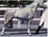
What’s wrong with the horse in the image?

A

Prepubic tendon rupture

8
Q

Which 3 muscles adduct the limb?

A
  1. Gracilis
  2. Pectineus
  3. Adductor
9
Q

If you needed to take a CSF sample where would you take it from?

A

Between the atlas and the occipital condyles (atlanto-occipital joint)

10
Q

What 8 muscles extend the hip?

A
  1. Biceps femoris
  2. Semitendinosus
  3. Semimembranosus
  4. Superficial gluteal muscle
  5. Middle gluteal muscle
  6. Deep gluteal muscle
  7. Adductor
  8. Gracilis
11
Q

What muscles make up the hypaxial muscles?

A

Longus capitis

Longus colli

12
Q

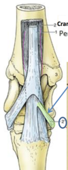
Name the joints shown in the image:

A

From most proximal to distal

Tibiotarsal joint

Proximal intertarsal joint

Distal intertarsal joint

Tarsometatarsal joint

13
Q

Which ribs are considered “true ribs” and why?

A

Ribs 1-9

Because they connect to the sternum (vertbrosternal)

14
Q

What muscle forms the cranial boarder of the femoral triangle?

A. Sartorius

B. Vastus medialis of the quadriceps femoris

C. Semimembranosus

D. Adductor

A

A. Sartorius

15
Q

Which 3 muscles insert at the trochanteric fossa of the femur?

A
  1. Internal obturator
  2. Gemelli
  3. External obturator
16
Q

What muscles make up the transversospinalis system?

A

Splenius

Semispinalis capitis: 2 parts

  1. Biventer cervicis
  2. Complexus
17
Q

Which two muscles originate at the pelvis symphysis via the symphysial tendon?

A
  1. Gracilis
  2. Adductor
18
Q

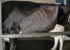
What is wrong with the horse in the following pictures?

A

Chronic obstructive pulmonary disease due to sustained involvement of the abdominal muscles during forced expiration

19
Q

Which vertebra is known as the anticlinal vertebra?

A

T11 - the spine is vertical

20
Q

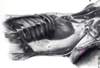
Which tendon is highlighted in green?

A

Cunean tendon

21
Q

Which abdominal muscle is the most medially located?

A

Transversus abdominis

22
Q

Which two muscles extend the stifle?

A
  1. Tensor fascia latae
  2. Quadriceps femoris (all heads)
23
Q

What is the dorsal boundary of the pelvic inlet?

A

Sacral promontory

24
Q

Which three muscles flex the tarsus?

A
  1. Cranial tibial
  2. Long digital extensor
  3. Fibularis longus
25
What articulates with the ilium to complete the pelvic girdle?
The auricular surface of the wing
26
What two spaces can be used for epidurals?
The sacrococcygeal space and the lumbosacral space
27
What are the two types of herniated discs?
Compressive and concussive Type I Hansen's disc herniation (ruptured through into the spinal cord) Type II Hansen's disc herniation (bulging)
28
The heads of ribs 1-10 articulate with the **cranial** or **caudal** costal fovea of the **same** vertebrae?
Cranial
29
Which ribs articulate with the cartilage of the cranial rib? what are they called?
Ribs 10-12 Vertebrocartilaginous
30
Which two muscles insert at the greater trochanter of the femur?
1. Middle gluteal muscle 2. Deep gluteal muscle
31
What is the name of the second pair of foramina on the atlas?
Lateral vertebral foramina
32
Which two muscles originate at the lateral supracondylar tuberosity of the femur, insert at the tuber calcanei and extend the tarsus?
1. Grastrocnemius 2. SDF
33
Which two muscles originate at the sacrotuberous ligament?
1. Biceps femoris 2. Superficial gluteal muscle
34
Which two muscles extend and abduct the hip, and rotate the pelvic limb medially?
1. Middle gluteal 2. Deep gluteal
35
What are considered the epaxial muscles?
Iliocostalis system Longissimus system Transversospinalis system
36
The nuchal ligament is not present in which animal(s)?
The cat and the pig
37
What animal contains an accessory ligament of the femoral head? A. Canine B. Feline C. Horse D. Cow E. C and D
C. Horse only
38
Whta is commonly known as the "yes" joint?
Atlanto-occipital joint
39
The tendon of the lateral head of the DDF passes over the _________ of the calcaneus where it is held down by the _______ retinaculum
Sustentaculum tali; flexor retinaculum
40
What is commonly known as the "no" joint?
The atlanto-axial joint
41
What's wrong with the horse in this image?
Upward fixation of the patella
42
The cunean tendon is a reference point of which joint?
Distal intertarsal joint
43
Which 5 muscle tendons contribute to the common calcanean tendon?
1. Biceps femoris 2. Semitendinosus 3. Gracilis 4. Gastrocnemius 5. SDF
44
The piriformis muscle is considered part of what muscle? A. Superficial gluteal B. Middle gluteal C. Deep gluteal
B. Middle gluteal
45
Which 3 muscles originate at the ischiatic tuberosity?
1. Biceps femoris 2. Semitendinosus 3. Semimembranosus
46
Which 5 muscles can flex but not extend the stifle?
1. Semitendinosus 2. Gracilis 3. Gastrocnemius 4. SDF 5. Popliteus
47
The crural retinaculum holds down the tendon of the following two muscles: A. Cranial tibial and long digital extensor B. Fibularis longus and long digital extensor C. Fibularis longus and cranial tibial D. None of the above
A. Cranial tibial and the long digital extensor
48
Which tendon can be surgically cut to reduce pressure from arthritis of the distal hock joints?
Cunean tendon
49
Which 2 muscle tendons form the main central component of the common calcanean tendon?
1. Gastrocnemius 2. SDF
50
The ________ is a ring of fibrocartilaginous tissue that forms the outer portion of each intervertebral disc
Annulus fibrosus
51
What muscle forms the caudal boarder of the femoral triangle? A. Iliopsoas B. Pectineus C. External obturator D. Adductor
B. Pectineus
52
What is the femur held in place by? Hint: two ligaments
Ligament of the femoral head and the transverse acetabular ligament
53
Rib #8 will articulate with which vertebrae?
Rib #8 will articulate with the cranial costal fovea of vertebra 8 and the caudal costal fovea of vetebra 7
54
Which 2 muscles originate at the ilium?
1. Quadriceps femoris - rectus femoris 2. Sartorius
55
What is wrong with the horse in the image?
Ruptured peroneus tertius - note flexed stifle and extended hock
56
What is the most superficial of the abdominal muscles?
External abdominal oblique
57
In which orientation does the external abdominal oblique muscle extend?
Caudoventral orientation (craniodorsal → caudoventral)
58
In which orientation does the internal abdominal oblique m. fibers run?
Cranioventrally (caudodorsal→ cranioventral)
59
Which muscle is a flexor of the vertebral column?
Rectus abdominis
60
The _________ mm. and the ________ \_\_\_\_\_\_\_\_ m. form a sheath around the rectus abdominis m. (rectus sheath)
The abdominal oblique mm. and the transversus abdominis m
61
The inguinal canal allows the passage of which structures out of the abdomen?
Vaginal process (f)/ tunic(m), genitogermoral nerve, external pudendal a. and v., cremaster m.
62
What makes up the cranial, medial and lateral and caudal boundaries of the deep inguinal ring?
**Cranial boundary:** Caudal border of the internal abdominal m. **Medial boundary:** The rectus abdominis m. **Lateral and caudal boundary:** The inguinal ligament
63
In the horse, the **right** or **left** paralumbar fossa is used to access the cecum?
The right
64
In the cow/bull, the **right** or **left** paralumbar fossa is used to access the rumen?
The left
65
In the horse and ox, what is the deep fascia of the trunk that consists largely of elastic tissue and is used to support the heavy weight of the abdominal viscera?
The tunica flava abdominis
66
What structure is being outlined in the picture?
The paralumbar fossa