Lymphoid Images Flashcards
(35 cards)
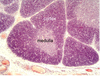
Identify

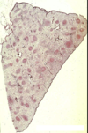
Lymph node
- Capsule: dense irregular connective tissue
- Cortex: sinuses and lymphatic nodules
- Medulla: dense lymphoid tissue and sinuses

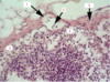
Identify

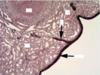
- Capsule: dense irregular CT
- Subcapsular sinus: loose lymphoid tissue, has a few reticular cells and many lymphocytes within
- Cortex (just beneath capsule, consists of sinuses and lymphatic nodules)

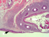
Identify
Lymph Node
- Afferent lymphatic vessel (within capsule)
- Valve (control the flow of lymph through the node)
- Capsule
- Subcapsular sinus

Identify
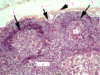
Lymph node
- Cortical lymphatic nodules
- Subcapsular sinus
- Capsule
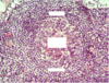
- Cortical lymphatic nodules
- Germinal center
- Cortex

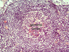
Idenitfy
Lymph Node: high power of secondary lymphatic nodule
Germinal Center
Mantle: concentrically arranged small lymphocytes, surrounds germinal center
Identify

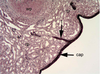
Dlt: dense lymphoid tissue (surrounds sinus)
Ms: medullary sinus (loose lymphoid tissue)
Reticular cells and lymphocytes within sinus
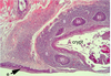
Identify
Lymph node
Efferent lymphatics: notice valves in most of these vessels

Identify

Lymph node
Post capillary venules with tall endothelium
Identify

Lymph node
Capsule
Subcapsular sinus
Cortex
Identify

Fx of HEV: cell adhesion molecules, play a role in lymphocyte recirculation

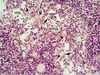
Identify

Perfused spleen
Capsule
Red pulp
White pulp
Trabeculum (contains a blood vessel)

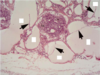
Identify

Perfused spleen
White pulp
Red pulp contains (s) splenic sinuses and (c) splenic cords (between sinuses)

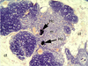
Identify
Perfused spleen
Red pulp (surrounds white pulp)
Lymphatic nodule
Germinal center
PALS (periarterial lymphatic sheath): have central arteries within, populated by T lymphocytes, irregulr shaped portion of white pulp
PALS + Nodules = white pulp

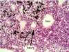
Identify

Perfused spleen
White pulp surrounded by red pulp
Central arteries within white pulp

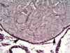
Identify

Fetal thymus
Cortex and medulla of each lobule

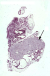
Identify

Thymic lobule
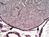
Identify

Fetal thymus
Hassall’s corpuscle

Identify

Fetal thymus
Hassall’s corpuscles (one has been keratinized)

Identify

Adult thymus
Cortical areas (c)
Medullary areas (m)
2 Hassall’s corpuscles that have calcified
Surrounded by connective tissue
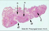
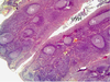
Identify

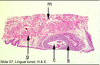
Tonsil (palatine - don’t need to know)
Dlt: dense lymphoid tissue - what tonsils are composed of
ln: lymphatic nodules - lie beneath epithelium
e: stratfied squamous epithelium
c: crypts - penetrate into the tonsil

Identify

Tonsil
Crypt (epithelial invagination)
Lymphatic nodules with germinal centers
Identify
(Optional)
Unperfused spleen
Red pulp with sinuses in the subcapsular region
Capsule around outside
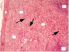
Identify
(Optional)
Unperfused spleen
White pulp surrounded by red pulp (congested with blood)
White pulp - lymphatic nodule with germinal center, also in the form of PALS

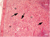
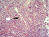
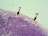
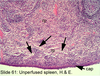
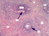
Identify
(Optional)
Spleen
Capsule on left
Sinuses within red pulp “stained” with hemolyzed blood (arrows)
2 areas of white pulp