Magnetic Resonance Imaging Flashcards
(51 cards)
What causes a nucleui to spin?
Spin is a fundamental property of nature like electrical charge or mass, comes in multiples of 1/2 and can be either + or -. Individual unpaired electrons, protons, and neutrons each possesses a spin of 1/2, i.e nucleus with either an odd atomic number or an odd mass number has an angular momentum, or spin angular momentum.
Two or more particles with spins having opposite signs can pair up to eliminate the observable manifestations of spin. An example is helium. In nuclear magnetic resonance, it is unpaired nuclear spins that are of importance.
Mention an important effect spin has on for instance protons
A proton has the property called spin. Think of the spin of a proton as a magnetic moment vector, causing the proton to behave like a tiny magnet with a north and south pole.
When the proton is placed in an external magnetic field B0, the spin vector of the particle aligns itself with the external field, just like a magnet would. There is a low energy configuration or state where the poles are aligned N-S-N-S and a high energy state N-N-S-S.
Explain Magnetization
The 1H nuclei in the human body, when placed in a magnetic field, B0 , will build up a magnetization M.
At 1T, the excess of magnetic moments of the 1H nuclei aligned with the external magnetic field are 10 ppm. They produces a net magnetization, M.
Explain Magnetization Torque
If the Magnetization is brought out of the alignment with B0, it will experience a torque which acts to return the net Magnetization to alignment
Explain Lamor’s equation
When placed in a magnetic field of strength B0, a particle with a net spin can absorb a photon of frequency f . The frequency f depends on the gyromagnetic ratio y (gamma) of the particle.
f = y B0
For hydrogen, y= 42.58 MHz / T.
Lamor with gradient vector:
f(r)=y(B0+G•r)
where G is the gradient vector and r is the position vector.
The Larmor equation is important because it is the frequency at which the nucleus will absorb energy. The absorption of that energy will cause the proton to alter its alignment and ranges from 1-100 MHz in MRI. The equation states that the frequency of precession of the nuclear magnetic moment is directly proportional to the product of the magnetic field strength (B0) and the gyromagnetic ratio (y).
Explain when Resonance/Lamor’s frequency are applied
A particle can undergo a transition between the low and high energy states by the absorption of a photon. A particle in the lower energy state absorbs a photon and ends up in the upper energy state. The energy of this photon must exactly match the energy difference between the two states. The energy, E, of a photon is related to its frequency, f , by Planck’s constant (h = 6.626x10⁻³⁴ J s).
E = fh
In NMR and MRI, the quantity f is called the resonance frequency and the Larmor frequency.
Explain Precession
Precession is a wobbling motion that occurs when a spinning object is the subject of an external force.
Relevant to MRI, the proton of a hydrogen nucleus spins around its axis giving it an angular moment (quantum mechanics). Through the protons positive charge and its spin it generates a magnetic field and gets a magnetic dipole moment (MDM) parallel to the rotation axis. If placed in a magnetic field the magnetic dipole moment will precess about the direction of the magnetic field with an angular frequency (Larmor frequency). The Larmor equation dictates that the frequency of the precession at higher field strengths is higher.
When does a photon gets absorbed by for instance a proton affected by an external a magnetic field?
When the energy of the photon matches the energy difference between the two spin states an absorption of energy occurs.
In what frequency range does the frequencies of the photons for clinical MRI occur?
In the NMR experiment, the frequency of the photon is in the radio frequency (RF) range. In NMR spectroscopy, is between 60 and 800 MHz for hydrogen nuclei. In clinical MRI, is typically between 15 and 80 MHz for hydrogen imaging.
What is the Magnetic flux density and the frequency of radio waves for a basic MRI setup?
- Magnetic flux density 1.5 T
- Linear magnetic field gradients
- Radio waves ca 63 MHz at 1.5 T (wavelength ca 0.5 m in tissue)
Explain which tissues MRI versus CT is specifically good at detecting.
Structures with high density appear bright – CT is very good at depicting bone.
MRI provides very good soft tissue contrast.
Describe the effects of external magnetic field and thermal energy.
Thermal energy
Chaos (at body temperature) The orientation of the spins of the 1H nuclei are randomized, scrambled by molecular thermal motion
External magnetic field - Order
The magnetic field tends to line B0 up the magnetic moments of the 1H nuclei.
Thermal equilibrium
The balance between chaos and order.
M
At 1T, the excess of magnetic moments of the 1H nuclei aligned with the external magnetic field are 10 ppm. They produces a net magnetization, M.

Explain the Motion of Magnetization
(Magnetization, Torque, Spin Angular Momentum, Precession, Relaxation)
Motion of the magnetization, M

Magnetization
The 1H nuclei in the human body, when placed in a magnetic field, B0 , will build up a magnetization M
Torque
If M is brought out of the alignment with B0 , it will experience a torque which acts to return M to alignment.
Spin Angular Momentum
The gyroscopic properties of the 1H nuclei prohibits M to simply flip back
Precession
and the torque results in a precession of M.
Relaxation
M will eventually relax and return to thermal equilibrium
Explain Resonance.
Resonance

If impulses (or ”pushes”) are applied synchronous with the inherent motion of the system the resonance condition is fulfilled and energy is transferred to the system
Examples:
- The motion of the swing increases in amplitude
- M is brought into precession at increasing angles to B
Explain the differences between Precession and Resonance (alternative view)
Nuclear precession is a long-term, spontaneous phenomenon, unaccompanied by energy exchange.
Nuclear resonance is a short-term, induced phenomenon, involving energy exchange between precessing spins and their environment.
Spins wobble (or precess) about the axis of the BO field so as to describe a cone. This is called precession. Spinning protons are like dreidles spinning about their axis. Precession corresponds to the gyration of the rotating axis of a spinning body about an intersecting axis.
Resonance: exchange of energy at a specific frequency between the spins and the radio-frequency pulses
Briefly explain T1 and T2 relaxation.

T1 relaxation
The build up of the magnetization towards thermal equilibrium Synonyms:
- spin-lattice relaxation
- longitudinal relaxation
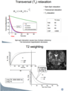
T2 relaxation
The decay of induction (signal) towards zero.
Synonyms:
- spin-spin relaxation
- transversal relaxation
Explain the Bloch Equations
Bloch Equations
In physics and chemistry, specifically in nuclear magnetic resonance (NMR), magnetic resonance imaging (MRI), and electron spin resonance (ESR), the Bloch equations are a set of macroscopic equations that are used to calculate the nuclear magnetization M = (Mx, My, Mz) as a function of time when relaxation times T1 and T2 are present.
What does TE and TR stand for?
- TE: Time to echo (i.e. Time to signal recording)
- TR: Repetition time

What is the difference between Gradient Echo and Spin Echo?
Gradient Echo and Spin Echo
- Spin Echo signal is generated by 2 RF-pulses
- Gradient Echo signal is generated by 1 RF-pulse + gradient reversal
- Spin Echo (but not Gradient Echo) compensates for field inhomogeneties.

Which component of the nuclear magnetisation induces signal in the MR scanner?
MR Scanner
Only the transversal component of the nuclear magnetisation induces signal in the coil
Records the MR signal at echo time (TE)
MR signal from tissue -> Greyscale in the image
How does the conversion between longitudinal and transversal magnetization occurs?

How does T1-weighted images of the brain appear?
T1-weighted (T1; short TR and short TE):
- Provides good contrast between gray matter (dark gray) and white matter (lighter gray) tissues, while CSF is void of signal (black).
- Water, such as CSF, as well as dense bone and air appear dark.
- Fat, such as lipids in the myelinated white matter, appears bright.
- Contrast between the neocortex and white matter is best.
- Contrast between some subcortical gray matter nuclei and white matter is fine but not as good as between cortex and white matter. These nuclei, such as the caudate and putamen, tend to have more white matter fibers and vascular infrastructure than other gray matter regions, increasing the brightness (i.e., lighter gray, more similar to white matter).
- Pathological processes, such as demyelination or inflammation, often increase water content in tissues, which decreases the signal on T1; white matter disease often shows up as darker areas in the lighter gray-colored white matter. (Extensive white matter disease on T1 (left); Moderate white matter disease on T1 (left)) Due to a better measure of water content, T2-weighted images are more sensitive to subtle white matter alterations.
Source: http://fmri.ucsd.edu/Howto/3T/structure.html

How does T2-weighted images of the brain looks like?
T2-weighted (T2; long TR and long TE):
- Provides good contrast between CSF (bright) and brain tissue (dark). Some T2 sequences demonstrate additional contrast between gray matter (lighter gray) and white matter (darker gray).
- Water, such as CSF, appears bright, while air appears dark.
- Fat, such as lipids in the white matter, appears dark.
- Pathological processes, such as demyelination or inflammation, often increase water content in tissues, which increases signal on T2; white matter disease often shows up as brighter areas, (Extensive white matter disease on T2 (right); Moderate white matter disease on T2 (middle)) which makes subtle changes easier to detect.
- source: http://fmri.ucsd.edu/Howto/3T/structure.html

How does PD-weighted images look like?
Proton density-weighted (PD; long TR and short TE):
- Provides good contrast between gray (bright) and white (darker gray) matter, with little contrast between brain and CSF.
- Water varies in signal, with CSF often gray while other fluids may be of higher signal intensity; air appears dark.
- Fat, such as lipids in the white matter, is relatively bright, although gray matter appears brighter than white matter.
- Subcortical nuclei and neocortex tend to be more similar in intensity than on T1.
- Pathological processes, such as demyelination or inflammation, often increase water content in tissues, which increases signal on PD; white matter disease often shows up as brighter areas but with signal different from CSF, unlike T2 (Moderate white matter disease on PD (right)) .
- Source: http://fmri.ucsd.edu/Howto/3T/structure.html


