MBB Pathology/Imaging Flashcards

(79 cards)

1
Q

Describe what you see here; what condition is this

A

rounded atrophic fibers and lymphocytic infiltrate; top right is macrophages attacking muscle fiber; polymyositis

2
Q

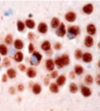
describe the constellation of findings seen and for what disease

A

a) balooning neuron

b and c) neurofibrillary tau tangles

d) astrocytic plaques

corticobasal degeneration

3
Q

Describe the difference between hemorrhagic infarct and a intraparenchymal hemorrhage and how they would look micro/macroscopically

A

Hemorrhagic infarct has brain tissue with blood infiltrating, tissue is becoming necrotic, wil lsee petechiae and neutrophils etc.

intraparenchymal hemorrhage is when blood spills out and pushes brain tissue out of the way and takes up space, so microscopically you would only see blood and fibrin

4
Q

what is this

A

medulloblastoma

5
Q

What is going on here, what is causing it

A

central herniation of temporal lobes, brainstem bein displaced downward, severing basilar pontine arteries, fatal (overall cause is edema)

6
Q

What is going on in this image? what condition is this common in

A

asymmetrical atrophy; corticobasil degeneration

7
Q

What is this? What conditions is this common in

A

lewy bodies, Parkinson’s disease, Lewy body dementia

8
Q

What is this, what is the most common cause, is it fast or slow growing

A

subdural hemorrhage, trauma, slow growing

9
Q

describe what you see; what condition is this common in

A

ballooned neuron; corticobasil degeneration

10
Q

Name 3 possible causes of this

A

high grade glioma (GBM)

cns lymphoma

rlly bad MS

11
Q

describe what you see; what type of cancer is this; what age group is involved

A

dense eosinophilic rosenthal fibers; pilocytic astrocytoma children

12
Q

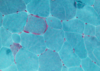
Describe the process going on here from left to right and what kind of process is this?

A

myopathic process

1) left most: muscle fiber necrosis
2) middle: attempts at regeneration
3) fibrosis

13
Q

What is going on here; what condition would this be seen in

A

alpha synnuclein cytoplasmic inclusions in GLIAL cells; multiple system atrophy

14
Q

What are yo ustaining for here; what disease is this common in

A

staining for ubiquitin/huntington protein inclusions ; huntington disese

15
Q

describe what you see; what disease is this

A

neuronal loss, reactive (alzheimers type 2 ) astrocytes; metabolic gliosis (thing in the center) –> wilson’s disease
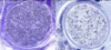
16
Q

what cancer is this

what demographic is it common in

A

medulloblastoma children

17
Q

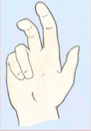
describe the symptoms; what syndrome do they belong to

A

plexiform neurofibroma on peripheral nervous sytem

cafe au lait spot

neurofibromatosis 1

18
Q

What is this? Name some causes?

A

interparenchymal hemorrhage, htn most common, also arteriovenous malformation, lesion, amyloid

19
Q

describe the two images; what condition is this

A

swollen neuron aka pick cell

status spongiosis aka vacuolization of the cortex

pick’s disease

20
Q

what is this, where does it come off of

A

vestibular schwannoma/acoustic neuroma; nerve VIII at the cerebellopontine angle

21
Q

describe what yo usee; what cancer is this

A

vascular tumor, vacuolated stroma, hemangioblastoma from von hippel lindau

22
Q

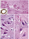
Describe these findings; what condition is this associated with

A

different manifestations of tau protein accumulations

left is neurfibrillatory tangles

bottom left is tufted astrocytes

right (c shpaed) is glial inclusions (also neuronal inclusions)

PROGRESSIVE SUPRANUCLEAR PALSY

23
Q

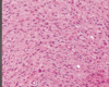
Describe what is happening here: what condition is this

A

perifascicular atrophy; dermatomyositis

24
Q

describe each of the three images; what disease is this

A

top- cerebral amyloid angiopathy (amyloid depositing in leptomeningeal and intraparenchymal arteries)

bottom left- granulovaculoar degeneration (clear cytoplasmic inclusions with basophilic granules)

bottom right- hirano bodies, glassy eosinophilic bodies made of actin

alzheimers

25
What virus is responsible for this? what is happening
polio virus; microglia are surrounding a dying neuron and trying to eat it (neuronophagia, microglial nodule)
26
what are these; what condition is it common in
glial cytoplasmic inclusions, multiple system atrophy
27
describe what you see; what condition is this
rounded atrophic fibers, lymphocytic infiltrate; purple cytoplasmic vacuolar inclusions; inclusion body myositis
28
what do you see; what is it called; what conditions is this associated with
acellular region then pallisating region, verrucay bodies; schwannoma and neurofibromatosis 2
29
describe what you see on the left and bottom right image; what are they called; what condition are they common in
left: dense eosinophilic center with clearing around it, lewy body right: lewy neurites
30
What process is going on here for what disease
in parkinsons alpha synnuclein can accumulate so much that the neuron dies and then this image shows microglia coming in to eat the dead neuron
31
What type of astrocytoma is this ? how do yo uknow
diffuse, only hypercellcularity and atypia
32
describe what you see
diffuse enhancing lesion; lymphoid cells cns lymphoma
33
What neuropathy causes this? why does this happen
radial nerve neuropathy; loss of extension of the fingers and wrist
34
what is going on here; what condition is this
atrophic muscle fibers and a few normal/hypertrophic fibers; werdnig hoffman disease/spinal muscular atrophy
35
What is happening here; what condition is this
onion bulb; attempt at remyelination after demyelination (right pick has schwann celsl surrounding); chronic inflammatory demyelinating polyneuropathy can also happen in charcot marie tooth
36
Describe what you see; what cancer is this and what condition can this occur
large astrocytes in sheaths, subepyndemal lgiant cell astrocytoma (SEGA); tuberous sclerosis
37
what is the arrow pointing to? wha is it made of what condition is this
bunina body, tdp43, amyotrophic lateral sclerosis
38
describe what you see; what is this
cystic; pilocytic astrocytoma
39
describe what you see; what hiv associated condition is this
calcifications; toxoplasmosis
40
what type of hemorrhage is this what condition is commonly associated with this and what age group
lobar hemorrhage, amyloidosis, old people
41
what neurodegenerative disease is this? what are these called
pick's disease; pick bodies made up of tau 3r
42
Describe what you see, what is this called, what can cause this
optic disc borders aren't sharp, blood vessels runcated; papilledema; increse intercanial pressure due to brain tumor
43
What type of astrocytoma is this ? how do yo uknow
gbm; has pseudopalisating necrosis and endothelial proliferation
44
What neuropathy causes this? Why do the fingers look like this
ulnar nerve neuropathy; inability to flex MCP (lumbricals) and inability to extend IP (interossei)
45
what is this called? what group of disorders is this common in
ragged red fibers (accumulation of abnormal mitochondria), mitochondrial myopathies
46
Describe what you see what tumor is this what population is affected what symptoms would yo uexpect
small blue cells, high nuclear to cytoplasmic ratio, can be arranged n lines, can have homer wright rosettes; can have drop mets medulloblastoma; homer wright rosettes
47
What are the signs of what the arrows are pointing to; what is it indicative of
insular ribbon sign and hyperdense MCA sign; infarct of MCA
48
what is this called? What condition is this common in
hummingbird sign (midbrain atrophy), progressive supranuclear palsy
49
What kind of rash is this; what is this common in? what else can concomittantly happen with this
maculopapular rash, neisseria meningitidis, DIC and thrombocytopenia
50
what do you see; what cancer is this
basiloid cells surrounding a cyst wet keratin (pink) purle= calcifications carniopharyngioma
51
Describe each of the three images, what staining it is and whether they belong to a certain condition
top left= normal staining for dystrophin (brown) top right: staining for duchenne muscular dystrophy; no dystrophin is present bottom right: staining for becker muscular dystrophy, reduced dystrophin is present
52
what are the triangular shaped blobs; what is wrong with this pic; what condition is this
neurons; loss of neurons; als
53
what type of tumor is this how can you tell
meningioma, dural tail, hyperostosis
54
what type of astrocytoma is this? how do you know?
anaplastic, has mitoses
55
Describe whaty you see; what condition is this seen in? what else coudl you stain for
neuronal loss and subsequent astrocytosis (small purple things); huntington's disease; can also stain ubiquitin to look for huntington protein inclusions, but not really necessary
56
What part of the body is this? what is happening
muscle, angulated atrophic fibers on the left, nuclear cluster on the right (due to atrophy of the muscle, nuclei are now closer together); denervation injury
57
what are C and U; what is different about them, what condition is this
lateral and anterior corticospinal tracts; demyelinated; ALS
58
what is going on here; what condition is this
lipid accumulation in the muscle fibers: carnityl palmitoyl transferase deficiency
59
What is this? what most commonly causes this? is it fast or slowly growing?
epidural hematoma, trauma to middle meningeal artery, fast growing
60
describe what you see ; What type of cancer is this?
cuboidal to elongated ependymal cells surrounding a stromal core; myxopappilary ependymoma
61
describe what you see; what hiv associated condition is this
enahncement of basal ganglia with sparing of thalamus; cryptococcus meningitis
62
what is this called; what imaging modality is this; what condition is this common in
hot cross bun sign (in pons); mri; multiple system atrophy
63
describe the two pictures, which cancer is this, what population is it most common in
whorling pattern and psamomma bodies; meningioma, older women (can be associated in chidlren if NF2 Chromosome 22q)
64
What i happening here; what condition is this
glycogen filled muscle fibers; myophosphorylase deficiency, can stain with PAS; mcardle's
65
describe the muscle on the left vs the muscle on the right. what condition is this
left is mroe normal, right is atrophic; amyotrophic lateral sclerosis
66
What are the two types of pathology in this image nd for what disease
diffuse plaque top (no neurites around) neuritic plaque bottom (amyloid center with dystorphic neurites around) alzheimers
67
what is this? what is the most common cause
subarachnoid hemorrhage, aneurysm (a. comm artery and p comm artery most common)
68
What neuropathy causes this and why?
median nerve neuropathy; inability to flex 1 and 2 fingers (lumbricals)
69
describe the abnormality seen here what cancer is this
perivascular pseudorosette ependymoma
70
Describe what is happening; what is the condition what age group is this common in
arteries and veins in clsoe proximity with brian tissue in between; arteriovenous malformation, dangerou young women
71
What is this before and after pictures showing?
loss of axons in diabetic neuropathy
72
what do you see what is this
fried egg= oligocytes oligodendroglioma
73
What do you see; what is this
loss of grey white matter interface, edema, mass effect; hSV encephalitis
74
what is this
meningioma
75
what type of neurodegenerative disease is this associated with
alzheimers, neurfibrillatory tangles
76
What part of the brain is this? what is change d from the normal view; what disease is this implicated in
dark= locus ceruleus, right picture it is less, parkinson's disease
77
What is happening here; what condition is this common in
atrophy of the caudate; huntingtons
78
describe the constellation of findings; what disease is this
decreased substantia nigra, discoloration of putamen; multiple system atrophy
79
describe this; what is the only condition this can be
cytoplasmic tdp 43 accumulations (should be nuclear)- frontotemporal dementia with tdp43 pathology