Mediastinum 2 Flashcards
(36 cards)
Where does the vagus nerve (R/L) enters the superior mediastinum?
They enter posterior to the respective sternoclavicular joint and branchiocephalic vein.
Where does the right vagus nerve enters the thorax, and what does it give rise to?
It enters anterior to the right subclavian artery and it gives rise to the right recurrent laryngeal nerve.
The right recurrent laryngeal nerve innervates the _______
Larynx.
True or False:
The right vagus nerve passes anteriorly to the right brachiocephalic vein, superior vena cava, and root of the right lung.
False, it passes posteriorly.
Going from superior to inferior, the right vagun nerve is part of what plexuses?
First right pulmonary plexus and then esophageal plexus and cardiac plexus.
Where does the left vagus nerve enters the mediastinum?
It enters between the left common carotid artery and left subclavian artery.
What nerves does the left vagus nerve give rise to and where?
The left vagus nerves gives rise to the left recurrent laryngeal nerve when it curves medially at the inferior border of the aorta.

What does the phrenic nerve innervates and where does it enters through?

- Innervation:
- Diaphragm with motor and sensory fibers
- Pericardium and mediastinal pleura with sensory fibers.
- Enters between the subclavian artery and the origin of the branchiocephalic vein
What helps you distinguish the phrenic nerve from the vagus nerve?
The phrenic nerve pass anteriorly to the roots of the lungs while the vagus nerve pass posteriorly to the roots of the lungs.
True or False:
The right phrenic nerve passes along the right side of the right brachiocephalic vein, superior vena cava, and pericardium over the right atrium.
True
True or False:
Most branching of the phrenic nerves for distribution to the diaphragm occurs on the diaphragm’s inferior (abdominal) surface.
True
True or False:
Left phrenic nerve crosses the left surface of the arch of the aorta posterior to the left vagus nerve.
False, it passes anterior to the left vagus nerve
What does the recurrent laryngeal nerves innervate?
They innervate all intrinsic muscles of the larynx, except cricothyroid
If you have an injury to the recurrent laryngeal nerves, what is affacted?
- voice
- It may be involved in bronchiogenic or esophageal carcinoma
- enlargement of mediastinal lymph nodes
- aneurysm of the arch of the aorta.
At what level does the trachea ends?
It ends at the level of the sternal angle by diving into right and left bronchi and superior to the level of the heart.
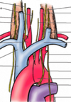
Where does the esophagus enters the superior mediastinum?
It enters the superior mediastinum between the trachea and the vertebral column, where it lies anterior to the bodies of the T1-T4 vertebrae.
True or False:
The esophagus is compressed posteriorly by the root of the right lung.
False. The esophagus is compressed anteriorly by the root of the left lung.

True or False:
Inferior to the arch, the esophagus again inclines to the left as it approaches and passes through the esophageal hiatus in the diaphragm.
True.
Where does the esophagus enters the diaphragm and at what level of the vertebral column?
Enters the esophageal hiatus at the T10 level, anterior to the aorta.
The esophagus is compressed by what structures? and how can they be observed?
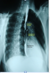
A. Compressed by:
- arch of the aorta
- left main brochus
- diaphragm
B. Can be observed in a oblique chest radiograph, using barium.
Why are impression on the esophagus by adjacent structures is of clinical interesr?
Because since there is a slower passage of substance at these sites, this impression indicate where swallowed foreign obects are most likely to lodge and where a stricture may develop after the accidental drinking of a caustic liquid such as lye
What are the borders of the anterior mediatinum?
Space between the body of the sternum and the transverse thoracic muscles anteriorly and pericardium posteriorly
True or False:
The anterior mediastinum is continuous with the superior mediastinum at the sternal angle and is limited inferiorly by the diaphragm.
True
What can you find in the anterior mediastinum of infants and children?
- loosse CT (sternopericardial ligaments)
- fat
- lymphatic vessels
- branches of internal thoracic vessels
- inferior part of thymus


