Medical Flashcards
(10 cards)
What is the Pathophysiology of Nausea and Vomiting
Pathophysiology of nausea and vomiting. Vomiting is caused by noxious stimulation of the vomiting center directly or indirectly via 1 or more of 4 additional sites: the gastrointestinal (GI) tract, the vestibule system, the chemo-receptor trigger zone, and higher centers in the cortex and thalamus. Once receptors are activated, neural pathways lead to the vomiting center, where emesis is initiated. Neural traffic originating in the GI tract travels along afferent fibers of cranial nerves IX
What is the Pathophysiology of Seizures
A “seizure” is a paroxysmal alteration of neurological function caused by the excessive, hyper-synchronous discharge of neurons in the brain. “Epileptic seizure” is used to distinguish a seizure caused by abnormal neuronal firing from a non-epileptic event, such as a psychogenic seizure. “Epilepsy” is the condition of recurrent, unprovoked seizures. Epilepsy has numerous causes, each reflecting underlying brain dysfunction. A seizure provoked by a reversible insult (e.g., fever, hypoglycemia) does not fall under the definition of epilepsy because it is a short-lived secondary condition, not a chronic state.
What is the definition of Status epileptics
A seizure that lasts > 5 minutes, or multiple seizures without returning to a normal level of consciousness between episodes.
What is the definition of
Genralized Seizures
➢ Tonic-Clonic
➢ Absence (petite mal)
➢ Myoclonic
➢ Tonic
➢ Clonic
➢ Atonic
Partial
➢ Simple
➢ Complex
➢ Partial with secondary generalization
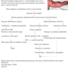
Generalized Seizures.
Generalized - thought to be caused by nearly simultaneous activation of the entire cerebral cortex. Begins with an abrupt loss of consciousness.
Tonic-Clonic
- LOC, tonic phase (rigidity) followed by clonic phase (symmetrical rhythmic jerking of trunk and extremities). Usually lasts 60-90secs.
Absence (petite mal)
- LOC with no loss of postural tone. Very short in
duration.
Myoclonic
- brief shock like muscular contractions that may be generalized or
limited to one of the extremities.
Tonic
- prolonged strained contraction of the body with deviation of the head
and eyes.
Clonic
- repetitive clonic jerks without the tonic phase.
Atonic
- sudden loss of postural tone which may be associated with a brief LOC.
Partial
electrical discharges within a localized region of the cerebral cortex.
Simple
- localized seizure activity with no effect on conscious state. May
involve any part of the body; motor or sensory.
Complex
- localized seizures, which have an effect on conscious state and
motion.
Partial with secondary generalization
- simple partial seizure which progresses to a generalized seizure. In this case the partial seizure is of sensory kind and is often referred to as an aura.
What is the Pathophysiology of Anaphylaxis
A second exposure to a previously encountered antigen. Antigen presenting cell, bind with helper T cell, helper T cell then bind with B cell to create IgE antibody. IgE bind to mast cell and basophil = degranulation. This degranulation is the release of mediators into the blood stream (histamines, prostaglandins and bradykins.) This results in: Vasodilation, increased permeability of capillaries and smooth muscle contractions.
Airway: mucosal plugging, rhinitis and mucosal oedema. Expiratory wheeze, smooth muscle contraction leads to bronchospasm.
Skin and Cardiovascular: redness, urticaria sin rash (caused by histamine). Vasodilation and increased capillary permeability + movement of fluid in the interstitial space. Angioedema and hypotension.
GIT = nausea, vomiting diarrhoea from smooth muscle contraction
What is the Pathophysiology of Meningococcal Septicaemia

What is the Pathophysiology of Autonomic Dysreflexia
Autonomic dysreflexia is a condition that emerges after a spinal cord injury, usually when the injury has occurred above the T6 level.
Cutaneous or visceral stimulation below the level of the spinal cord injury, initiates afferent impulses that elicit reflex sympathetic nervous system activity. The sympathetic response leads to diffuse vasoconstriction, typically to the lower two-thirds of the body, and a rise in blood pressure. In an intact autonomic system, this increased blood pressure stimulates the carotid sinus leading to a parasympathetic outflow slowing the heart rate via vagal stimulation and causing diffuse vasodilation to balance the original increased sympathetic response. However, in the setting of a spinal cord injury, the normal compensatory parasympathetic response cannot travel below the level of the spinal cord injury, and generalized vasoconstriction continues below the level of injury leading to systemic hypertension. The compensatory parasympathetic response leads to bradycardia and vasodilation, but only above the level of the spinal cord injury.
The most common stimuli are distention of a hollow viscus, such as the bladder or rectum. Pressure ulcers or other injuries such as fractures and urinary tract infections are also common causes. Sexual intercourse can also be a stimulus.
What is the Pathophysiology of a Stroke.
Stroke is a neurological disorder characterized by blockage of blood vessels. Clots form in the brain and interrupt blood flow, clogging arteries and causing blood vessels to break, leading to bleeding. Rupture of the arteries leading to the brain during stroke results in the sudden death of brain cells owing to a lack of oxygen.
Stroke is defined as an abrupt neurological outburst caused by impaired perfusion through the blood vessels to the brain. It is important to understand the neurovascular anatomy to study the clinical manifestation of the stroke. The blood flow to the brain is managed by two internal carotids anteriorly and two vertebral arteries posteriorly (the circle of Willis). Ischemic stroke is caused by deficient blood and oxygen supply to the brain; hemorrhagic stroke is caused by bleeding or leaky blood vessels.
Ischemic occlusions
contribute to around 85% of casualties in stroke patients, with the remainder due to intracerebral bleeding. Ischemic occlusion generates thrombotic and embolic conditions in the brain. In thrombosis, the blood flow is affected by narrowing of vessels due to atherosclerosis. The build-up of plaque will eventually constrict the vascular chamber and form clots, causing thrombotic stroke. In an embolic stroke, decreased blood flow to the brain region causes an embolism; the blood flow to the brain reduces, causing severe stress and untimely cell death (necrosis). Necrosis is followed by disruption of the plasma membrane, organelle swelling and leaking of cellular contents into extracellular space, and loss of neuronal function. Other key events contributing to stroke pathology are inflammation, energy failure, loss of homeostasis, acidosis, increased intracellular calcium levels, excitotoxicity, free radical-mediated toxicity, cytokine-mediated cytotoxicity, complement activation, impairment of the blood–brain barrier, activation of glial cells, oxidative stress and infiltration of leukocytes.
Hemorrhagic stroke accounts for approximately 10–15% of all strokes and has a high mortality rate. In this condition, stress in the brain tissue and internal injury cause blood vessels to rupture. It produces toxic effects in the vascular system, resulting in infarction. It is classified into intracerebral and subarachnoid hemorrhage. In ICH, blood vessels rupture and cause abnormal accumulation of blood within the brain. The main reasons for ICH are hypertension, disrupted vasculature, excessive use of anticoagulants and thrombolytic agents. In subarachnoid hemorrhage, blood accumulates in the subarachnoid space of the brain due to a head injury or cerebral aneurysm
Explain the different types of Hemorrhagic strokes.
Types of Haemorrhagic strokes
Subarachnoid hemorrhage
is considered a stroke when it occurs spontaneously (not result from external forces and head trauma).
A spontaneous hemorrhage in the brain usually results from:
Sudden rupture of an aneurysm in an artery in the brain
Congenital aneurysms
Secondary to prolonged hypertension (occurs when an artery branches in a weakened area of artery’s wall)
Rupture of an abnormal connection between arteries and veins (arteriovenous malformation AVM)
Inflamed artery (Septic emboli) travels to an artery that supplies the brain, and causes inflammation and as a result the inflamed artery may weaken and rupture
Intercerberal hemorrhage
Spontaneous bleeding into the brain parenchyma results from rupture of small penetrating arteries in the brain. Degenerative changes in the vessel wall may be associated with advancing age, chronic HTN, diabetes, and other vascular risk factor and It usually occurs at or near the bifurcation of affected arterioles.
The exact cause of brain damage following intracerebral hemorrhage is unknown. It is thought that ICH may result in brain injury by following mechanisms:
Neuronal ischemia following decreased blood flow to the area surrounding the clot
Overexpression of matrix metalloproteinases (MMPs), which may result in the breakdown of the blood brain barrier and edema
Explain the effects of Increased ICP and how cushings traid occurs
Increased ICP
Intracranial hypertension is primarily due to brain injury caused by cerebral ischemia. Cerebral ischemia is the result of decreased brain perfusion secondary to increased ICP. Cerebral perfusion pressure (CPP) is the pressure gradient between mean arterial pressure (MAP) and intracranial pressure (CPP = MAP - ICP). CPP = MAP
A increase in ICP occurs from bleeding in the brain. When bleeding occurs ICP rises, When ICP rises the CSF will shift allowing for more space in the cranium for blood. The brain stops getting vital oxygen causing cereberal agitation (hypoxia) thus causing a decrease in CPP. The body recognizes this and intiates a sympathetic response, causing vasoconstriction (trying to increase blood flow to Isheemic areas of the brain). By doing this MAP is increased (which increase blood-flow to the brain). The brain now getting more blood , however it is still leaking into the craium, causing a further increase in ICP. Once the space is filled the brain it will begin to be pushed downward into the foramen magnum (brain herniation). This will lead to Cushings triad/ reflex.
Cushings Triad
The Cushing response refers to the changes the body experiences to compensate for rising intracranial pressure. Cushing’s triad of signs includes hypertension, bradycardia and apnea/ cheynne stokes respirations.
with increases in intracranial pressure, the Cushing response begins with a rise in systolic blood pressure, widening pulse pressure, bradycardia and irregular breathing (chyennes stokes). Left uncorrected, the heart rate will increase, breathing will become shallow with periods of apnea, and the blood pressure will begin to fall. Eventually the patient will develop an agonal rhythm. Brain stem activity will cease when herniated, and the patient will experience cardiac and respiratory arrest.


