Melanocytic lesions Flashcards
(21 cards)

JUNCTIONAL NEVI
- Uniformly pigmented (quite flat)
- Light to dark brown in colour
- Melanocytes located in the dermoepidermal junction
INTRADERMAL NEVUS
- Skin-coloured papule, often seen on the face
- Can resemble a basal cell carcinoma
- Melanocytes located in the dermis

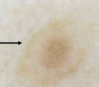
COMPOUND NEVUS
- Pigmented papule, often seen on face or body
- Melanocytes located in the dermoepidermal junction and dermis
- Can contain skin coloured areas within it (variation in pigment but uniform in size)

CONGENITAL NEVI
- Onset at birth or within first year of life (95% of the time they don’t change)
- Typically larger in diameter compared to other nevi, but can be macular
- Some have a more warty or papillomatous appearance
- Stable over time
Describe the size classifications of congenital nevi
- Small = <1.5cm
- Medium = 1.5-19.9cm
- Large “Giant” = >20cm
What is the concern regarding giant congenital hairy melanocytic nevi?
Malignant transformation risk
What is required for a diagnosis of congenital melanocytic nevus syndrome?
What is the genetic mutation?
≥1 giant congenital melanocytic nevus at birth +/- neuromelanosis
Genetic mutation: somatic mutation in NRAS
Can be associated with dysmorphic features of a prominent forehead and short nose

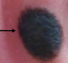
BLUE NEVUS
- Macule or papule
- Uniform, blue colour (can have some subtle white areas, like central pallor)
- Often seen on hands, feet, face or scalp
- If seen in an older person, treat with more suspicion (activating mutations GNAQ, GNA11)
- There is a cellular variant (nodule or plaque) that has had metastatic behaviour described

HALO NEVUS
- White halo around central symmetrical nevus
- Consider melanoma if multiple present
- Associated with vitiligo
- Concerning if older onset +/- asymmetrical nevus within it or mole changing rapidly
What are the 4 stages of a halo nevus?
- Stage 1: nevus surrounded by a rim of hypopigmentation
- Stage 2: nevus turns pink
- Stage 3: nevus disappears, leaving depigmented area
- Stage 4: re-pigmentation over months to years

SPITZ NEVUS
- Pink papule, symmetrical + lacking in pigment
- Concerning if older onset
- Epithelioid cells on histology
SPINDLE CELL NEVUS OF REED - SPITZ NEVUS VARIANT
- Deeply pigmented, flatter lesion
- Spindle cells on histology
- History to distinguish from melanoma (more inclined to excise if >12yo)

MEYERSON’S NEVUS
- Patch of eczema around nevus → pink, inflamed area circumferentially
- Central nevus is symmetrical, usually solitary
- Settles with topical steroids (moderate potency)
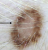
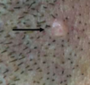
ECLIPSE NEVUS
- Pigmented rim surrounding uniformly lighter centre
- Typically occurs on scalp
EN COCARDE NEVUS
- Bull’s eye appearance (pigmented symmetrical rim with intervening lighter area and pigmented centre)
- Often co-occurs with eclipse nevi

NEVUS SPILUS
- Speckled appearance, resembling splatter from paintbrush
- Need to be self-monitored as have the potential to change

NEVUS OF OTA
- Seen in darker skin types traditionally
- Unilateral tan/grey/brown mottled macule
- Females > males
- Typically V1 or V2 distribution, often involves the sclera
- Regular eye examinations are important

BECKER’S NEVUS
- Unilateral, pigmented, hair-bearing, usually affecting the shoulder
- Males > females
- Onset in 2nd or 3rd decade
What are the features of Becker’s nevus syndrome?
- Becker’s nevus
- Hypoplastic breast
- Shortened arm
- Accessory nipple
What are the features of an atypical nevi?
- >5 mm in diameter
- 2 tone in appearance
- Irregularity in shape

COMBINED NEVI
- Combination of blue and common melanocytic nevus
- Often excised due to atypical appearance → DDx includes melanoma
