Microscopic UA Flashcards
(36 cards)
When is microscopic examination typically performed?
On urine specimens that….
- Show some abnormalities on the physical and chemical analysis (UA w/ reflex to microscopic)
- Come from patients with known renal disease (e.g. chronic kidney disease)
- Microscopic specifically ordered by the clinician (complete UA)
What is microscopic examination used to identify?
- Cells
- Casts
- Crystals
What is normal sediment?
- Red Blood Cell (RBC) count: small amount is normal
- White Blood Cell (WBC) count : small amount is normal
- Epithelial cells: occasionally can be normal, can indicate contamination
- Bacteria: should NOT be present
- Casts: Should NOT be present
- Crystals: should NOT be present
Is CKD reversible?
NO. CKD is rarely reversible
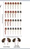
CKD pathophysiology
- Rarely reversible
- Progressive decline in renal function, even after the cause has been removed
- Reduction in renal mass → hypertrophy of the remaining nephrons
- Hyperfiltration of remaining nephrons
- GFR in these nephrons is at a supranormal level…overall GFR still declines
*Once once nephron fails, the rest will fail over time due to increased stress on other nephrons which will result in a decreased GFR
What are associated conditions with RBCs found in the urine?
- Infection (pyelonephritis, cystitis)
- ureterolithiasis
- GU malignancy (bladder cancer)
- Renal cyst
- Acute kidney injury (AKI)
What are associated conditions with WBCs found on microscopy?
- Infection (pyelonephritis, cystits, urethritis)
- Renal inflammatory processes (interstitial nephritis)
What conditions are associated with Renal tubular epithelial cells found on microscopy?
***Always abnormal****
- AKI (acute tubular necrosis, interstitial nephritis)
- Nephrotic syndrome
What conditions are associated with squamous epithelial cells found on microscopy?
Contaminated catch
What conditions are associated with oval fat bodies found on microscopy?
- Nephrotic syndrome
- Autosomal dominant polycystic kidney disease
Graph of major causes of hematuria by age and duration

What do “dysmorphic RBCs” indicate on a urine microscopy report?
injury or pathology on the glomerular level
What are Urinary casts?
- Urinary proteins form fibrils that attach to the epithelial cells lining the tubule lumen as:
- Urine in tubules becomes very concentrated (ex: dehydration)
- Urine flow ceases (stasis)
- Urinary pH is very low
- Urinary salt concentration is high
- Fibrils may intertwine to form casts
- Casts may in turn entrap chemicals or formed elements that are present in the urine
- Casts are eventually washed out of their point of origin and appear in the urine
***Urinary casts are unique to the kidneys
How do urinary casts appear?
They appear as cylindrical, cigar-shaped bodies that represent molds or “casts” of the lumen of the renal tubule in which they were formed
- Distal convoluted tubule
- Collecting duct
What is the major protein constituent of normal urine and that often forms the commen matrix of casts?
Tamm-Hosfall glycoprotein is the major protein constituent
- It is a major defense protein of urothelium against bacteria
- Reagent chemical strips do not detect this protein (dipstick wont turn positive), because it is so heavily glycosylated
what is an element of the urine that is truly unique to the kidney?
Urinary casts!
Casts are the only formed element of urine that is truly unique to the kidney
ex: WBCs can be from many different things/organs but WBC casts are ONLY from the kidney
What conditions are associated with hyaline casts seen on microscopy?
nonspecific
may be “normal”- due to dehydration, concentrated urine
*If this is due to dehydration/concentrated urine then you would also see a high specific gravity and the color of the urine would be amber or dark yellow
What conditions are associated with white blood cell casts seen on microscopy?
- infection (pyelonephritis)
- AKI/renal inflammatory processes (interstitial nephritis)
What conditions are associated with red blood cell casts seen on microscopy?
- AKI (glomerulonephritis)
- Nephritic syndrome
What conditions are associated with granular casts seen on microscopy?
AKI (acute tubular necrosis
What conditions are associated with renal tubular epithelial cell casts seen on microscopy?
- AKI (acute tubular necrosis, interstitial nephritis)
- Nephritic syndrome
What conditions are associated with waxy casts seen on microscopy?
nonspecific
CKD
What conditions are associated with broad casts seen on microscopy?
CKD


