Midterm #1 Flashcards
(111 cards)
1
Q
Organization of the Circulatory System
A
- Left ventricle is the main one that pumps blood throughout body.
- Right ventricle goes to lungs
- Makes it so that oxygenated and non-oxygenated don’t mix
- Pressure
- Right has less pressure when contracted/relaxed (24/8 mmHg)
- Easy to push blood through little cappilaries, not need as much pressure
- Need low pressure in pulmonary capillaries because thin epithelium separating air and blood. Too much pressure, fluid would leave and you would essentially drown
- Pulmonary edema
- Goes along with heart failure and other cardiovascular situations
- Right has less pressure when contracted/relaxed (24/8 mmHg)
- Left has more pressure (120 mmHg/80)

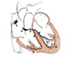
2
Q
Flow of Blood in Circulatory System: Figure
A

3
Q
Chambers of Heart: Shape and Wall Thickness
A
- Atria: thin walled
- Store up blood preparatory for ventricle filling
- Stretchy
- Ventricular filling
- A lot of the blood is “sucked in” (3/4)
- When atria contract, top off the filling of the ventricle (1/4)
- Ventricles
- Right ventricle thinner than left ventricle
- Left is thicker and circular
- Create tension for systemic circulation
- Circular cross section allows muscle contraction to provide efficient pressure
- Contracts like squeezing fist
- Right ventricle
- Shape to move volumes of blood
- Outer moves towards the inner septum

4
Q
Left and Right: Veins/Arteries
A
- Veins are thin, blue, larger, compliant (stretchy, ability to accommodate blood)
- Right Atrium
- Vena cava (superior and inferior)
- Coronary sinus
- Left Atrium
- 4 pulmonary veins
- Right arteries
- Pulmonary trunk
- Left arteries
- Aorta
- Lots of elastin, less compliant than veins, important for blood pressure. Expands when put blood into in and then springs back
5
Q
Heart Valves
A
- Through atrioventricular valve into the ventricles
- Right is tricuspid, left is mitral valve
-
Passive valves
- open and close because of pressure
- Flaps that are called leaflets “cuspid”
- Blood flowing opens the leaflets
- Blood flowing backward, closes the leaflets
- Fibrous connective tissue
- Supports valves
- Separate atria and ventricle
-
AV Valves
- Mitral valve
- Left atrioventricular valve
- Leaflets extends down, and when they closed they touch each other
- When open, create a funnel
- Larger than aortic and pulmonary valves
- Have connective tissue strands attached to leaflets to prevent leaflets from being blow back
- Chordae tendineae
- Connected to mounds of tissue known as papillary muscles
- When leaflets bulge backwards; prolapsed valve
-
Pulmonary valve and Aortic Valve
- Blood balloons them down and pushed them together to prevent backflow

6
Q
Aortic and Pulmonary Valves: Figure
A

7
Q
AV valves (triscuspid and mitral): Figure
A

8
Q
Echocardiograms
A
- Transducer eliciting ultrasound
- Beam of ultrasounds sweeps around
- Goes into heart and reflects off of structures and bounces back to sensor
- Measures the time it takes to bounce back
- Makes what looks like triangular slice through heart
- Can add doppler to measure blood flow
- Sound toward you, beams compressed, higher pitched
- Sound away from you, beams less compressed, lower pitched
- Insufficiency: when blood squirts backwards out of valve.
9
Q
Normal Heart Sounds: S1, S2
A
- Valve snaps such and then vibrates tissues to produce sound
- Known as “lub” and “dup”
- S1, S2 ….. S1, S2 …… S1, S2
- S1 at start of ventricular contraction (systole)
- Ventricle continue contraction
- At the very moment that ventricle begins relaxation, pulmonary valves close
- S2, pulmonary and aortic valves close
- Sounds at start of contraction and start of relaxation
10
Q
Times of systole and diastole
A
- Time between S1 and S2 is systole
- Time between S2 and next S1 is diastole
11
Q
Split Sounds
A
- Normally S1 is both close at same time and S2 is where pulmonary and aortic close at same time
- S2 split, asymmetry and not close at same time
- A little bit of splitting if inhale very deeply (subtle in health person)
- Bundle branch block
- Ventricles contracting out of synchrony
12
Q
S3, S4
A
- S3 occurs during diastole
- Rapidly filling of ventricles
- Ventricles vibrate
- Weak S3 in small kids
- In elderly with expanded ECF volume
- Occurs in CHF
- Volume overload, ventricles become too weak, during filling, ventricles vibrate during filling.
- S3 will be more prevalent “lub dup dup” sound
- S4 just before S1 (and after S3)
- Atria contract and complete ventricle filling
- If stiff ventricles due to heart disease (diastolic HF), when atria contract, get vibrating ventricles
- Not mutually exclusive “lub dup dup dup” (gallop sound)
13
Q
Laminar and Turbulent Flow
A
- Sound hearing from blood pressure and valve abnormalities is from turbulent flow
- Laminar flow
- Cell in middle of tube will stay in middle of tube
- Fluid moves in smooth layers/sheets through tube
- Most efficient way to move fluid through a tube, silent
- Normal flow through cardiovascular system
- Turbulent flow
- Move fluid through faster and faster, fluid will start bonking around everywhere
- Laminar flow pattern breakdown
- Creates noise

14
Q
Stenosis
A
- Narrowing
- If valve leaflets don’t open fully
- Channel that blood flows through is narrower than normally
15
Q
Isufficiency (Regurgitation)
A
- Valve leaflets don’t close fully
- Blood squirts backwards through the hole
- Be able to go through and determine if murmur is systolic or diastolic for either stenosis or insufficiency
- Ex: Aortic stenosis.
- Valve leaflets don’t open fully
- Aortic open at the beginning of systole
- Get murmur right after AV valve close and at start of systole
- “Lub shhhh dup”
- Diastolic murmur will be “lub dup shhh”
- Ex: Aortic stenosis.
16
Q
Senile Aortic Stenosis
A
- Aortic valve is in a stressful position
- HTN can put stress on aorta
- Get fibrosis, prevent leaflets from opening fully
- Inflammation for long periods can cause calcification
17
Q
Bicuspid Aortic Valve
A
- In middle age have to be replaced
- Life expectancy is normal
- Genetic
- More prone to stenosis (fibrosis and calcification)
18
Q
rheumatic fever (heart disease)
A
- After a person gets strep throat
- Only 1-2% who get strep throat
- Ab against streptococcus will also attack valve system in the heart
- Especially the mitral valve
- Mitral stenosis
- Causes left atrial pressure to rise
- Pulmonary edema
- Shortness of breath: dyspnea
- Can progress to congestive heart failure
19
Q
infective endocarditis
A
- Happens when get bacteria in the blood
- Colonize leaflets of valves as go through circulatory system
- Usually after invasive medical procedure
- Hospital IV
- Dentistry (occasionally)
- IV drug abuse
- Clots around the leaflets
- Vegetations, big floppy thing (goobers) sticky around leaflet
- Can break down chordae tendineae
- Can cause aortic or mitral insufficiency
- Mitral insufficiency: pulmonary edema
- Exercise intolerance because unable to increase cardiac output
20
Q
Artificial Valves
A
- Bileaflet totally artificial valve made from carbon fibers, last longer, more likely to form clots
- Biological (from animal or cadaver), not last as long, less problems associated with them
- The endothelium is gone and cross-link all proteins, no live cells, cross linked collagen so that there is not immulogical problem
- Trans-catheter Aortic Valve replacement (TAUR)
- Balloon at end of catheter that is threaded into position.
- Balloon expanded and then opens up to push damaged out of place
- Less invasive.
21
Q
Coordination of the Heart Beat
A
- Some heart muscle is myogenic: able to begin contractions by itself
- Heart still beat even when nerves to it are severed
- In early embryonic development, all cardiac fibers are myogenic
- As develop, only some specialized tissue retain this
- Any injured tissue can cause beating on its own
-
Intercalated discs that connect cells and there are gap junction ion channels
- Action potentials are able to jump from cell to cell
- Atrial and ventricular muscle cells are separated by fibrous tissue
22
Q
SA Node
A
- # 1; sinoatrial node
- Shaped like a dime, can’t see it in dissection of heart without special techniques
- Have myogenic property
- The natural pacemaker of the heart
- 100 bpm without any other hormones, nervous input, etc
- Parasympathetic nerves lower the heart beat
- Conducts over the atria
- Then flows to AV node

23
Q
AV Node
A
- # 2; atrioventricular node
- Looks like the SA node
- Delayed in AV node
- AP leave the AV node and enter 3, 4, 5
- If SA node is out of commission, this one comes into effect
- Has inherent rhythm of 60 bpm
- Since SA node makes AP at a higher rate than AV node
- Muscle has long refractory periods and the AV node is reset so that it won’t do its own heartbeat
24
Q
AV Bundle (Bundle of His)
A
- # 3
- Picks up action potential and muscle fibers goes through the layer separating ventricles
- Big cells and rapidly conduct action potential quickly
- Quickly through everything

25
Right and Left Bundle Branches
* #4
* Drive heartbeat at 30 bpm

26
Purkinje Fibers
* #5, dropped of on the lower **inner** surface of ventricle
* Then outwards and downward through the thick ventricular walls.
* Then conducts up outer wall of ventricle
* This is usually when the ventricle contracts

27
Ventricle Action Potential
* Lots of different ion channels
* Voltage gated ion channels
* Up sweep of AP (A) is by the fast Na+
* Lidocaine will block this
* Action potential has to act a long time (B), not in neuronal action potentials
* Ca++ channel that is slower opening and slower closing
* Really positive equilibrium potential
* Creates the plateau
* Also need slow K+ channels, (C) like neuronal action potential
28
SA Node Action Potential
* No fast Na+ channels
* Do have slow Ca++ and K+ channel
* Slower action potential
* Pattern of the injured cardiac muscle cell
29
Pacemaker Potential
* Doesn’t stay at resting membrane potential
* Starts creeping up
* Closing of slow K+, first part of pacemaker potential
* Opening of “funny “ Na+ channel, open with repolarization rather than with depolarization.
* Open slowly
* Calcium channels that open at the same time as well
30
Change heart rate by changing slope of pacemaker potential
* Speed heart rate by make pacemaker potential reaching threshold faster
* Slow heart rate by make pacemaker potential reach threshold slower
* Things altering slope of pacemaker potential:
* Ach (acetylcholine)
* Autonomic neural transmitters cause slow postsynaptic potential
* 7TMD Receptor binding Ach, Trimeric G protein, gamma and opens K+ channel
* Norepinephrine
* 7TMDR, Trimeric G protein, opens Ca++ (Na+)
* Depolarize faster and increase heart beat
31
Adenosine
* Paracrine and drug
* Works through trimeric proteins an opens K+
* Reduces excitability and reduces heart beat
32
Refractory Period
* Period of time in which ion channels aren’t back to normal configuration
* Can’t have action potential during that time
* Really long in cardiac muscle
* Max heart rate of 190 bpm
* Long refractory period, after ventricle contract allows time for ventricle to relax
* Can’t get a steady contraction (tetanus), one action potential after another
33
Action Potentials: Graphs

34
Basis of Lead II Waveform in Electrocardiogram
* P wave is action potential moving through the atria
* QRS wave, the action potential moving through the bulk of the ventricle
* T wave, repolarization, positive because not occurring in the same direction as the depolarization
35
First Degree AV Block
* Can’t get through the AV node
* Rather vulnerable part of heart
* Prone to not working, small cells/muscle fibers
* Long time between P and QRS wave
* Slowed conduction velocity, action potential still goes through though
* Due to heart disease or benign
* Transient ischemia
* Athlete, trained heart pumping a lot of blood, needs less bpm, slowed by vagus nerve, ach opens K channels, slows the conduction of the heart
* Drugs that can cause this as well; Beta-blockers, Calcium channel blockers, digoxin
* Reduce excitability of the heart
36
Second Degree AV Block
* P interval gets longer until QRS wave missing
* Some of the QRS wave are missing
* Can’t get through the AV node at times
* Circumstances like the first degree
37
Third Degree AV Block
* Don’t see QRS right after P
* See QRS that is big and weird
* Action potential never gets through AV node
* Other specialized tissue will then cause the heart to beat
* AP starts somewhere other than SA node; ectopic focus
* Instead of going out through ventricular wall quickly, get a right then left contraction, abnormal flow over heart
* Causes a prolonged and misshaped QRS
* 30 bpm, person is barely getting enough blood flow to keep themselves going
* Has serious heart disease, perhaps from a myocardial infarction (MI)
38
Premature Atrial Contraction
* Instead of waiting normal interval, get a P-QRST stuck in right away
* From an ectopic focus somewhere in the atria that all of a sudden makes an action potential
* Could be from heart disease
* Could also be benign, actually fairly common
* Know that it is in the atria because the QRST is normal, ventricular tissue getting activated normally
* Might not have symptoms
* May have palpitation:
* Extra beat causes a refractory period, causes a delay before the next heart beat
* During pause, ventricle fills more fully, so it pumps stronger and person may feel it
* May have this in older people during stress test; not a good sign
39
Premature Ventricular Contraction
* Ectopic focus in a ventricle
* QRS wave is prolonged and misshaped; action potential not all of a sudden dropped to bottom of both ventricles
* Get a pause because next SA node contraction falls in the refractory period
* During a stress test; not a good sign, shows damages ventricular muscle
* QRS waves can be either positive or negative
* If see both, then there are two ectopic focuses going on
* start on different sides of the heart
40
Bundle Branch Block
* Would see normal rhythm but WRS would be distorted in shape and prolong. However QRS wave is occurring in regular intervals
* Result that both ventricles are not contracting in synchrony
* Split heart sounds.
41
Sinus Bradycardia
* Normal ECG with a really slow heart rate
* Less and 50 bpm
* Athlete can wake up at 40 bpm, not the same thing
* Need a pacemaker in this case
* Eldery, hypothyroidism, cardiovascular disease, drugs (beta blocker, CCB, digoxin)
* Fatigue, start fainting (syncope)
42
supraventricular tachycardia
* P waves begin before T wave done
* AV node and higher in heart driving the heart beat
* Really fast heart beat, faster than 100 bpm
* May have episodes of it, or can be a persistent thing
* Less caffeine, stress reduction, etc.
* Paroxysmal; all of a sudden, for a period of time, then goes back to normal
* Increase pumping of heart and changes in blood vessels (need to go hand in hand)
* Increase pumping and no changes in blood vessels, ventricles not pump properly, may feel woozy and faint
43
AV Node Reentry
* Most common circumstance that causes supraventricular tachycardia
* Parts of AV node not working properly
* AP goes fast through some pathways and slower through other pathways in AV node
* AP in slow pathway goes into the fast pathway, out of refractory period and causes another AP
* Goes around and around and around
44
Accesory Pathway an Supraventricular Tachycardia
* AP potential hits an **accessory pathway**
* Scrap of muscle tissue that connects atria and ventricles
* Not normally there
* Causes the action potential to loop AP in circular motion back through atria and ventricles
* Need to destroy that tissue; ablations that heats up tissue with radiofrequency wave that cooks it.
* **Wolff-Parkinson-White Syndrome**
45
Ventricular Tachycardia
* Bad in any circumstance
* Ectopic focus in ventricle that is going off constantly
* ICU ward, having a heart attack
* Hearts racing because chunk of damage to ventricular damage (MI)
* Genetic causes with abnormal ion channel that makes ventricular fibrillating (myopathy)
* Will lapse into ventricle fibrillation; death seconds away
* ECG looks like villi
* Pacemaker with a defibrillator
* Defibrillator shocks the crap out of heart to wipe the slate clean
46
Atrial Fibrillation
* 10% of people over 80 have A-fib
* Atria get stretched out and get slow pathways
* AP gets into the left atrium
* Gets past refractory period
* AP never goes away
* Atria sitting there and quiver
* AP shows up at AV node and then contracts
* Random arrival of AP at the AV node
* Random heart rate
* No distinct P wave
* Hashed/wiggly line and AP at random times
* May or may not be symptomatic
* Fatigue
47
Atrial Fibrillation Treatments
* Rate control
* Beta Blocker (slower HR down)
* Rhythm control
* Block sodium channels to reduce excitability
* Anticoagulation
* Clot tends to form in the atria
* Clot in left atria, up into brain, stroke
* **Aspirin** and **clopidogrel** (lowest level and probably will go further; aspirin blocks TXA2 and clopidogrel blcoks ADP)
* **Warfarin**
* Safety net in the fact that it is easy to reverse the effects
* **Dabigatran**, etc.
* Direct thrombin inhibitor
* Can’t reverse effects quickly
* **Apixaban**, etc.
* Factor Xa inhibitor
* Can’t reverse effects quickly
* Ablation around pulmonary veins to get rid of the slow pathways
* Pacemaker
48
Ventricular Fibrillation
* Fatal within a minute
* Ventricles siting there and quivering, blood isn’t being pumped
* Lethal arrhythmias
* MI (heart attack, clot clogs coronary artery)
* Myopathy
* These cause ventricular tachycardia which can lapse into fibrillation
* Person needs pacemaker with defibrillator
49
Pharmacology for Arrhythmias
* Sodium Channel Blocker
* Lidocaine
* Flacainide
* Beta Adrenergic Blockers
* Propranol
* Metoprolol
* Prolong Repolarization (increase the refractory period)
* Amiodarone
* Block Calcium Channels
* Verapamil
* Open Potassium Channels
* Adenosine
50
Cardiac Cycle: Opening and Closing of Valves

51
Cardiac Output
* CO=HR\*SV
* Normal is 5 L/min, exercise 20 L/min, world class athletes are 35 L/min
52
Heart Rate
* Beats per minute
* Pacemaker potential: sodium, potassium and calcium channels
53
parasympathetic innervation and heart rate
* Ach
* Predominate effect on heart
* 100 bpm left to it’s own devices
* Normal is around 70 bpm due to Ach release
* Work through trimeric G protein to open potassium channels
* Ach makes pacemaker potential go up more slowly to increase the refractory period, slows down the heart.
54
sympathetic innervations and epinephrine and heart rate
* Norepi, Calcium and sodium
* Beta receptor
* Also epinephrine
55
Stroke Volume: Sympathetic Inervation and Epinephrine
* More calcium stored and released
* Ventricles contract more forcefully
* Ejection fraction goes up (EF)
* Highest EF is when someone is exercising vigorously
56
Stroke Volume:
## Footnote
Increased blood in central veins and increased atrial pressure
* Causes an increase in EDV
* More ATP expended
* This causes an increase in stroke volume
* Stretch cardiac muscle further so that ventricles contract more forcefully.
* Greater stretch, more ATP energy expended
* Ventricles fill more fully
57
Stroke Volume: End Diastolic Volume
* End diastolic volume=100 mL, because that is how much is in ventricle when done filling
* Stroke volume=70 mL
* Therefore EF=70/100=0.7
58
Frank-Starling Mechanism
* Heart muscle contract more forcefully when stretch it
* Increased EDV causes Increase SV
59
What causes changes in stroke volume?
* Posture
* Muscle Contraction
* FS important to keeps pumping of ventricles pumping exactly the same
* Premature Heart beat if not
60
Posture: Changes in stroke volume
* Gravity causes blood to pool in leg veins
* Right ventricular stroke volume is lower
* If change to laying volume, stroke volume increases
61
Muscle Contractions: Changes in stroke volume
* Locked knees, cause blood to pool in leg veins
* Stroke Volume is decreased
* Veins that go through muscle get contracted with muscle contracts-“muscle pumpin”
* Increases stroke volume
62
Keep pumping of the two ventricles pumping exactly the same!!!
* If right ventricle pumping 1% more than left ventricle (0.7 ml/beat goes into pulmonary from systemic circulation)
* Blood accumulates in pulmonary veins
* Get pulmonary edema, lungs fill up with fluid
* Increase SV in right side, increase pressure in pulmonary veins, increases stroke volume on left side
63
Premature Heart Beat: Changes in stroke volume
* Ectopic focus makes the premature heart beat
* Pause before next SA node action potential
* Delay causes ventricle to fill more fully, heart will have a stronger stroke volume
64
Central Venous Pressure
* "Venous Return"
65
Aortic Pressure
* "After load"
* Effect of dialation of arterioles
* Influences the aortic pressure
* Raise aortic pressure makes it harder to left ventricle to pump blood into aorta (decreases SV)
* Decrease aortic P, Increase in SV
* CO and blood vessels have to change together when making changes in cardiovascular system
66
Systolic Failure
* Decrease in the ejection fraction
* Causes:
* MI
* Myopathies
* Alcoholism, valve problem, etc.
67
Systolic Failure: sequence of events; Law of Laplace
* MI causes decreased EF (gets below 0.5, 0.3 is bad, 0.1 can be shock)→Increased blood in central veins (Except Frank-Starling effect to come to the rescue)
* Since ventricle is weakened, the Frank Starling effect is weakened
* Increases the EDV (ventricle fills fine, but the SV will not increase), ventricle starts dilating
* If know tension in walls can calculate the pressure on the inside
* Law of Laplace
* Proportional to tension, inversely proportional to radius
* P=T/R
* Dilating ventrical needs more tension, starts failing.
* Increased wall tension sets in motion an abnormal response
68
Increased Tension Causes Abnormal Response
* Hypertrophy
* Muscle cells increase in size, but abnormally
* Get **fetal isoforms** of contractile proteins
* Capillary growth doesn’t keep up
* Collagen Damage
* Abnormal stretch causes **collagen damage**
* **Fibrosis**
* **Abnormal Regulation**
69
Abnormal Regulation from Increased Wall Tension
* This is where drug treatments revolve around
* Constant sympathetic drive in the heart creates an abnormal situation
* Kidneys release renin
* Poor renal perfusion
* Normally regulates ECV; may see poor renal perfusion as low ECV/plasma volume
* Renin acts on angiotensinogen→angiotensin I (not very potent)
* ACE converts angiotensin I→angetension II (very potent)
* ACE and angiotensiongen are always in the blood
* Constricts arterioles
* Increase the ECV
* Volume overload causes congestive heart failure

70
Treatments for Systolic and Diastolic Failure
* ACE inhibitor
* Diuretic
* Vasodilation of arterioles
* Decreasing the afterload
* Beta Adrenergic I blockers
* Decreases the counter-productive constant sympathetic input
* Aldosterone
* Saves sodium, expands ECV
* Treatments: aldosterone antagonist
* Also helps abnormal hypertrophy
* Diuretics
* Furosemide (Lasix)
* Pacemaker with defibrillator
* Cardiac transplant
71
Diastolic Heart Failure
* Nothing wrong with EF
* The problem is in the filling
* The ventricles become too stiff
* Decreased compliance
* Cardiac output goes down
* HTN (longstanding) can causes this
* Valve problems can cause this
* Hypertrophy
* Wall thickness increases
72
Pressure, Flow, and Resistance
* Hydrostatic Pressure
* The pressure from weight of water
* Pressure in ankle 100 mmHg more when standing
* Wall tension
* Arterial pressure
* Elastostatic pressure (“Linder’s Name”)
* Resistance to Flow
* Factor that determines how much flow given the pressure
* Determined by diameter of pipe
* 1 L/min, halve the pipe and get 1/16 L/min
* Constriction of smallest arterioles determines the flow
73
Structure of Arteries
* Elastic arteries
* Aorta and major branches
* Lots of elastin in the walls
* Muscular arteries
* Like radial artery
* Media tunica has smooth muscle rather than elastin
74
Role of Elastic Arteries
* Expands/stretches when blood is pumped into it
* Stores energy during systole
* Give energy during diastole
* Smooth it so that pressure doesn’t have huge swings
75
Compliance: Effect of Age
* Change the pressure and see change of volume in aorta from autopsy and see what happen with push fluid in
* Made plot with pressure of X and volume on Y

76
Mean Arterial Pressure
* Average pressure over time
* Balloon with spout on two sides
* C.O=How fast pump water in it
* Spout=diameter of arteriole
* Lump together all arteriole effects=total peripheral resistance
* Dilation; TPR decreases

77
Pulse Pressure
* High SV increases pulse pressure
* Reduced compliance increases pulse pressure
* High pulse pressure in elderly due to reduced compliance
78
Atherosclerosis
* Places that are more likely to happen (distal aorta, common carotids, coronary arteries)
* Get cholesterol in blood that gums up artery, not a good analogy
79
Atherosclerosis: Sequence of events
* Something wrong with endothelium and tunica intima
* Inflammation
* Accumulation of oxidized LDL
* Macrophages phagocystoze the cholesterol

80
Something wrong with endothelium and tunica intima in atherosclerosis
* Places where there is a lot of flow stress
* Structurally intact (nothing you can see histologically)
81
Inflammation in atherosclerosis
* Cells are recruited
* Macrophages
* Statin drugs have an anti-inflammatory angle to them (as well as cholesterol reducing)
82
Accumulation of oxidized LDL in atherosclerosis
* Gets into the tunica intima
* Especially small particles
* ApoB accumulation (only find on LDL, NOT ON HDL)
83
Macrophage phagocytized the cholesterol in atherosclerosis
* Have a protein that would normally make to HDL
* In atherosclerosis, macrophage starts to loose motility
* Bind ApoB (LDL)
* Don’t effectively transfer to HDL
* Macrophages start accumulating cholesterol
* Become “foam cells”
* “Fatty streak” where starting to get atherosclerosis
84
Why atherosclerosis?
* Increases small LDL
* Motility problems
* General inflammation
85
Smooth Muscle and Atherosclerosis
* Move into the tunica intima and start synthesizing fibrous connective tissue
* **Growth factors** making them do this
* Makes a **fibrous plaque**
* Initially soft
* Cells in middle not have capillaries to them
* Get extracellular lipids
* Makes **cholesterol crystals**
* **Thick cap**=lots fibrous tissue between blood and necrotic region
* With time thick cap gets calcification
86
Occludes vessels start to get **symptoms**
* Claudication (BV to legs)
* Angina pectoris in coronary arteries
* Syncope in carotid arteries
* Weakened wall
* Aneurysm
* Stress test
* ECG changes
* Look between S and T wave
* Tends to shift with not enough blood flow
* Visualize vessels with an angiogram
87
Fibrosis plaque with thin cap
* May rupture
* Exposes **extracellular lipids**
* Promotes clotting
* Clot forming
* Can cause **MI**, **stroke**
88
Atherosclerosis Risk Factors
* HTN damages all parts of CV system
* Diabetes
* Smoking
* Hyperlipidemia
* Increased LDL (ApoB)
* Lower this with statin drugs
* Low HDL
* High TAG (VLDL)
* Saturated, Trans Fats
89
Framingham Risk
* Calculates risk of having heart problems soon
* Blood pressure
* Diabetes
* Smoking
* LDL, HDL
* Gender, Age
* High enough risk and taking statin
90
Drugs for Atherosclerosis
* Statins
* inhibit HMG-CoA reductase, the rate-limiting enzyme in cholesterol synthesis
* Ezetimibe
* inhibit cholesterol absorption in small intestine
* (PCSKa)
* Ab that blocks enzyme that degrades LDL receptors
* Undergoing clinical trial
* Degrade LDL receptors
* (Fibrates)
* bind to the nuclear receptor PPAR-alpha
* Increase HDL and lower TAG
* (Niacin)
* Increase HDL by preventing breakdown
* (CETP inhibitors)
* Prevents transfer of HDL to LDL
* increases reverse cholesterol transport
* anacetrapib and evacetrapib
91
Arterioles: Structure
* Determine the TPR
* Control the distribution of blood flow
* Less than half a millimeter
92
Control of arterioles: autonomic nervous system
* Sympathetic nerves
* Skin, gut, kidneys
* Alpha receptors
* Too much sympathetic action to skin is Raynaud’s
* Strongly vasoconstrictor the arterioles
* Then vasodilation that causes pain
* Drugs: Calcium channel I, alpha blockers
* NO (nitric oxide) releasing nerves
* Cause vasodilation
* Penis arterioles, gut
93
Control of arterioles: paracrines
* Inflammatory paracrine
* NO
* vasodilation
94
Control of arterioles: hormones
* Angiotensin II
* Vasopressin
* Powerful constriction of blood vessels
* Important during hemorrhage to keep up blood pressure
95
Control of arterioles: local chemical factors
* Local metabolic
* Increase in Co2, increase in K, osmolarity
* Osmolarity because metabolism makes big molecules into little molecules
* Important in muscle/exercise
* So brain doesn’t have to think about it.
96
Capilaries: Structure
Endothelium
97
Capilaries: Permeability
* Permeability
* Anything smaller than a blood protein
* Exception: less permeable in brain
98
Fluid balance across capillary wall osmotic effect of blood proteins
* Blood protein osmotic effect opposes the blood pressure
* Osmosis when solute is not permeable, water is permeable (diffusion of water)
* Some leaves capillaries and goes to lymphatic system
* Proteins that are blood proteins
* Albumin

99
Edema
* Common medical symptom
* When fluid leaves the capillaries
* Causes:
* Increases capillary permeability (inflammatory paracrine)
* Diabetic retinopathy
* Decrease in blood proteins (hemorrhage, protein starvation)
* Increases in ECF (CHF)
* Increase in venous pressure
* Blocked lymphatics
* Hepatic portal vein damage/blockage
* Ascites
100
Veins: Structure
* Thinner walled (thin tunica media)
* High compliance
* Larger
* Anastomoses
* Many pathways for blood to get back to the heart
* When veins divide but then come back together
* Valves
* Important in muscles that squeeze blood back to heart
* Amount of blood: 75% of blood in systemic circulation
* Can change amount of blood in veins easily
* Get away with changes in blood volume because of vein compliancy
101
Veins: sympathetic innervation
* Contract the veins
* Smooth muscle in the adventitia
* Angiotension II causes vein constriction
* Constriction doesn’t change the TPR
* Changes how much blood available for circulation
102
Regulation of Arterial Pressure
* Cardiac output and total peripheral resistance
* Carotid baroreceptor reflex
* Hormones

103
Carotid Baroreceptor Reflex
* Short term regulation
* Most sensitive
* Understand what carotid massaging does in ER
* Increases parasympathetic effects to the heart

104
Regulation of Arterial Pressure: Hormones
* Angiotensin II
* Vasopressin
* Increase TPR
* Important for supporting blood pressure when loose fluid volume
105
Hypertension
* \>140/\>90; HTN1
* 120-139/80-89; Prehypertension
* With drugs trying to get pressure under 150
106
Primary HTN
* **essential hypertension**
* Not clear what causes it
* 95% HTN
* Increase CO that goes with it at the beginning of HTN case (young)
* The whole MAP goes up
* Established HTN (old), CO is normal and increased TPR
* Usually the systolic is higher
107
Secondary HTN
* Because of some other disease process
* Kidneys; control ECF, release renin, etc.
* Hormone: adrenal medulla tumor
* Only 5% of cases
108
HTN: Treatments
* Lifestyle (1st)
* Weight, aerobic exercise, fruits and vegetables, sodium (\<2 grams, HTN \<1.5g)
* (Less salt causes more renin excretion)
* Thiazide Diuretic
* Beta blocker
* ACE inhibitors
* Angiotensin receptor blocker
* Calcium channel blockers
109
Standing, Walking
* Counteract pooling of blood in legs by contracting muscles
* When start walking SV goes up due to Frank-Staring.

110
Standing, Walking Figure

111
Effects of Training
* Will look at in the respiratory section
* World class athletes have higher VO2max because they have a higher stroke volume.
