Midterm #2 Flashcards
(172 cards)
MHC 1: what they bind
MHC 2: what they bind
- MHC 1: bind peptides derived from proteins made in the cell itself
- MHC 2: bind and display peptides from protein that has been phagocytized
Purpose of MHC Molecules
- allow certain cells of the immune system to examine them via T cell receptors
MHC 1: Structure
- two polypeptide chains
- first is long and consists of an intracellular domain, a transmembrane domain, and three extracellular domains
- second polypeptide chain is short and consists of one domain

What should you notice?

Notice:
- peptide nestled in the top of the molecule
- in this context that the TCR receptor binds its specific peptide antigen
- Domains based on beta sheets
- homologous with the domains from which antibodies and T cell receptors are built
- alpha helices that create the groove in which the peptide is bound
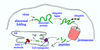
Steps from protein in cell to being expressed in MHC I molecule on the surface of the cell.
- Marked for destruction by ubiquitin
-
Ubiquitin ligases have the inherent ability to recognize abnormal proteins
- Virally infected/misfolded protein
-
Ubiquitin ligases have the inherent ability to recognize abnormal proteins
- Protein degraded to peptides by proteosome
- typically 9 amino acids
- peptides are transferred into the rough ER via a TAP transporter
- Meanwhile, an MHC I molecule is synthesized and placed in the membrane of the rough ER
- peptide binds in the groove in an MHC I molecule
- combination moves through the Golgi apparatus and into a secretion vesicle
- Exocytosis of the secretion vesicle places the MHC I molecule with its peptide on the surface of the cell
- peptide and MHC I molecule are now in position to be recognized by a T cell receptor on a T cell
MHC II: Structure
- also has two polypeptide chains
- each polypeptide chain consists of an intracellular domain, a transmembrane domain, and two extracellular domains
- domain structure is similar to the MHC I molecule.
- big difference, however, is that a peptide from a phagocytized protein is bound the the MHC II molecule on the surface of the cell

Steps in phagocystosed protein being expressed on MHC II molecule
- pathogen is phagocytized, winding up in a phagocytic vesicle
- lysosome with proteases fuses with the phagocytic vesicle, and the proteases digest the proteins into peptides
- Meanwhile the MHC II molecule is synthesized in the rough ER
- vesicle with the MHC II molecule now fuses with the vesicle containing the peptides, and a peptide bind to each MHC II molecule
- Exocytosis again places the MHC molecule and its peptide on the surface of the cell
- peptide and MHC II molecule are now in position to be recognized by a T cell receptor on a T cell

Do you remember the term for the general type of molecule used by phagocytes to recognize a newly encountered foreign molecule? In other words, what is the general type of molecule used to recognize an antigen before a specific immune response has had a chance to make antibodies or T cell receptors?
A good term is innate receptors. One important group is the toll-like receptors. Another is the mannose receptor, which recognizes a repeating carbohydrate pattern
Do you recall the term for the type of molecule that specifically binds the peptide displayed on an MHC molecule?
T cell receptor
Dendritic Cells: Role and Action
- distributed throughout the body
- phagocytosis is their key process
- not primarily for the purpose of destroying microbes
- capture antigens and transport them to lymphoid tissue
- facilitate the development of an immune response
- Display peptides in MHC II molecule
Helper T-Cell: Role and Action with dendritic cells
- In lymphoid tissue
- check to see if their T cell receptors specifically bind the peptide displayed in the MHC II molecule on dendritic cell
- certain dendritic cells also have a mechanism for displaying peptides on MHC I molecules
Two Common Features of T-Cells
- All T cells have T cell receptors
- remain attached to the membranes of the T cells
- always recognize peptide antigens presented on MHC molecules
*
CD4+ T-Cells
- bind peptides displayed on MHC II molecules
- Only with phagocytic cells
- dendritic cells (or macrophages)
- B cells
Activated T-Cells
- T-Helper Cells
- T cell recognizes its specific peptide antigen presented on a dendritic cell or B cell
TH1 vs. TH2 Cells: Tendency to Form
TH1 tend to form when:
- lots of strong stimulation by the phagocytized antigen
- lots of activation of the innate immune system
TH2 tend to form when:
- weaker, more prolonged stimulation
- less activation of innate mechanisms
Important Role of TH1
- travel around the body to macrophages that have phagocytized the antigen
- bind and release IFN-gamma
- increases the fusion of lysosomes with phagosomes
Why aren’t macrophages always active?
- Killing mechanisms can damage the body as well
Which of the following do you suppose is treated sometimes with IFN-gamma?
a. hepatitis A
b. hepatitis B
c. multiple sclerosis
d. chronic granulomatous disease
e. rheumatoid arthritis
D: You have got it! The interferon-gamma would be expected to stimulate exactly the process that is weak in macrophages in this disorder.
Helper T Cells and “helping B cells”
- Help B cell’s respond to antigens
- Many antigens cannot by themselves cause a specific B cell to divide into a clone of antibody secreting plasma cells
- B Cell Divide into Clone:
- Phagocytose antigen and display peptides on MHC II
- activated helper T must bind to the cell
- T helper release appropriate cytokines
TH1 vs TH2: Types of antibodies made
- cytokines secreted are different for different types of helper T cells
- TH1
- Heavy chain switching to make IgG
- Good opsonin
- TH2
- IgE
- IgM
- IgA (MALT)
what term refers to the type of antigen that can activate a B cell without necessarily requiring help from helper T cells?
multivalent
When do CD8+ T-Cell Divide into Clone?
- following its encounter with another cell displaying its specific peptide on an MHC I molecule
- In a lymph node this initial cell is likely to be a dendritic cell
- Termed Cytotoxic T-Cell
Cytotoxic T-Cells
- look for ordinary cells in the body displaying the specific peptide on MHC I molecules
- Virally infected cells
- Must undergo apoptosis
How Cytotoxic T-Cells Induce Apoptosis
- TCR to the peptide in the MHC I
- forms an adhesion complex with the infected cell
- Releases secretion vessicles
- Secretion vessicles contain perforin
- forms channels in the infected cell
- Also contain granzyme
-
proteases that activate certain caspases
- Set of enzymes that trigger apoptosis processes
-
proteases that activate certain caspases
- Fas ligand, can also activate caspases via another pathway