Midterm III - Helminthology 1 [INCOMPLETE] Flashcards

1
Q
A

Trematoda: Egg

2
Q
A

Trematoda: Miracidium (released from egg)

3
Q
A

Trematoda: Sporocyst

4
Q
A

Trematoda: Sporocyst containing rediae

5
Q
A

Trematoda: Cercaria

6
Q
A

Trematoda: Cercaria

7
Q
A

Trematoda: Full cercaria

schistosoma

Typical form of the blood flukes

8
Q

[White spheres]

A

Trematoda: Metacercariae

9
Q
A

Fasciola hepatica (Common liver fluke)

Haemorrhagic tracks in a sheep’s liver

10
Q
A

Fasciola hepatica (Common liver fluke)

Haemorrhagic tracks in a sheep’s liver

11
Q
A

Fasciola hepatica (Common liver fluke)

Identifying features:

  • Dorsoventrally flattened/Leaf-like
  • Grey-brown
  • Conical head shape (with shoulders)
12
Q
A

Fasciola hepatica (Common liver fluke)

Identifying features:

  • Dorsoventrally flattened/Leaf-like
  • Grey-brown
  • Conical head shape (with shoulders)

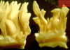
[See the yellow eggs]

13
Q
A

Fasciola hepatica (Common liver fluke)

Identifying features:

  • Dorsoventrally flattened/Leaf-like
  • Grey-brown
  • Conical head shape (with shoulders)

[Notice the yellow eggs]

14
Q

*MIDTERM*

A

*Fasciola hepatica (Common liver fluke)*

Identifying features:

  • Digested blood in the intestine = Liver fluke
15
Q
A

Fasciola hepatica (Common liver fluke)

Identifying features:

  • Cross-section showing intestine with digested blood
16
Q
A

Fasciola hepatica (Common liver fluke)

Identifying features:

  • Enlarged gall bladder & bile duct
17
Q
A

Fasciola hepatica (Common liver fluke)

Identifying features:

  • Enlarged bile duct wall
18
Q
A

Radix labiata

Occasional host of Fasciola

19
Q
A

Galba truncatula

Most common host for Fasciola & Calicophoron spp.

20
Q
A

Rumen fluke

Either Paramphistomum or Calicophoron spp.

21
Q
A

Rumen fluke

Either Paramphistomum or Calicophoron spp.

  • 1 large ventral sucker; 1 smaller oral sucker
22
Q
A

Rumen fluke

Either Paramphistomum or Calicophoron spp.

Identifying features:

  • 1 large ventral sucker
  • 1 smaller oral sucker
  • Genital opening in the centre
23
Q
A

Rumen fluke [Ventral end] Cross section

Either Paramphistomum or Calicophoron spp.

24
Q
A

Planorbid snail

Intermediate host for Paramhistomum spp.

25
Effect of young rumen flukes moving in the s. intestine mucosa
26
Developing rumen flukes on the abomasal wall
27
Rumen fluke [Among ruminal papillae] Either *Paramphistomum* or *Calicophoron spp.*
28
Rumen fluke: Non-embryonated egg Either *Paramphistomum* or *Calicophoron spp.*
29
*Dicrocoelium dendriticum* (Lancet fluke)
30
*Dicrocoelium dendriticum* (Lancet fluke)
31
*Dicrocoelium dendriticum* (Lancet fluke)
32
*Dicrocoelium dendriticum* (Lancet fluke)
33
*Dicrocoelium dendriticum* (Lancet fluke): Egg **Hard & thick shell** - Detect with **floatation**
34
*Dicrocoelium dendriticum* (Lancet fluke): Eggs
35
*Dicrocoelium dendriticum* (Lancet fluke): Cercaria Found in slime balls on grass
36
*Dicrocoelium dendriticum* (Lancet fluke): Metacercaria
37
*Dicrocoelium dendriticum* (Lancet fluke) Chronic dicrocoeliasis: Thickened bile ducts
38
*Dicrocoelium dendriticum* (Lancet fluke) Chronic dicrocoeliasis: Thickened bile ducts
39
*Dicrocoelium dendriticum* (Lancet fluke) **This can be identified by:** * Presence of eggs
40
How to distinguish between Liver fluke and Lancet fluke?
* **Liver fluke:** Visible digested blood in the intestine * **Lancet fluke:** Visible eggs present
41
*Moniezia benedeni* **Identifying features:** * Each proglottis has **2** sets of genitals which open on both sides
42
*Moniezia benedeni:* Egg **Identifying features:** * Quadrangular egg shape * Oncosphaera is eccentric
43
*Moniezia expansa:* Egg **Identifying features:** * Triangular egg shape * Oncosphaera is central
44
*Anoplocephala perfoliata:* Egg ooooor monezia expansa? heste egg er brødskive form **Identifying features:** * Semicircular shape * Contains 6 hooked oncosphera inside
45
*Dipylidium caninum (Flea/cucumber tapeworm)*: Mature segment **Identifying features:** * Genital openings on both sides (impressions on the surface) * 2 x Ovaries in the centre * Testicles fill the central part of the proglottis
46
*Dipylidium spp.:* Cocoon **Identifying features:** * Oncospheres fill the centre
47
*Diphyllobothrium spp:* Eggs **Identifying features:** * Resemble the features of Trematode eggs * No oncosphera
48
*Taenia spp.:* Rostellum **Identifying features:** * Strong hooks on the rostellum [Ø T. saginata] * Impressions of 2 x suckers (acetabuli)
49
*Taenia spp.:* Mature segment **Identifying features:** * Mass of scattered testicles * 2 longitudinal excretory canals * 2 genital ducts parallel to the genital opening
50
*Taenia spp.*: Gravid segment **Identifying features:** * Branches of uterus are filled with white eggs
51
*Taenia spp.:* Egg **Identifying features:** * Thick refractive eggshell * Six-hooked oncosphere (not all hooks are visible) * Colour: White-brown
52
*Taenia spp.*: Cysticerci ## Footnote *Cut surface of skeletal muscle*
53
*Taenia multiceps*: Coenurus cerebralis larva White solices can be seen on the opened wall
54
*Echinococcus granulosus*: Adult **Identifying features:** * Scolex with a rostellum & suckers * Short neck * Immature segment & mature segment * Gravid segment * Centrally-aggregated eggs
55
*Echinococcus hyatidosus* **Identifying features:** * Spherical protoscolices attached to the wall of inner broad capsules
56
Difference between a *Cysticercus pisiformis* & an *Eccinococcus interlocularis* necropsy specimin.
* *Cysticercus pisiformis:* Lesions appear "**on**" the liver * *Eccinococcus interlocularis:* Lesions appear inside the liver
57
*Cysticercus bovis*
58
* Cysticercus pisiformis* * Note the lesions appear "***on**" *the liver*
59
*Mesocestoides lineatus*
60
*Cysticercus tenuicollis*: On a liver
61
*Taenia pisiformis*: Gravid segment
62
*Taenia saginata:* Egg
63
*Cysticercus:* Heart muscle
64
*Taenia /cysticerus pisiformis* Haemorrhagic tracks in the liver
65
Mature cysticerci on the peritoneal surface cysticercus pisiformis
66
* Cysticercus cellulosae or bovis* * taenia solium or saginata* In the heart muscle
67
*Cysticercus cervi*
68
*Echinococcus multilocularis* larva: *Echinococcus alveolaris*
69
Taenia-type eggs
70
*Echinococcosis interlocularis* Note the cysts are **in** the liver
71
*Echinococcus hyatidosus:* Larvae
72
*Taenia pisiformis*
73
*Taenia pisiformis*
74
*Taenia pisiformis:* Eggs
75
*Taenia saginata:* Eggs
76
*Cysticercus tenuicollis* | (Taenia hydatigena larval form)
77
*Strobilocercus fasciolaris* | (*Taenis taeniaeformis* larval form)
78
* Cysticercus longicollis* * (Taenia crassiceps larval form)*
79
*Coenurus cerebralis* | (*Taenia multiceps* larval form)
80
*Echinococcus granulosus*
81
*Echinococcus granulosus*