Mitral Valve Disease Flashcards

(63 cards)

1
Q

mitral valve

  • discuss its anatomical structure and location in the heart.
  • under physiological conditions - when is it open and how wide does it open?
A
  • structure: a bicuspid valve made of 2 leafelets
    • its opening is enclosed by a fibrous ring
    • its leafelets are anchored by chordae tendinae to papillary muscles on the left ventricular wall
  • location: in beween the LA and LV
  • open: during diastole
    • this allows for filling of the left ventricle
    • normally opens to a cross sectional area of 4-6 cm2
2
Q
  • cross sectional area of mitral valve
    • what is normal?
    • when do we start to see blood flow impairment?
    • when does the valvular area become “critical”?
  • how do we determine this area?
A
  • normal cross sectional area: 4-6 cm2
  • blood flow impairment seen at < 2 cm2
    • symptoms start to present at this area
  • valve area becomes critical < 1 cm2

cross sectional area determined with echocardiogram.

3
Q

what is mitral stenosis?

A

decreased pliability of the mitral valve leafelets that impedes blood flow from the LA to LV

4
Q

what are the major causes of mitral stenosis?

A
  • rheumatic fever - #1 cause
  • congenital
  • aging with annular severe calcification
  • SLE (lupus)
  • RA (rheumatoid arthritis)
5
Q

discuss the etiology of rheumatic fever

  • what sex/demographics does it effect most
  • what valvular diseases can it lead to
A
  • # 1 cause of mitral stenosis worldwide
    • 40% of valvuar manifestations of RF are mitral stenosis, and the remainder are mitral regurgitation or aortic valve disease.
      • theses valvular manifestations usually present a few years - 20 years after initial infection
  • demographics: most prevalent developing countries
  • sex: seen more in females > males
6
Q
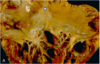
  • discuss the major gross morphological changes seen in rheumatic valvular disease?
  • what changes are characteristic of early vs end stage rheumatic valve disease?
A

i believe these are referring to changes in the mitral valve*

  • major changes:
    • thickening of valve leaflets
    • fusion of comissures
    • chorda tendinae shortening/thickening
  • stage-specific features:
    • early
      • leafelets flap open in a “curved over” manner due to restriction of motion at their tips.
        • this causes an opening snap on ascultation
    • as diease progresses:
      • symmetric fusion of comissures results in a small, central oval orifice during diastole
    • end stage:
      • ​leafelets can be so fibrotic/stiff they cannot open/shut
7
Q

pathogenesis of rheumatic valve disease

A

it is an abrnomal immune response to group A streptococcal pharyngitis

8
Q

what criteria must be met to diagnose rheumatic valve disease

A
  • must see 2 major criteria OR 1 major + 2 minor criteria
    • major criteria
      • carditis
      • polyarthritis
      • erythema migratorum
      • subcutaneous nodules
      • chorea (uncontrolled movement)
    • minor criteria
      • fever
      • arthralgia
      • previous rheumatic fever/known rheumatic heart disease
9
Q

what clinical presentations might indicate carditis?

A
  • pleuritic chest pain
  • friction rub
  • heart failure

carditis is one of the major criteria for dx of rheumatic valve disease

10
Q

what valves are most/least likely to be effected by rhuematic valve disease

A
  • most likely = mitral/aortic valve
  • least likely = pulmonic valve
11
Q

what physiological changes are seen in mitral stenosis

A
  • mitral stenosis = impaired flow from LA to LV during diastole. blood backs up in LA, doesn’t make it to LV. this leads to:
    • LA pressure increase
    • LA enlargement
    • LV filling dimished
      • this decreases end diastolic volume (preload) –> decreases stroke volume –> thus decreases cardiac output
        • (CO = HR x SV)
12
Q

clinical presentation of mitral stenosis

A
  • mild vs moderate
    • mild stenosis = typically asymptomatic
    • moderate stenosis (valve area 1-2 cm2) = exertional dyspnea and fatigue
  • other clinical manifestations:
    • chest pain
    • atrial fibrillation/palpatitations
    • stroke (18 x higher risk in these pts)
    • right sided heart failure
      • presents w/ edema, ascities, liver failure
    • hoarseness (ortner’s dynrome)
    • dysphagia
    • hemoptysis from ruptured bronchial veins
13
Q

what two cinical manifestations of mitral stenosis are due to LA enlargement?

A
  • orntner’s syndrome: hoarseness due to the LA enlarging and putting pressure on the recurrent laryngeal nerve
  • dysphagia: trouble swallowing due to LA enlarging and impinging on the esophagus
14
Q

what kind of heart failure is seen in mitral stenosis & why?

what other clinical manifestations typically present alongside this heart failure?

A
  • right sided heart failure
    • blood back up into LA backs up into lung and then creates a huge afterload that the right heart must overcome.
    • right sided HF can present with:
      • edema*
      • JDV*
      • ascites
      • liver failure
15
Q

discuss key exam findings indicative of mitral stenosis

  • what sounds will heard on ascultation?
  • other findings
A
  • ascultation
    • murmurs/sounds:
      • loud S1 (mitral valve closure)
      • opening snap (mitral valve opening) after S2 (aortic valve closure)
      • low-pitched diastolic flow murmur
        • caused by turbulent blood flow thru valve
    • i_rregular heart rhythm_
  • other findings:
    • signs of right sided heart failure
      • periperal edema
      • JVD
    • stroke symptoms:
      • facial drooping
      • weakness on one side of the body
16
Q

define the clinical significance of the “opening snap” heard on ascultation of a pt with mitral stenosis

A
  • opening snap
    • often heard in the early phase of rheumatic valve diease
    • this sound is caused by the restriced motion of the diseased valve leafelets during mitral valve opening, which occurs after S2 (aortic valve closure)
      • the shorter the interval between S2 and the “opening snap” the more severe the mitral stenosis
17
Q

label these components of a stethoscope. what is purpose/proper orientation of each component

A
18
Q
  • when ausculating a patient with possible mitral stenosis
    • what part of the stethoscope should you listen with and where on the heart should you place it?
    • how should the patient be positioned?
A
  • listen to apex of heart with the bell of the stethoscope
  • place patient in left lateral decubitus
19
Q

what is the “standard” diagnostic test for mitral stenosis

A

echocardiogram

20
Q

echocardiogram for dx of mitral stenosis

  • what types of echocardiograms can be done and in what circumstances?
  • what can an echocardiogram tell you?
A
  • echocardiogram = standard diagnostic test for mitral stenosis
    • two possible views:
      • transthoracic - usually preferred
      • transesophageal (TEE) - preferred when 1. it is difficult to view valves or 2. we’re assessing for vegetations/thrombus
    • echocardiogram is key to tell you:
      • degree of stenosis
        • mild: mitral valve area > 1.75 cm2
        • moderate: mitral valve 1.25 -1.75 cm2
        • severe: mitral valve < 1.0 cm2
      • pressures across valves/heart chambers
21
Q

other than an echocardiogram - what other diagnostics tests can be done to assess mitral stenosis and under what circumstances?

A
  • EKG - useful if there is an irregular rhythm. could show
    • LA enlargement
    • right axis deviation
    • RBBB
  • cardiac catheterization - used if echocardiogram is undiagnostic. can tell you pressure in LA/LV
  • exercise test stress - to induce exertional symptoms
22
Q

when to do a cardiac catheterization in diagnosis of mitral stenosis/what can it tell you?

A
  • do if echocardiogram is diagnostic
  • can tell you pressure of LA/LV
23
Q

when do an EKG if the diagnosis of mitral stenosis/what it can tell you

A
  • do if there is an i_rregular rhythm_
  • can show:
    • LA enlargement
    • right axis deviation
    • RBBB
24
Q

how to treat asymptomatic mitral stenosis

A
  • generally, no treatment.
  • unless:
    • there is severe pulmonary artery HTN OR new onset atrial fibrillation, in which case:
      • control HR with beta blocker/CCB
      • and give warfarin (anti-coagulant) if their INR is 2-3
    • they need a percutaneous balloon mitral valveotomy
25
how to treat heart failure secondary to mitral stenosis
give diuretics
26
how to treat mitral stenosis in patients with _exertional symptoms_ (what methods are preferred vs not preferred)
* NOT preferred: percutaneous balloon mitral valcotomy * preferred: * **open/closed surgical mitral valvotomy** * **open heart mitral valve replacement** * mechanical mitral valve: * these patients are given lifelong warfarin (comoudin) anticoagulant * bioprosthetic mitral valve: * for patients older than 65 or who can't take anti-coagulants
27
* when are valve replacements done to treat mitral stenosis? * what are the _two types of valves_ that can be used in an open heart mitral valve replacement? * what are the pros and cons of each?
* done in mitral stenosis patients with _exertional symptoms:_ * valve replacements: * **mechanical mitral valve:** * **​**this valve has a longer lifepan than bioprosthetic valves * these patients are given lifelong warfarin (comoudin) anticoagulant * **bioprosthetic mitral valve:** * **​**dont require anti-coagulants * for patients 65 or older OR who cannot take anticoagulants
28
_complications_ of mitral stenosis
aka, the clinical manifestations of mitral stenosis. this list should be familiar. * LA enlargement * stroke/thrombus formation * pulmonary hypertension * right ventricular hypertrophy * right sided heart failure * peripheral edema * JVD * ascites * hepatic congestion/cirrhosis
29
what is mitral regurgitation
the inability of the mitral valve to fully close, resulting in a backflow of blood int the left atrium
30
primary vs secondary mitral regurgitation
* Primary: resulting from detect of valve & its components * Secondary: regurgitation resuling from _progressive enlargement of LV_ that ends up causingcausing mitral annular dilation
31
what are the two types of non-infective endocarditis? what do they have in common?
* marantic endocarditis and libman sacks disease * both involve **sterile** vegetations
32
33
* marantic endocarditis * describe its vegetations * effects what valves * is associated with what underlying diseases/populations
* marantic endocarditis = a non-infective endocardtitis * vegetations * sterile * small (1-5 mm) * typically occur along the lines of closure * are non-invasive: _do NOT cause inflammatory response._ * **mitral & aortic valve** * **associated w/ underlying systemic hypergoabulable state** * **​**cancers patients, pts with catheters, sepsis, anti-phopholipid disorder * can cause systemic emboli --\> stroke
34
* libman sacks disease * describe its vegetations * what valves does it effects * what underlying diseases/populations is it associated with?
* libman-sacks = a non-infective endocarditis * vegetations * small (1-4 mm) * single or multiple **sterile, pink vegetations** * **warty appearance** * tend to extend into endocardium/chordae tendinae (not confined to closure lines like marantic\*) * tend to cause _intense inflammation!_ (unlike marantic\*) * **mitral & tricusupid valve** * associated with: * **SLE (systemic lupus erythematous):** see in young females, causes strokes * **anti - phospholipid syndrome:** seen mostly on aortic/mitral vavlve
35
what valvular disease is shown here?
marantic endocarditis (non-infective) note the **vegetations confined to the closure lines**
36
what valvular disease is shown here?
libman sacks disease (non-infective endocarditis) note the **pink, warty vegetations** extending onto chordae tendinae/other valve components
37
physiological presentation of _acute_ mitral regurgitation * note the volume/pressure status of the heart chambers * note cardiac ouput status
* regurgitation of blood from LV to LA during _systole_ increases LA pressure and volume. since not all of LV blood is getting to the aorta, the _end diastolic volume_ in the _LV is decreased_ * **_increased_ LA volume and pressure** * **​**can cause pulmonary edema * **_decreased_ LV volume -**-\> **therefore _low_ cardiac ouput**
38
discuss the physiological presentation of _chronic_ mitral regurgitation * note the volumes/pressures of the four heart chambers * note the status of the cardiac ouput
* regurgitation of LV blood into LA _during systole_ increases LA pressure/volume. *over time,* accumulated LA blood + new blood from venous return increases the point of also increasing end diastolic volume in LV. * **i**_ncreased_** LA volume and pressure** * **_increased_ LV volume and pressure** * **​cardiac output _maintained_**
39
clinical presentation of acute mitral regurgitation
* **acutely ill and often in cardiogenic shock\*** * **​**due to _low cardiac ouput_ unique to acute MR. * this would be indicated by *dyspnea, hypotension, tachycardia, cool extremities, diaphoresis* * **chest pain** * **acute pulmonary edema**
40
clinical presentation of chronic mitral regurgitation
* _very similar to that of mitral stenosis_ due to LA volume "back up" the lung & right heart * dyspnea * peripheral edema * JVD * ascites * palpitations/atrial fibrillation * stroke
41
what are the key exam findings pertinent to mitral regurgitation? * describe sounds heard on ascultation * other clinical findings
* ascultation * _sounds/mumurs:_ * **S1 decreased** (soft mitral valve closure sound) * **S2 is split** (delay between seminlunar valve closure) * occasional S3 heard - *would sound like kentucky* * **_systolic murmur:_** created by blood flow thru mitral valve during sysole when it _should be shut._ * ​_holosystolic murmur_ in chronic MR * _early systolic murmur_ in acute M * _possible laterally displaced PMI_ * *seen chronic MR, due to enlarged LV* * other exam findings * may see signs of right heart failure - ascites, JVD * may see signs of _cardiogenic shock_ - hypotension, tachy, cold skin - if acute MR
42
* .discuss the _systolic murmur_ heard in mitral regurgitation * how is it different beween acute vs chronic MR? * do we hear this in mitral stenosis?
* acute vs chronic MR: * acute MR --\> **early systolic murmur** * chronic MR **--\> holosystolic murmur** * heard throughout systole (the _whole time_ between S1-S2) * radiates to the axilla * do not hear a systolic murmur in mitral stenosis. * in MS, the mitral valve closes sufficiently during systole but _does not open sufficiently_ in diastole. thus, in MS we hear a **_diastolic murmur_ -** created by turbulent blood flow during diastole
43
diagnostic testing for mitral regurgitation * what is the "standard"? what are other useful tests and in what circumstances? * compare/contrast this to dx of mitral stenosis
* like in mitral stenosis: * echocardiogram is the diagnostic standard * TTE - sufficient in most circumstances * TEE - for detailed valvular anatomy/presence of thrombi * EKG - if you suspect rhythm abnormality * cardiac catheriziation - done if echocardiogram does not give definitive diagnosis * unlike mitral stenosis: * **consider chest x-ray** to assess _fluid overload/overall heart size_ * _don't_ need to do an exertional test
44
how to treat severe acute mitral regurgitation
* **most important: treat shock** * treat underlying cause of MR * consider surgery
45
how to treat _chronic asymptomatic_ MR
* control HTN * medications: warfarin if a-fib present * monitor w/ echocardiogram every 6-12 months * consider surgical repair if: * a-fib present * of EF \< 60% or LV end-diastolic volume of 4- mm * pulmonary HTN present
46
how to treat _chronic symptomatic_ mitral regurgitation?
* *just like with asymptomatic chronic MR*: * control HTN * give warfarin if they have a-fib * specific tx for primary vs secondary chronic symptomatic MR: * **primary MR:** * **tx = valve surgery:** * **_repair:_** **open vs mitral cip** * mitral clip: * not as effective as open * but minimally invasive * *is thus good for high risk patients who cannot undergo open heart surgery as it can reduce symptoms enough to improve quality of life* * open alfari stitch repair * **_replacement:_ mechanical vs bioprosthetic valve** (same as MS) * recall that mechanical valves have a longer lifespan but require lifelong anti-coagulants * **secondary MR:** * **​no surgery** * since secondary MR is due to _LV enlargement rather than a defect of the valve itself_, repair/replacement does not fix the underlying problem
47
what is mitral valve prolapse?
myxomatous degermation of the mitral valve that causes leafelets to _bulge back into_ the _left atrium_
48
symptoms of mitral valve prolapse
* symptoms * Palpitations * Syncope * Chest pain not associated with CAD
49
exam findings of mitral valve prolapse * what will the ausculation show? * other exam findings? * what diagnostic test is _standard_ to dx for mitral valve prolapse?
* ascultation: * could show **mid-systoic click** followed by **late-peaking systolic murmur** * other exam findings: typically normal unless MR (mitral regurgitation) is also present * diagnostic standard: echocardiogram * (like with all valvular diseases)
50
what is the treatment for mitral valve prolapse?
* typically no treatment _unless MR_ is also present * if there is treatment: * possible beta-blockers - *if palpitations* * baby aspirin - *to reduce stroke risk* * if surgery: * done when pt's have severe MR symptoms * men typically have surgery more (if someone)
51
triscuspid valve * describe its anatomy * where in the heart is it found * when during the cardiac cycle is it typically open? * what is its normal valvular area?
* three valves * between RA and RV * open during systole * normal valvular area: 4-6 cm2
52
* describe the disease incidence of the tricuspid valve relative to the other valves * what diseases most commonly effect the tricuspid valve and in what populations?
* lower incidence of disease than in aortic/mitral valves * triscupid valve disease is most commonly **seen IV drug users**, and is usually one of the two: * rheumatic heart disease * infective endocarditis (IE)
53
tricuspid stenosis * incidence * causes
* incidence: rare * causes: * rheumatic heard disease - most common * other causes: * carcinoid heart disease * myxoma/metastasis * vegetations obstructing RV inflow * typically from pacemaker leads
54
carcinoid heart disease * cause * pathogenesis * gross presentation * associated clinical presentaiton * what markers aid in diagnosis of this cardinoid heart disease?
* cause/pathogenesis/presentations: * cardinoid tumors (though, typically not GI carcinoid tumors) * _bioactive products_ released from these these cardinoid tumors - serotonin (5-HT), kallikrein, bradykinin, histamine, prostaglandins - cause **"carcinoid syndrome"**- which can lead to the following: * _clinical presentation:_ * nausea/vomitting/diarrhea * cramps * _episodic skin flushing_ * _gross cardiac presentation:_ * **​plaque-like thickening** of the endocardium & valves of the heart. specifiaclly, the **right heart valves- pulmonary & tricuspid** * markers: * plasma levels of _serotonin_ and _5-hydroindoleacetic acid_ (urinary serotonin) correlete to severity of the lesions
55
_clinical presentation_ resulting from carcinoid heart disease
* symptomatology resulting from carcinoid heart disease is largely due to the **increased systemic venous pressure** resulting from increased **_right sided pressure_** * **​peripheral edema** * **abdominal distention** * **RUQ pain/fullness** * **neck pulsation**
56
exam findings seen with carcinoid heart disease: * auscultation * other exam findings
* _right sided_ heart failure findings: * **JVD with cannon-a waves** * hepatomegaly * ascites * peripheral edema * auscultation: * murmurs: * **low pitched diastolic mumur** at the **_lower left sternal edge_** * _​_due of turbulent flow thru tricuspid valve during diastole (tricuspid located at left lower sternal border) - all physicians take money * **increase with inspiration** * often heard alongside _mitral stenosis_ murmurs
57
what is mitral regurgitations and its primary causes?
* inability of the leafelets the fully close * caused by same causes as carcinoid heart disease: *
58
* what are the diagnostic tests to order for carcinoid heart disease and what key things will they show?
* echocardiogram - standard * shows **tricuspid valve pathology:** thickened leafelets, decreased mobiliby, doming * EKG: * **possible** **peaked P-wave in lead II** * **​**due to RA enlargement * atrial fibrillation * Chest X-ray: * **RA enlargement - seen as** **prominence of right heart border**
59
what is the treatment regimen for _carcinoid hear disease:_
* first: _relieve venous congestion_ * use **diruetics** or **nitrates** (vasodilators) * next: dubulk obstructive tumors/myoxomas in the heart * consider the following: * control a-fib * anti-coagulants if needed * surgical valve replacement: * do if * mean diastolic pressure gradient **\> 5mmHg** & **tricsupid orifice \< 2.0 cm2** * or if concurrent mitral valve repair is happening * do NOT do balloon valvoltomy
60
what is tricuspid valve regurgitation and its common causes?
* inability of valve leafelets to close * causes: * most are same as tricuspid stenosis/carcinoid heart disease: * rheumatic heart disease * IE * myxomatous disease * carcinoid heart * could also be dilation due to --\> pulmonary HTN --\> putting pressure on RV --\> RV overload opens trisupid valve
61
clinical presentation of tricuspid regurgitation
* Symptoms often associated with left sided heart disease or pulmonary disease * Dyspnea * Paroxysmal nocturnal dyspnea * Peripheral edema * Abdominal distension * Peripheral edema * Chest pain
62
_exam findings_ for **tricsupid regurgitation** * what will be seen on ascultation * other findings
* ascultation: * murmurs * **_low intensity holocystolic murmur:_** * due to open tricsupid valve when it should be closed during systole * heard at _left sternal edge/subxiphoid area_ * may hear S3 * **prominent cv wave** * signs right sided heart failure * JVD * ascites * hepatomegaly * perpheral edema
63
treatment for tricuspid valve?
* medications * treat HF with diuretics * treat underlying cause (COPD, pulmonary HTN, ect) * surgery: * **surgical tricsupid annuloplasty** is the go-to * a "semi-prosthetic" ring * valve replacement is rare * but it if is, use **bioprosthetic valve** * this is because a bioprosthetic valve doesnt require anti-coagulants, and the tricuspid valve is _fairly prone to thrombosis_