Mod VI: Congenital Heart Disease Flashcards
(90 cards)
Congenital Heart Disease
Congenital Heart Diseases are present in about what percentage of newborn infants?
1%
Congenital Heart Disease
What are the causes of Congenital Heart Disease?
Idiopathic
Genetic
Environmental
(rubella 1st trimester, lithium, FAS)
Congenital Heart Disease
What are risk factors for Congenital Heart Disease?
Parent with CHD
Prematurity
Multiple gestations
Noncardiac congenital anomalies (Down’s syndrome)
Congenital Heart Disease
Signs & Symptoms of Congenital Heart Disease in infants are:
Tachypnea
Failure to gain weight
Tachycardia (>200)
Heart murmur
Congestive heart failure
Hypoxemia
Cyanosis
Congenital Heart Disease
Signs & Symptoms of Congenital Heart Disease in children are:
Dyspnea
Failure to grow
Decreased exercise tolerance
Heart murmur
Congestive Heart Failure
Cyanosis
Clubbing of digits
Squatting (To increase SVR)
HTN
Chest pain
Congenital Heart Disease
T/F: Most Congenital Heart Diseases are diagnosed prior to birth
True
Congenital Heart Disease - Diagnosis
T/F: Congenital Heart Disease is apparent during first week of life in 50% of afflicted neonates and before 5yrs in all remaining
True
Congenital Heart Disease - Diagnosis
What’s the initial diagnostic test recommended for CHD?
US Echocardiography
Congenital Heart Disease - Diagnosis
Test that demonstrates valvular dysfunction and septal defects
Doppler US
Congenital Heart Disease - Diagnosis
Tests that demonstrate anomalies involving great vessels
CT scan - MRI
Congenital Heart Disease - Diagnosis
What’s the most definitive diagnostic technique for CHD?
Cardiac catherization
Congenital Heart Disease
Problems afflicting patients with Congenital Heart Disease include:
Pulmonary vascular disease & associated PHTN
Congestive heart failure
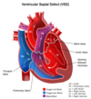
Infective endocarditis (VSD/PDA)
Requires prophylaxis antibiotics
Hypertension (Coarctation)
Polycythemia (HCT > 65%)
Physiologic response to chronic hypoxemia - Increases risk for thromboembolism
Coagulation defects
Deficiency in VT K clotting factors - Defective PLT aggregation
Brain abscess development
Problems Afflicting Patient with Congenital Heart Disease
Congenital Heart Disease a/w Infective endocarditis (VSD/PDA) Requires Prophylaxis with which drugs?
Antibiotics
Problems Afflicting Patient with Congenital Heart Disease
Polycythemia (HCT > 65%) a/w Congenital Heart Disease is a physiologic response to:
Chronic hypoxemia
Problems Afflicting Patient with Congenital Heart Disease
Polycythemia (HCT > 65%) a/w Congenital Heart Disease increase risk for:
Thromboembolism
Problems Afflicting Patient with Congenital Heart Disease
Coagulation defects a/w Congenital Heart Disease are a consequence of:
Deficiency in Vit K clotting factors
Defective PLT aggregation
Pathophysiology of Congenital Heart Disease
T/F: Management of anesthesia for patients with CHD requires a thorough knowledge of the pathophysiology of each cardiac defect
True
However, this is confusing due to complexity of lesions
Utilization of a structured approach that emphasizes ratio of pulmonary blood flow & systemic blood flow based on resistance in these vascular beds is helpful
Pathophysiology of Congenital Heart Disease
Important pathophysiologic questions w/ CHD include:
Is there on obstruction?
Is there a shunt?
Pathophysiology of Congenital Heart Disease
What are the effects of R side obstruction?
Blood unable to go from RV to lungs
↓ pulmonary blood flow => hypoxemia/cyanosis
Blood does not get oxygenated
Pathophysiology of Congenital Heart Disease
What are effects of L side obstruction?
Blood unable to flow from LV to systemic circulation
Tissues organs do not get perfused
↓ systemic blood flow => hypoperfusion/acidosis/shock
Pathophysiology of Congenital Heart Disease
How can shunt be defined?
Mixing of pulmonary/systemic circulations
(or mixing of oxygenated and de-0xygenated blood)
Pathophysiology of Congenital Heart Disease
What determines the direction of of shunt?
Ratio of pulmonary blood flow (Qp) / systemic blood flow (Qs)
Qp:Qs
Pathophysiology of Congenital Heart Disease
Qp:Qs < 1 means:
Pulmonary blood flow < Systemic blood
Instead of flowing to the lungs, blood is flowing to the left side
[R to L shunt]
Blood flowing directly to the left fails to be oxygenated
This leads to hypoxemia and cyanosis
Ineffective pulmonary blood & mixing systemic/pulmonary circulations => hypoxemia/cyanosis
Pathophysiology of Congenital Heart Disease
Qp:Qs > 1 means:
Pulmonary blood flow > Systemic blood
[L to R shunt]
Volume/pressure overload of R ventricle => CHF
Pulmonary overcirculation => Pulmonary HTN/ ↑ PVR