Module One Flashcards
(9 cards)

Late 3rd Week
Cut edge of amniotic membrane
Arrow points to primitive node at junction of primitive streak and posterior end of neural groove.
Neural groove bounded by neural plate which will form neural folds.
Mesoderm forms by ectodermal–>mesenchymal transition in primitive groove.
Primitive pit anterior to primitive node–>notochordal process

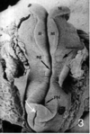
Late 3rd Week
Widened neural folds of head plate flare and form a shelf
X=Prochordal plate
BP=brain plate unsegmented
Fb=forebrain
Primitive pit is visible
Early 4th Week
Rapid anterior growth leads to small primitive streak (arrow)
NT=neural folds which have elevated and begin to approach over NG (neural groove)
Somites can be distinguished by looking for horizontal separating grooves. Fusion of neural folds begins here.
PN=posterior neuropore which will be the last place fusion occurs. Most common site for spina bifida.
M=midbrain
H=hindbrain
Bulging areas next to hindbrain are developing cardiac process and 1st mandibular branchial arches

Early 4th week
Triangle at junction of forebrain and midbrain
Hindbrain marked by square
Forebrain extends over prochordal plate and what will become the stomodeum (arrow)
1st branchial arch (circle) begins to form below midbrain near origin of CN V
Pericardial cavity and embryonic heart visible beneath arch

Early 4th Week
Cervical fold has taken place
FB=forebrain
MB=midbrain
HB=hindbrain
Stomodeum and foregut have come into more adult anatomical relationship
Buccopharyngeal membrane shown by arrow separating stomodeum from foregut

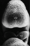
Early-Mid 4th Week
Anterior folding has begun
Within cardiac bulge heart has begun to beat
Neural folds of forebrain (telencephalon and diencephalon) and of membrane have started to fuse
Hindbrain folds still wide apart
After fusion in anterior, single broad and smooth surface called frontonasal proces FNP
Mandibular process of first arch is now visible and lies between FNP and cardiac bulge

Early to mid 4th week
Dorsal aspect
MB and HB neural folds still widely separated
Neuroectoderm medially and integumental ectoderm laterally

Mid 4th Week
Fusion of brain plate nearly complete
1st branchial arch has enlarged and extends forward beneath FNP
Slit like invagination is stomodeum (primitive oral cavity)
Heart has been removed but cut edge of pericardium obscures opening of stomodeum in center
Mid to Late 4th Week
Midline fusion completed and FNP smooth
Stomodeum deep cavity bordered by FNP and mandibular processes (Mnd) below
Mnd merge in v shape groove
Maxillary processes (mx) bound the stomodeum laterally and appear as slight bulges between Mnd and FNP
