More diseaess-Kerr, Dry mouth Flashcards
(100 cards)
Erythroplakia
Treatment
○ Biopsy required for diagnosis
○ If a source of irritation can be identified and removed, biopsy may be delayed for 2 weeks to allow lesion to heal
○ Complete excision
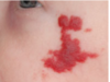
What is this clinical presentation?

Erythroplakia.
Well-circumscribed red patch on the
posterior lateral hard and soft palate
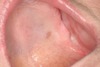
What is this clinical presentation?

Erythroplakia.
Erythematous macule on the right
floor of the mouth.
Biopsy–
Turned out to be early invasive squamous cell
carcinoma.
What is this clinical presentation?

Smokeless tobacco keratosis/TOBACCO POUCH KERATOSIS
Smokeless Tobacco–related Gingival Recession.
Extensive recession of the anterior mandibular facial gingiv
What is this clinical presentation?

Smokeless tobacco keratosis/TOBACCO POUCH KERATOSIS
Tobacco Pouch Keratosis, Severe
What is this clinical presentation?

Smokeless tobacco keratosis/TOBACCO POUCH KERATOSIS
Tobacco Pouch Keratosis, Mild. A soft, fissured,
gray-white lesion of the lower labial mucosa located in the area of
chronic snuff placement.
Smokeless tobacco keratosis
Treatment:
typically resolves weeks after cessation
○ if persists 6+weeks -> biopsy to rule out dysplasia + SCC
What is this clinical presentation?

Pemphigus Vulgaris.
. Multiple erosions affecting the
marginal gingiva.
What is this clinical presentation?

Pemphigus Vulgaris.
Multiple erosions of the left
buccal mucosa and soft palate.
What is this clinical presentation?

Pemphigus Vulgaris.
Large, irregularly shaped ulcerations
involving the floor of the mouth and ventral tongue.
What is this clinical presentation?

Pemphigus Vulgaris.
What is this clinical presentation?

Pemphigus vulgaris
● Multiple, chronic, mucocutaneous ulcers
● Many patients also have
● Relatively non‐specific
● Very superficial, only in epithelium
● Occur on any mucosal surface: oral, ocular, nasal, GI, esophageal,
genital
What is this clinical presentation?

Pemphigus vulgaris
PV Lesions can affect
virtually any mucosal
surface (oral, nasal,
ocular, pharyngeal,
esophageal, genital)
What is this clinical presentation?

Pemphigus vulgaris
usually suffer from Desquamative
gingivitis (DG)
More superficial erosion of the marginal gingiva, typically with an
intense erythema and inflammation, and very often in the absence of
local factors that would typically cause a gingivitis
o Hurts to brush their teeth
Immediately look for areas where there are no local factors and look for
inflammation there
o To check the possibility of systemic factors causing local
gingivitis
What is this clinical presentation?

Pemphigus vulgaris
Combination of PV
inflammation and
gingival inflammation
accumulating local
factors can result in
advanced loss of
attachment and tooth
loss
Pemphigus vulgaris
Etiology
Pemphigus vulgaris is not fully understood.
Experts believe that it’s triggered when a person who has a genetic tendency to get this condition comes into contact with an environmental trigger, such as a chemical or a drug.
In some cases, pemphigus vulgaris will go away once the trigger is removed.
Pemphigus vulgaris
Treatment
Treatment has 3 stages:
● Stage 1: Control
○ Suppress inflammation / lesion activity with Systemic Corticosteroid: Remains initial / 1st‐line treatment…
○ Then quickly add steroid‐sparing agents (mycophenolate mofetil) to minimize dose and duration of corticosteroid treatment as well as improve disease control
● Stage 2: Consolidation
○ Reducing auto‐antibody production with the addition of Immunosuppressants
○ Assessed by the lack of development of NEW lesions
● Stage 3. Remission / Maintenance:
○ achieving complete remission of lesion activity OFF medication is the GOAL
○ When lesion activity OFF medications cannot be achieved, principle of MINIMALLY effective therapy is the goal, typically with combination of immunosuppressant medications
○ RITUXIMAB has become the FIRST CHOICE treatment after
○ the consolidation phase to achieve DISEASE REMISSION
● TOPICAL / INJECTABLE CORTICOSTEROID MEDICATIONS
○ o Can be used to help control limited number of lesions resistant to systemic therapy: it treats ONLY the disease
○ outcome (lesions) and not the systemic illness / pathologic antibody production
○ ex:clobetesol 0. 05% , halbetesol 0.05% (most potent)
What is this clinical presentation?

Mucous membrane pemphigoid
What is this clinical presentation?

Mucous membrane pemphigoid
SEVERE/HIGH RISK FORMS OF MMP
▪ Ocular
▪ Esophageal
can
result in functional
blindness
What is this clinical presentation?

Mucous membrane pemphigoid
Oral Hygiene: Plaque
related gingival
inflammation
contributing to
continued VB
desquamative
gingivitis
What is this clinical presentation?

Mucous membrane pemphigoid
REMEMBER:
▪ Plaque and calculus can be the consequence of painful MMP lesions
▪ When assessing MMP lesions/desquamative gingivitis, look for areas of intense inflammation WITHOUT local factors as evidence of VB disease
Mucous membrane pemphigoid
Etiology
Mucocutaneous autoimmune disease characterized by sub‐epithelial
blisters (bullae) which ruptures to form large, non‐healing ulcerations
Mucous membrane pemphigoid
Treatment
o Approach is similar to PV – but generally not as aggressive unless
hi‐risk areas ( ocular, esophageal ) where more intense immunosuppression indicated
▪ NON‐immunosuppressive treatments uniquely effective:
- *o** Dapsone
- *o Tetracycline + nicotinamide**
MMP & PV BIOPSY
take two different sites
○ For H&E, still must be perilesional
○ If you get only ulcer just because the clinician thinks
○ that is the pathology → there is no epithelium!
○ The sample is useless and no diagnosis can be made