Morphological plan Flashcards
(39 cards)
Evolution of the upper limb
Most mammals are Quadrapedal which means that they use all 4 limbs for getting around.
The upper limbs sits right under the trunk and this is really important for locomotion when running as well climbing.
An example of quadrupeds include the large cats.
However, men have evolved and we are are Bipedal.
we freed our upper limbs for other uses.
these changes have evolved due to environmental changes like hunter gatherers, making stone tools, carrying our young ) . we are not the only bipedal.
so during human evolution we have gone from being quadrupeds to bipeds.
Quadrupeds
animals hat has all four limbs specialized for walking.
Bipeds
an animal that uses two legs for walking.
Anatomical evolution:
Changes to the foot, hip, knee, vertebral column, skull
- we now have Longer clavicles
- Pronation/Supination of the limbs (putting up and down)
- we have developed an Opposable thumb which is important for gripping objects
*All of these has enabled us to mechanically interact with the environment
Design of the Upper limb
- 32 bones
- 57 muscles
- brachial plexus
- major vessels
Arrangement of upper limb
- shoulder region
- Arm
- Elbow
- forearm
- wrist
- hands
skeleton of the upper arm
1. Pectoral girdle = is the name for the scapula and the clavicle
2. Humerus (the pectoral girdle articulates with the humerus which then articulates with the 2 forearm bones )
3. Radius
4. Ulna (the radius and ulna articulates with the carpal bones)
5. Carpal Bones (there are 8 of them)
6. Metacarpals (5)
6. Phalanges (14)

Scapular and humerus
- The scapula has an inferior and superior angle which one can palpate on themselves.
- There is also a prominent spine of the scapula that sits posteriorly and you can also feel this on yourself
There are 2 bonny prominences.
1. Acromion- which is continuous with the spine of the scapula
2. Coracoid- this translate to crows head, it does look like a crows head.
- we have the glenoid fossa which articulates with the large rounded head of the humerus.
- we have two dorted line on the humerus. one is the anatomical neck and the other is the surgical neck. the surgical neck is more prone to fractures.
- between the two necks we have two tubercules; a greater tubercle and a lesser tubercle and they are groups that muscles are attached.
-DELTOID TUBEROSITY- this is where the deltoid muscle attaches
- There is a LATERAL EPICONDYLE located om the lateral side of the lower humerus
- There is a MEDIAL EPICONDYLE

Tuberosity vs tubercles
- A tuberosity is a large roughened area about the mid-shaft for bones.
- Tubercles tend to sit near the ends of bones. tend to be smaller and more rounded nodules.
The bones of the forearms
they are;
- Radius (Laterally)
- Ulna (Medially)
*At the proximal end, there is a bonny point called the OLECRANON. This is the bony part you feel behind your elbow joint is the olecranon of the ulnar.
*the rounded head of the radius articulates against the ulna and the rounded head is really important for the movement of pronation and supination.
*we can see 2 tuberosities towards theproximal end
1. A radial TUBEROSITY - its the biceps muscles that insert here.
2. A radial TIBEROSITY - a muscle in the forearm called brachialis ins erts into the ulna tuberosity.
- Between the 2 bones there is thin membrane called the Interosseous membrane. Interosseous just meaning between the bone. this membrane holds the 2 bones together.
- At the very distal end of the forearm we have 2 bony points. the STYLOID PROCESS of the radius, the brachioradialis muscle attaches unto this.
- we also have the STYLOID PROCESS OF THE ULNA.

Wrist and hand
- There are 8 carpal bones in two rows of 4.
- The metacarpals (Digits one is the thumb, digits 5 is referring to our small finger)

All the movements that can be performed by the upper limbs
- Flexion/Extension
- Abduction/Adduction
- Lateral/Medial rotation
- Pronation/Spination
- Circumduction
- Opposition / reposition
Joints of the upper limb
1. Glenohumeral joint
This is the joint of articulation between the glenoid fossa of the scapula with the head of the humerus.
- It’s a ball and socket joint which means that is highly mobile
- we can flex and extend this arm and adduct and abduct it. we can put these movements together to perform circumduction which makes the arm go in circles.
- we can also medially rotate the arm at the Glenohumeral joint.

Joints of the upper limb
2. Hinge Joint
This joint is at the elbow, just before the radius and ulnar bone.
-this joint allows extension, supination and pronation of the forearm

Joints of the upper limb
3. radiocarpal joint of the wrist
- Its made up of articulation between the radius and the proximal carpal row .
- this is the wrist joint
- we can extend, flex, abduct, abduction, the hand at the wrist joint
- Abduction is known as radial deviation
- we can also adduct, this is known as ulnar deviation
- you can put those together to circumduct your hand at the wrist joint

Joints of the upper limb
- METACARPOPHLANGEAL JOINTS
- There is not a lot of movement at the midcarpal joints . when you get to the carpometacarpal joint which is the articulations between the distal carpal and the metacarpals, for digits 2, 3, 4, and 5. there is very limited movements and this contrast to digit 1 which is the joint that sits at the base of the thumb. this is a saddle joint that can be flexed, abducted, adducted, opposition (gripping) and reposition.
- The knuckles are the METACARPOPHLANGEAL JOINTS. we can flex, extend, adduct and abduct.
- we also have INTERPHALANGEAL JOINTS, there is 2 for digits 2, 3,4, and 5 but one one for digit 1. its a simple hinge joint that we can flex and extend.

Anterior vs posterior compartments of muscles in the arms and upper arms
- The anterior muscles compartments are flexor compartments. those muscles are there to flex the limbs.
- posteriorly, we have the extensor compartments of the forearm and those muscles function to extend the joints within the upper limb.

®Attachment of the upper limb to trunk
-Posteriorly, we have the superficial back muscles and these originate from the vertebral column.

Attachment of the upper limb to trunk
1. Pectoralis major; comes from sternum, the medial portion of the clavicle as well as the costal cartilages. this muscles inserts into the lateral lip of the bicipital groove of the humerus (anteromedial proximal humerus). this sits between the greater and lesser tubercle of the humerus. the pectoralis major is a power adductor, flexor, and medial rotator of the arm.
2. Pectoralis minor- originates from rib 3, 4, and 5 and inserts into the crows head, the choroid process of the scapula. it is a protractor that protracts the shoulder forward.
3. Serratus anterior- this muscle comes from the upper 8 ribs and passes posteriorly to inserts eventually into the medial edge of the scapula. this muscle is also a protractor of the shoulder and helps to stabilise the scapula.

Shoulder muscles
1. Deltoid muscle
- the main muscle of the shoulder.
origin; spine of the scapula, acromion and lateral part fo the clavicle
Insertion; deltoid tuberosity that sits halfway down the lateral side of the humerus.
function; a powerful abductor, bring the arm to the side.
this muscle is divided into the anterior portion, the middle part and a posterior part. these different parts allow the muscle to perform flexion, extension, medial and lateral rotation of the arm.
- the anterior part fo the muscle performs flexion of the arm and medial rotation
- the posterior part of the muscle performs extension of the arm and lateral rotation

Shoulder muscles
-If we take away the deltoid muscle, we’ll see another group of muscles called the ROTATOR CUFF MUSCLES.
-TERES MAJOR MUSCLE- teres just means cylindrical and that is the shape of the muscle.

Muscles of the arm
*FLEXOR MUSCLES
-brachi means arm
1. BICEPS BRACHii; has 2 heads, a short head from the coracoid process, along with a head from just above the glenoid fossa and the muscle inserts into the radial tuberosity.
it is an important flexor of the arm and flexor of the forearm at the elbow joint.
2. Coracobrachialis muscle; it comes from the coracoid and inserts into the brachialis, the arm. this muscle allows you to flex your arm at the glenohumeral joints (shoulder joint).
3. BRACHILIS muscle - it is coming from the anterior part, the distal end of the humerus and inserts into the ulnar tuberosity and that muscle is a flexor muscle of the forearm the elbow joint.

Muscles of the arm
*EXTENSOR MUSCLE
TRICEPS BRACHII
The triceps have 3 head, a long head, a lateral and a medial head.
these muscles come together to insert into the Olecranion of the ulna at the back of the elbow.
-This muscle is a powerful extensor of the arm at the glenohumeral joint and extensor of the forearm at the elbow joint

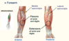
Muscles of the forearm
- The anterior muscles perform flexion of the wrist/digits of the hand.
- most of the flexors come from the medial epicondyle
- in the posterior compartments, we have the muscles that extend the wrist/ digits. most of these come from around our lateral epicondyle
-medial epicondyle is the origin of most of our flexor muscles of the hands and fingers, lateral epicondyle is the origin of most of our extensor muscles of the hand and the fingers.
-BRACHIO RADIALIS; this muscle comes from the humerus, the lateral side and insets into the radio stylus process, the bony prominence of the lateral side of the forearm right down towards the wrist. this muscle enables you to flex your forearm at the elbow joint when you are half pronated. you can call this the beer-drinking muscle. its the one that enables you pick up a big glass and take it towards your mouth.