MORPHOLOGY Flashcards

(52 cards)

1
Q

EMBRIOLOGY

Respiratory (laryngotracheal) Diverticulum

A
  • week 4
  • from ventral wall of the foregut (endoderm)
  • lower respiratory tract formation (trachea, bronchi and lungs)
  • endoderm → respiratory epithelium
  • mesoderm → muscles + cartilages + connective tissues
2
Q

EMBRIOLOGY

Development of Respiratory Diverticulum

A
  1. Respiratory Diverticulum enlarges → lung bud (distal portion)
  2. Bifurcation → lung bud + 2 bronchial buds
  3. Tracheoesophageal Septum
  4. Divisions → bronchial tree formation (month 6)
    • main bronchi
    • secondary bronchi
    • terciary bronchi (bronchopulmonar)
3
Q

EMBRIOLOGY

What is the critical time for lung formation?

A

25 - 28 week

  • pneumocytes (types I and II) are formed
  • surfactante production is possible
  • premature fetus at this time can survive (intensive care)
4
Q

EMBRIOLOGY

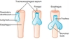
Clinical Correlate: Tracheoesophageal Fistula

A
  • malformation of the tracheoesophageal septum
  • 90% → esophagus + distal third of the trachea
  • esophageal atresia + polyhydramnions
  • symptoms
    • regurgitation of milk
    • cyanosis after feeding
    • abdominal distention after crying
    • pneumonitis (reflux into lungs)
5
Q

EMBRIOLOGY

Clinical Correlate: Pulmonary Hypoplasia

A

Congenital Diaphragmatic Hernia

(herniation of abdominal contents into thorax)

or

Potter’s Sequence

(bilateral renal agenesia → no urine → oligohydramnio → increase of fetal thorax pressure)

6
Q

ANATOMY

Upper Respiratory Tract

A
7
Q

ANATOMY

Trachea

A
  • hollow tube
    • 10 cm length
    • 2 cm diameter
  • bifurcation at the carina
  • C-shaped hyalin cartilage rings
    • 16 - 20 rings
    • anterior wall
    • rings are interconnected by smooth muscle
8
Q

ANATOMY

Bronchi

A

Bronchial Tree

  1. Primary Bronchi (enter the lung)
  2. Secondary Bronchi (lobar)
    • 2 left lung
    • 3 right lung
  3. Tertiary Bronchi (segmental)
    • 10 segmental bronchii each lung
9
Q

ANATOMY

Lung: surfaces and regions

A

Costal

(convex + smooth + related to chest wall)

Mediastinal

(concave + related to mediastinum and heart)

Diaphragmatic

(base + concave)

Apex

(4 cm above first rib + crossed by subclavian vessels)

10
Q

ANATOMY

Right lung is superior than the left lung

True or False?

A

TRUE

liver presence in the right

11
Q

ANATOMY

Cardiac impression in the left lung is more pronuced than the right lung

True or False?

A

TRUE

12
Q

ANATOMY

Lung: lobes and fissures

A
  • RIGHT LUNG
    • superior lobe
      • horizontal fissure
    • middle lobe
      • oblique fissure
    • inferior lobe
  • LEFT LUNG
    • superior lobe
      • oblique fissure
    • inferior lobe
    • lingula (corresponds middle lobe)
13
Q

ANATOMY

Lung Projections: fissures

A
  • Oblique Fissure
    • 5th intercostal space → 6th costal cartilage
    • both lungs
    • midclavicular line
  • Horizontal Fissure
    • only right lung
    • 5th intercostal space → 4th costal cartilage
14
Q

ANATOMY

Lung Projections: lobes

A

superior → above 4th rib (anteriorly)

middle → below 4th rib (anteriorly)

inferior → below 6th rib (posteriorly)

15
Q

ANATOMY

Lung: segments

A
16
Q

PLEURAL CAVITY

Pleura

A
  • mesodermal-derived membrane (serous)
  • double-layered membrane
    • friction-reducing movements
    • parietal layer
    • visceral layer
    • pleural cavity → potencial space
17
Q

PLEURAL CAVITY

Parietal Pleura

A

Costal Parietal Pleura

(lateral → ribs + intercostal space)

Diaphragmatic Parietal Pleura

(inferior)

Mediastinal Parietal Pleura

(medial → reflects to become visceral pleura at hilum)

Cervical Parietal Pleura

18
Q

PLEURAL CAVITY

Visceral Pleura

A

Tightly invest the surface of the lungs

fissures + lobes

19
Q

PLEURAL CAVITY

Pleural Innervation

A
  • Parietal Pleura
    • somatic sensory innervation
    • intercostal nerve → costal + diaphragmatic pleura
    • phrenic nerve → diaphragmatic + mediastinal pleura
  • Visceral Pleura
    • visceral sensory innervation
    • autonomic nerves
20
Q

PLEURAL CAVITY

Pleural Cavity and Pneumothorax

A
  • Cavity
    • potencial space
    • closed space + small amount of serous fluid
    • negative pressure
  • Pneumothorax
    • introduction of air into the pleural cavity → lost negative pressure → lung collapse
    • open pneumothorax x tension pneumothorax
21
Q

PLEURAL CAVITY

Pleural Reflections

A
  • pleural reflection = parietal pleura changing the direction from one wall to another wall
  • 2 rib interspaces separated parietal pleura from visceral pleura
22
Q

PLEURAL CAVITY

Pleural Recesses

A

potencial spaces not occupied by long tissue (except deep inspiration)

costodiaphragmatic

+

costomediastinal

23
Q

ANATOMY

Lymphatic Drainage

A
  • Plexus
    • superficial (below visceral pleura)
    • deep (into the lungs, drains through pulmonary nodes)
  • Lymph Nodes
    • bronchopulmonary (hilar) → both deep and superficial plexus
    • tracheobronchial → bifurcation of the trachea
    • bronchomediastinal (nodes and trunk)
24
Q

ANATOMY

Clinical Correlate: pleurisy

A
  • inflammation of parietal pleura
  • sharp pain uppon respiration
    • costal inflammation → dermatome pain
    • mediastinal irritation → shoulder dermatomes (phrenic nerve // C3-C5)
25
ANATOMY Clinical Correlate: Pancoast Tumor
Apex + can result in thoracic outlet syndrome and horner syndrome
26
ANATOMY Clinical Correlate: aspiration a foreign body
RIGHT PRIMARY BRONCHUS (shorter + wider + more vertical) ⇒ **posterior basal segment of right inferior lobe** (more often // if patient was in vertical position)
27
ANATOMY Clinical Correlate: breath sounds
**Superior Lobes** above 4th intercostal space + anterior chest wall **Middle Lobe** below 4th intercostal space (right) + medially toward sternum + anterior chest wall **Inferior Lobes** posterior chest wall
28
ANATOMY Clinical Correlate: which lobe of the left lung drains also **across the midline** to right bronchomediastinal nodes?
LOWER LEFT LOBE important to metastasis of lung cancer
29
HISTOLOGY Air-Blood Barrier
* 500 ml of air per breath * 120 square meters of air-blood barrier * important * susceptible to infections and pollution * receive all cardiac output * metabolism (endothelium) - lypoprotein and prostaglandin * production of ACE (angiotensin conversor enzyme)
30
HISTOLOGY Trachea
* Mucosa * pseudostratified epithelium * lamina propria (vessel, immune cells and connective tissue) * muscularis mucosa (thin smooth cells layer) * Submucosa * blood and lymphatic vessels * nerves * collagen fibers * Adventicia * several layers * losse connective tissue
31
HISTOLOGY Tracheal Epithelial Cells: Columnar Cells
* 200-300 cilia (microvilia) per cell * cilia → mucous movement (out to pharinx) * mucociliary scalator system
32
HISTOLOGY Tracheal Epithelium Cells: Goblet Cells
* secrete mucous (polysaccharide) with help from submucosal mixed glands * mucous * traps → viruses + bacteria + dust * absorbs → water-soluted gases (ozonio)
33
HISTOLOGY Tracheal Epithelium Cells: PNE cells
Pulmonary Neuroendocrine Cells * comparable to the endocrine cells in the gut * clusters + airway branch points * **APUD** (amino-precursor-uptake-decarboxylase) * **DNES** (diffuse neuroendocrine system) * **K cells** (kulchitsky) * **brush cells** * **sensory receptors** (synapses with intraepithelial nerves) * intermediate stages of goblet cells
34
HISTOLOGY Tracheal Epithelium Cells: Basal Cells
* stem cells * formation * goblet cells * ciliated cells * responsible for pseudostratified appearance
35
HISTOLOGY Bronchi
* pseudostratified epithelium (columnar ciliated cells + goblet cells + PNE cells + basal cells) * submucosa (glands with ducts to lumen) * cartilage plates + circular smooth-muscle (bound together by elastic fibers) * decrease of mucous production * - goblet cells * - submucosal glands
36
HISTOLOGY Bronchioles
* not pseudocolumnar epithelium → **simple columnar** * **​**ciliated cells * goblet cells * **clara cells** * wall * **no cartilage** * **no glands** * smooth-muscle fascicles + elastic fibers
37
HISTOLOGY Terminal Bronchiole + Respiratory Bronchiole
**Terminal Bronchiole** last conducting bronchiole **Respiratory Bronchiole** terminal bronchiole that is periodically interrupted by alveoli in the wall * ciliated simple columnar epithelium * no goblet cells * clara cells
38
HISTOLOGY Clara Cells
* bronchiolar **secretory** cells (non-ciliated) * 80% of terminal bronchiole epithelium * production of **serous fluid** similar to surfactant * **chloride ion** transport to the lumen * **detoxification** (increase with polutants like cigarette smoke) * **stem cells** * clara cells * ciliated cells
39
HISTOLOGY Alveoli
* alveolar sacs + ducts → basically alveoli * alveoli = 80-85% lung volume * 300 mi alveolu → 200 microns in diameter each
40
HISTOLOGY Alveoli: Pneumocytes
* Pneumocyte - Type I * "small alveolar cell" * major cell of the alveolar surface * gas exchange * 40% of the alveolar cells, BUT cover 90-95% of alveolar surface * Pneumocyte - Type II * 60% of the alveolar cells, BUT cover only 5% of alveolar surface * surfactant * stem cell → type I and type II
41
HISTOLOGY Surfactant
phospholipids + surfactant proteins * **decrease of surface tension** (phospholipids act like detergent) * prevent alveolar collapse (after expiration) * cycle of surfactant * most recycle by pneumocyte II * some undergoes phagocytosis by macrophages
42
HISTOLOGY Alveolar Wall
* Cells → pneumocytes + macrophage + mast cells + fibroblasts + myofibroblast + smooth-muscle cells * Fibers → collagen (type I and II) + elastic fibers * Blood-Gas Barrier (0,1 microns) * surfactant * pneumocyte type I (squamous epithelium) * shared basal lamina * capillary endothelium
43
HISTOLOGY Pores of Kohn
collateral ventilation 10-15 microns
44
HISTOLOGY Alveolar Macrophage
* derived from monocyte * resident alveolar macrophage * alveoli (1-3) * interalveolar septum * may pass through pores of Kohn * last defence of the lung * trap in mucous (go to pharinx) * enter lymphatic system
45
HISTOLOGY Clinical Correlate: Respiratory Distress Syndrome
* deficiency of surfactant * premature infants + infants of diabetic mother + prolonged intrauterine asphixia * treatment → thyroxine + cortisol
46
HISTOLOGY Clinical Correlate: Hyaline Membrane Disease
* surfactant deficiency + gasping + cyanosis * atelectasis * eosinophilic fluid
47
HISTOLOGY Clinical Correlate: Ineffective Mucosal Clearance
* **Cystic Fibrosis** * fluid is viscous * deficiency of chloride transport * Clara Cells * **Kartagener Syndrome** * cilia immotile → dynein deficiency * males → same problem at sperm predisponition to pneumonia (bacteria) and pneumoconiosis (dust)
48
HISTOLOGY Clinical Correlate: Squamou Metaplasia
columnar epithelium is sensitive to irritation → ciliated cells become taller + goblet cells increase in number → ciliated epithelium becomes squamous (reversible process)
49
HISTOLOGY Clinical Correlate: Bronchial Metastatic Tumor
arise from K cells | (Kulchitsky cells)
50
HISTOLOGY Clinical Correlate: COPD
Chronic Obstrutive Pulmonary Disease * affect bronchioles * emphysema * loss of elastic fibers * chronic airflow obstrution * irreversible * asthma * chronic process but reversible * narrowing of airways
51
HISTOLOGY Clinical Correlate: Premature Fetus and Diabetic Mother
Corticosteroids induce synthesis of sulfactant + high insulin levels (diabetic mother) antagonize the sulfactant production + diabetic mother = higher incidence of respiratory distress syndrome
52
HISTOLOGY Clinical Correlate: Alveolar Macrophage other names
**Dust Cells** (phagocyte dust and cigarette particles) + **Heart Failure Cells** (phagocyte blood cells that have scaped into alveolar space during congestive heart failure)