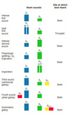
Mosby 14 Flashcards

(139 cards)

1
Q

Position of heart with respect to costal cartilages

A

3rd to 6th

2
Q

The area overlying heart

A

Precordium

3
Q

Upper portion of heart

Lower portion of heart

A

Base

Apex

4
Q

Relationship of heart position to tallness

A

The more tall the more central/vertical vs. left/horizontal

5
Q

Mirror image heart (right)

A

Dextrocardia

6
Q

Somach and heart on right side

A

Sinus inversus

7
Q

Apical pulse of left ventricle position

A

5th intercostal space @ midclavicular line

8
Q

Infancy

Size of left/right atrium

Extra connections

Arrangement of heart

A

Equal unlike adult

ductus arterisus, foramen ovale

More horiztonal than adults

9
Q

Pregnancy

Change in blood volume

Which part

When start/edn

Heart position change

A

Increases 50%

Plasma

Starting in first timester and peaking in 30th week, 3-4wks after delivery

Rotations towards horizontal axis

10
Q

Hemodynamic Changes during pregnancy

A
11
Q

Pressure or choking sensation substernally or into the neck

Cause?

A

Angina

strenuous physical activity, eating, exposure to intense cold, windy weather, or exposure to
emotional stress

12
Q

sudden, sharp, relatively brief pain that does not radiate, occurs most often at rest, and is unrelated to exertion and may not have a discoverable cause

A

precordial catch

13
Q

Ddx for chest pain

A

Cardiac
• Typical angina pectoris
• Atypical angina pectoris, angina equivalent
• Prinzmetal variant angina
• Unstable angina (acute coronary syndrome)
• Coronary insufficiency
• Myocardial infarction
• Nonobstructive, nonspastic angina
• Mitral valve prolapse
Aortic
• Dissection of the aorta
Pleuropericardial Pain
• Pericarditis
• Pleurisy
• Pneumothorax
• Mediastinal emphysema
Gastrointestinal Disease
• Hiatus hernia
• Reflux esophagitis
• Esophageal rupture
• Esophageal spasm
• Cholecystitis
• Peptic ulcer disease
• Pancreatitis
Pulmonary Disease
• Pulmonary hypertension
• Pneumonia
• Pulmonary embolus
• Bronchial hyperreactivity
• Tension pneumothorax
Musculoskeletal
• Cervical radiculopathy
• Shoulder disorder or dysfunction (e.g., arthritis, bursitis, rotator cuff injury, biceps tendonitis)
• Costochondral disorder
• Xiphodynia
Psychoneurotic
• Illicit drug use (e.g., cocaine)

14
Q

Anginal pain

Characteristic

A

Substernal; provoked by effort, emotion, eating; relieved by rest
and/or nitroglycerin; often accompanied by diaphoresis,
occasionally by nausea

15
Q

Pleural pain

Characteristic

A

Precipitated by breathing or coughing; usually described as
sharp; present during respiration; absent when breath held

16
Q

Esophageal Pain

characteristics

A

Burning, substernal, occasional radiation to the shoulder;
nocturnal occurrence, usually when lying flat; relief with food,
antacids, sometimes nitroglycerin

17
Q

Pain from a peptic ulcer

characteristics

A

Almost always infradiaphragmatic and epigastric; nocturnal
occurrence and daytime attacks relieved by food; unrelated
to activity

18
Q

Biliary pain

characteristics

A

Usually under right scapula, prolonged in duration; often
occurring after eating; will trigger angina more often than
mimic it

19
Q

Arthritis/bursitis

characteristics

A

Usually lasts for hours; local tenderness and/or pain with
movement

20
Q

Cervical pain

characteristics

A

Associated with injury; provoked by activity, persists after
activity; painful on palpation and/or movement

21
Q

Musculoskeletal (chest) pain

characteristics

A

Intensified or provoked by movement, particularly twisting or
costochondral bending; long lasting; often associated with
focal tenderness

22
Q

Psychoneurotic pain

characteristics

A

Associated with/after anxiety; poorly described; located in intramammary region

23
Q

DDx comparison

Angina pectoris

muscoskeletal

gastrointestinal

A
24
Q

HPI Chest pain description: onset and duration

A

sudden, gradual, or vague onset, length of episode; cyclic nature;
related to physical exertion, rest, emotional experience, eating, coughing, cold temperatures,
trauma, awakens from sleep

25
Chest pain description: character
aching, sharp, tingling, burning, pressure, stabbing, crushing, or clenched fist sign
26
HPI Chest pain description: location
radiating down arms, to neck, jaws, teeth, scapula; relief with rest or position change
27
HPI Chest pain description: Severity
interference with activity, need to stop all activity until subsides, disrupts sleep, how severe on a scale of 0 to 10
28
HPI Chest pain description: Associated symptoms
anxiety; dyspnea; diaphoresis; dizziness; nausea or vomiting; faintness; cold, clammy skin; cyanosis; pallor; swelling or edema (noted anywhere, constant or at certain times during day)
29
HPI Chest pain description: treatment
rest, position change, exercise, nitroglycerin, digoxin, diuretics, beta-blockers, angiotensin-converting enzyme (ACE) inhibitors, calcium channel blockers, nonsteroidal antiinflammatory drugs, antihypertensives
30
HPI Fatigue Associated Symptoms Medications causing
dyspnea on exertion, chest pain, palpitations, orthopnea, paroxysmal nocturnal dyspnea, anorexia, nausea, vomiting beta-blocker
31
HPI Cough character medications
dry, wet, nighttime, aggravated by lying down ACE inhibitors
32
HPI related to cariovascular (general)
Chest pain Fatigue Cough Difficulty breathing Loss of conciousnes
33
HPI Questions regarding difficulting breathing
aggravated by exertion (how much?) worsening or remaining stable lying down or eased by resting on pillows (how many? or sleep in a recliner?) paroxysmal nocturnal dyspnea
34
HPI Loss of consciousness' associated symptoms
palpitation, dysrhythmia, unusual exertion, sudden turning of neck (carotid sinus effect), looking upward (vertebral artery occlusion), change in posture
35
PMH Cardio
Cardiac surgery or hospitalization Rhythm disorder Acute Rheumatic feverl unexplained fever, swollen joins, Sydenham chorea (St. Vitus dance), abdominal pain, skin rash (erythema marginatum) or nodules Chronic illness: hypertension, bleeding disorder, hyperlipidemia, diabetes, thyroid dysfunction, coronary artery disease, obesity, congenital heart defect
36
FH Cardio
Long QT syndrome Diabetes Heart disease Dyslipidemia Hypertension Obesity Congenital heart defects
37
SH
Emplyoment w/ physical, env hazards (e.g. heat/stress) Tabacoo Nutritional status Diet Weight Alcohol Releaxation Hobbies Exercise Sexual activity Illegal drugs
38
Risk factors for cardiac disease
• **Gender** (men more at risk than women; women’s risk is increased in the postmenopausal years and with oral contraceptive use) • **Hyperlipidemia** • **Elevated homocysteine** level • **Smoking** • **Family** history of **cardiovascular disease**, diabetes, hyperlipidemia, hypertension, or sudden death in young adults • **Diabetes mellitus** • **Obesity**: dietary habits including an excessively fatty diet • **Sedentary lifestyle** without exercise • **Personality type**: intense, compulsive behavior with feelings of hostility; negative emotions, pessimistic attitude, failure to share emotions
39
Concers for infants related to cardiovascular disease
◆**Tiring easily** during feeding ◆ **Breathing change**s: more heavily or more rapidly than expected during feeding or defecation ◆ **Cyanosis**: perioral during eating, more widespread and more persistent, related to crying ◆ Weight gain as expected ◆ **Knee-chest** position or other position favored for rest ◆ **Mother’s health** during **pregnancy**: medications taken, unexplained fever, illicit drug use
40
Concers for chidren related to cardiovascular disease
◆ **Tiring during play**: amount of time before tiring, activities that are tiring, inability to keep up with other children, reluctance to go out to play ◆ **Naps**: longer than expected ◆ **Positions**: squatting instead of sitting when at play or watching television ◆ **Headaches** ◆ **Nosebleeds** ◆ **Unexplained joint pain** ◆ **Unexplained fever** ◆ **Expected height** and **weight gain** (and any substantiating records) ◆ **Expected physical** and **cognitive** **development** (and any substantiating records)
41
Concers for pregnant women related to cardiovascular disease
◆ **History** of **cardiac disease** or surgery ◆ **Dizziness** or faintness on standing ◆ Indications of heart disease during pregnancy, including progressive or severe dyspnea, progressive orthopnea, paroxysmal nocturnal dyspnea, hemoptysis, syncope with exertion, and chest pain related to effort or emotion
42
Concers for chidren related to cardiovascular disease
◆ Common symptoms of cardiovascular disorders: confusion, dizziness, blackouts, syncope, palpitations, coughs and wheezes, hemoptysis, shortness of breath, chest pains or chest tightness, impotence, fatigue, leg edema: pattern, frequency, time of day most pronounced ◆ If heart disease has been diagnosed: drug reactions: potassium excess (weakness, bradycardia, hypotension, confusion); potassium depletion (weakness, fatigue, muscle cramps, dysrhythmias); digitalis toxicity (anorexia, nausea, vomiting, diarrhea, headache, confusion, dysrhythmias, halo, yellow vision), interference with activities of daily living; ability of the patient and family to cope with the condition, perceived and actual; orthostatic hypotension
43
NIH recommendation for cholesterol total LDL LDL after MI
200 mg/dL 100 mg/dL 70 mg/dL
44
Lifestyle changes to improve cardio
LDL levels Diet Exercise No smoking Monitor BP, glucose, inflamatory, lipids anually
45
Non-cardio signs of heart failure
Crackles in the lugs Engorgement of liver Peripheral edema
46
Influencing factors during cardiovascular exam: General? Skin? Abdominal region? Eye?
◆ Effect of a barrel chest or pectus deformity ◆ Xanthelasma ◆ Funduscopic changes of hypertension ◆ Ascites or pitting edema ◆ Abdominal aortic bruit
47
What is the indication of downward displaced and stronger apical impulse?
Left vencticular hypertrophy
48
What is the indication of lift of apical impulse along sternal border
Righte ventricular hypertrophy
49
The point at which the apical impulse is most readily seen or felt
point of maximal impulse (PMI)
50
Cause Loss of thrust for apical impulse
overlying fluid or air or to displacement beneath the sternum
51
Cause for displacement to the right without a loss or gain in thrust for apical impulse
dextrocardia, diaphragmatic hernia, distended stomach, or a pulmonary abnormality
52
What is thrill Indication for what?
fine, palpable, rushing vibration, a palpable murmur, often, but not always, over the base of the heart in the area of the right or left second intercostal space indicates turbulence or a disruption of the expected blood flow related to some defect in the closure of one of the semilunar valves (generally aortic or pulmonic stenosis), pulmonary hypertension, or atrial septal defect
53
Why percussion is not useful for cardio? How heart borders can be estabilished then?
Heart is more maleable than chest and it will conform shape Chest radiograph
54
Five ascultatory areas
◆ **Aortic valve area:** second right intercostal space at the right sternal border ◆ **Pulmonic valve area:** second left intercostal space at the left sternal border ◆ **Second pulmonic area:** third left intercostal space at the left sternal border ◆ **Tricuspid area:** fourth left intercostal space along the lower left sternal border ◆ **Mitral (or apical) area:** at the apex of the heart in the fifth left intercostal space at the midclavicular line
55
Where is split S2 beast hear at
Pulmonic valve area: second left intercostal space at the left sternal border (during inspiration)
56
Categories by which heart sounds are classified
Pitch, intensity, duration, timing (in cardiac cycle)
57
Normal heart sounds S1 vs. S2 comparison
58
Where is S1 best heard? Comparison of sound S1/S2?
Toward apex S1 is lower in pitch and longer than S2
59
Location where sounds are heard the best 1st sound 1st sound split 2nd sound Physiological 2nd sound split 3rd sound (entricular gallop) 4th sound (atrial gallop) summation gallop
60
Reasons for lounder S1
Systole begins too early: Blood veolicty increased (anemia, fever, hyperthyrodism, anxiety, exercise) Stenosis of mitrial valve
61
Degree of opening of the S1 valve (and loudness of the S1) Decrease
Complete heart block Gross disruption of rhythm Increase of overlying, tissue, fat Pulmonary hypertension Fibrosis and calcification e.g. due to RF
62
S2 increase in loudness causes
``` Systemic hypertension (S2 may ring or boom), syphilis of the aortic valve, exercise, or excitement accentuates S2 ``` Pulmonary hypertension, mitral stenosis, and congestive heart failure accentuate P2 The valves are diseased but still fully mobile; the component of S2 affected depends on which valve is compromised.
63
S2 decrease in loudness
A shocklike state with arterial hypotension causes loss of valvular vigor. The valves are immobile, thickened, or calcified; the component of S2 affected depends on which valve is compromised. Aortic stenosis affects A2. Pulmonic stenosis affects P2. Overlying tissue, fat, or fluid mutes S2 giving
64
Which sound is heard later in physiologic splitting in S2?
Pulmonary valve
65
S3 "Ken-TUCK-y" Cause Timing Quality When lounder?
Vibration of ventricle during passive diastole Quiert, low pitched, difficult to hear Increased filling pressure or decreased ventricular compliance
66
S4 "TEN-nes-see" Cause Timing When louder?
vibration in the valves, papillae, and ventricular walls late diastole (presystole) \* can be confused with s1 elderly, increased resistance because of loss of complance of ventricular walls
67
What makes S3 and S4 easier to hear?
Increasing venous return (e.g. raising leg or grip hand)
68
69
Wide splitting causes
DELAYED PULMONIC CLOSURE right bundle branch block stenosis of pulmonary valve pulmonary hypertension delays emptying mitral regurgitation (cause early closure of aortic valve)
70
Fixed splitting causes
WHEN OUTPUT OF RIGHT VENTRICLE IS GREATER THAN OF THE LEFT Large atrial septal defect ventricular septal defect left-to-right shunting
71
Paradoxic (reversed) splitting
AORTIC VALVE IS DELAYED
72
Extra Heart Sounds examples
Pericardial friction rub
73
Murmur definition
Prologned extra sound during sys or diastole Due to disruption of the blood flow
74
Forward blood restriction in valve
Stenosis
75
Allowing backward flow of blood through valve
Regurgitation
76
First Heart Sound Preferable position of patient Area for auscultation Endpiece Pitch Effects of respiration External Influences Cause
77
Second Heart Sound Preferable position of patient Area for auscultation Endpiece Pitch Effects of respiration External Influences Cause
78
Third Heart Sound Preferable position of patient Area for auscultation Endpiece Pitch Effects of respiration External Influences Cause
79
Fourth Heart Sound Preferable position of patient Area for auscultation Endpiece Pitch Effects of respiration External Influences Cause
80
Quadruple rhythm Preferable position of patient Area for auscultation Endpiece Pitch Effects of respiration External Influences Cause
81
Summation gallop Preferable position of patient Area for auscultation Endpiece Pitch Effects of respiration External Influences Cause
82
Ejection sounds Preferable position of patient Area for auscultation Endpiece Pitch Effects of respiration External Influences Cause
83
Systolic Click Preferable position of patient Area for auscultation Endpiece Pitch Effects of respiration External Influences Cause
84
Open Snap Preferable position of patient Area for auscultation Endpiece Pitch Effects of respiration External Influences Cause
85
Classification of murmur: timing and duration
86
Classification of murmur: intensity
87
Classification of murmur: pattern
88
Classification of murmur: quality, location, radiation, respiratiory phase
89
Mitrial Stenosis Detection Findings Description
**DETECTION** Heard with bell at apex, patient in left lateral decubitus position **FINDING** Low-frequency diastolic rumble, more intense in early and late diastole, does not radiate; systole usually quiet; palpable thrill at apex in late diastole common; S1 increased and often palpable at left sternal border; S2 split often with accented P2; opening snap follows P2 closely Visible lift in right parasternal area if right ventricle hypertrophied Arterial pulse amplitude decreased **DESCRIPTION** Narrowed valve restricts forward flow; forceful ejection into ventricle Often occurs with mitral regurgitation Caused by rheumatic fever or cardiac infection
90
Aortic Stenosis Detection Findings Description
**DETECTION** Heard over aortic area; ejection sound at second right intercostal border **FINDINGS** Midsystolic (ejection) murmur, medium pitch, coarse, diamond shaped,\* crescendodecrescendo; radiates along left sternal border (sometimes to apex) and to carotid with palpable thrill; S1 often heard best at apex, disappearing when stenosis is severe, often followed by ejection click; S2 soft or absent and may not be split; S4 palpable; ejection sound muted in calcified valves; the more severe the stenosis, the later the peak of the murmur in systole Apical thrust shifts down and left and is prolonged if left ventricular hypertrophy is also present **DESCRIPTION** Calcification of valve cusps restricts forward flow; forceful ejection from ventricle into systemic circulation Caused by congenital bicuspid (rather than the usual tricuspid) valve, rheumatic heart disease, atherosclerosis May be the cause of sudden death, particularly in children and adolescents, either at rest or during exercise; risk apparently related to degree of stenosis
91
Subaortic Stenosis Detection Findings Description
**DETECTION** Heard at apex and along left sternal border **FINDINGS** Murmur fills systole, diamond shaped, medium pitch, coarse; thrill often palpable during systole at apex and right sternal border; multiple waves in apical impulses; S2 usually split; S3 and S4 often present Arterial pulse brisk, double wave in carotid common; jugular venous pulse prominent **DESCRIPTION** Fibrous ring, usually 1 to 4 mm below aortic valve; most pronounced on ventricular septal side; may become progressively severe with time; difficult to distinguish from aortic stenosis on clinical grounds alonea
92
Pulmonary Stenosis Detection Findings Description
**DETECTION** Heard over pulmonic area radiating to left and into neck; thrill in second and third left intercostals space **FINDINGS** Systolic (ejection) murmur, diamond shaped, medium pitch, coarse; usually with thrill; S1 often followed quickly by ejection click; S2 often diminished, usually wide split; P2 soft or absent; S4 common in right ventricular hypertrophy; murmur may be prolonged and confused with that of a ventricular septal defect **DESCRIPTION** Valve restricts forward flow; forceful ejection from ventricle into pulmonary circulation Cause is almost always congenital
93
Tricuspid Stenosis Detection Findings Description
**DETECTION** Heard with bell over tricuspid area **FINDINGS** Diastolic rumble accentuated early and late in diastole, resembling mitral stenosis but louder on inspiration; diastolic thrill palpable ov er right ventricle; S2 may be split during inspiration Arterial pulse amplitude decreased; jugular venous pulse prominent, especially a wave; slow fall of v wave **DESCRIPTION** Calcification of valve cusps restricts forward flow; forceful ejection into ventricles Usually seen with mitral stenosis, rarely occurs alone Caused by rheumatic heart disease, congenital defect, endocardial fibroelastosis, right atrial myxoma
94
Mitrial Regurgitation Detection Findings Description
**DETECTION** Heard best at apex; loudest there, transmitted into left axilla **FINDINGS** Holosystolic, plateau-shaped intensity, high pitch, harsh blowing quality, often quite loud and may obliterate S2; radiates from apex to base or to left axilla; thrill may be palpable at apex during systole; S1 intensity diminished; S2 more intense with P2 often accented; S3 often present; S3-S4 gallop common in late disease If mild, late systolic murmur crescendos; if severe, early systolic intensity decrescendos; apical thrust more to left and down in ventricular hypertrophy **DESCRIPTION** Valve incompetence allows backflow from ventricle to atrium Caused by rheumatic fever, myocardial infarction, myxoma, rupture of chordae
95
Mitral Valve Prolapse Detection Findings Description
**DETECTION** Heard at apex and left lower sternal border; easily missed in supine position; also listen with patient upright **FINDINGS** Typically late systolic murmur preceded by midsystolic clicks, but both murmur and clicks highly variable in intensity and timing **DESCRIPTION** Valve is competent early in systole but prolapses into atrium later in systole; may become progressively severe, resulting in a holosystolic murmur; often concurrent with pectus excavatum
96
Aortic Regurgitation Detection Findings Description
**DETECTION** Heard with diaphragm, patient sitting and leaning forward; Austin- Flint murmur heard with bell; ejection click heard in second intercostal space **FINDINGS** Early diastolic, high pitch, blowing, often with diamondshaped midsystolic murmur, sounds often not prominent; duration varies with blood pressure; low-pitched, rumbling murmur at apex common (Austin-Flint); early ejection click sometimes present; S1 soft; S2 split may have tambour-like quality; M1 and A2 often intensified, S3-S4 gallop common In left ventricular hypertrophy, prominent prolonged apical impulse down and to left Pulse pressure wide; waterhammer or bisferiens or Corrigan pulse common in carotid, brachial, and femoral arteries (see Chapter 15) **DESCRIPTION** Valve incompetence allows backflow from aorta to ventricle Caused by rheumatic heart disease, endocarditis, aortic diseases (Marfan syndrome, medial necrosis), syphilis, ankylosing spondylitis, dissection, cardiac trauma
97
Pulmonic Regurgitation Detection Findings Description
**DETECTION/FINDINGS** Difficult to distinguish from aortic regurgitation on physical examination **DESCRIPTION** Valve incompetence allows backflow from pulmonary artery to ventricle Secondary to pulmonary hypertension or bacterial endocarditis
98
Tricuspid Regurgitation Detection Findings Description
**DETECTION** Heard at left lower sternum, occasionally radiating a few centimeters to left **FINDINGS** Holosystolic murmur over right ventricle, blowing, increased on inspiration; S3 and thrill over tricuspid area common In pulmonary hypertension, pulmonary artery impulse palpable over second left intercostal space and P2 accented; in right ventricular hypertrophy, visible lift to right of sternum Jugular venous pulse has large v waves **DESCRIPTION** Valve incompetence allows backflow from ventricle to atrium Caused by congenital defects, bacterial endocarditis (especially in IV drug abusers), pulmonary hypertension, cardiac trauma
99
Innocent murmurs another name Population where heard Description
Still murmurs Younger (blood flowing from large chamber to blood vessels) They are usually grade I or II, usually midsystolic, without radiation, medium pitch, blowing, brief, and often accompanied by splitting of S2
100
Benign murmur
The result of a structural anomaly that is not severe enough to cause a clinical problem
101
DDx systomic murmurs Right-sided chambers
Inspiration-increase Expireation-decrease
102
DDx systomic murmurs Hypertrophic
Valsalva - Increase
103
DDx systomic murmurs Cardiomyopathy
Squatting to standing (rapidly for 30 seconds) - Increase Standing to squatting (rapidly) - Decrease Passive leg elevation to 45 degrees, patient supine - Decrease
104
DDx systomic murmurs Mitral regurgitation
Handgrip - Increase
105
DDx systomic murmurs VSD
Transient arterial occlusion (sphygmomanometer placed on each of patient’s upper arms and simultaneously inflated to 20 to 40 mm Hg above patient’s previously recorded blood pressures; intensity noted after 20 seconds) _ increase Inhalation of amyl nitrate (three rapid breaths from a broken ampule) (Not routinely recommended) - decrease
106
DDx systomic murmurs Aortic Stenosis
No maneuver distinguishes this murmur; the diagnosis can be made by exclusion
107
Irregular rate in repeated pattern likely cause
sinus dysrhythmia
108
Irregular rhytms possible cause
heart disease or conduction system impairment
109
Infancy purplish plethora symptom of
Polycythemia
110
Infancy ashen white color symptom of
Shock
111
Infancy central cyanosis symptom of
Congential heart disease
112
Cyanosis of hands and feet without central cyanosis Important?
Acrocyanosis Dissapears after few days/hours after birth
113
Cyanosis at birth suggestion
transposition of the great vessels, tetralogy of Fallot, tricuspid atresia, a severe septal defect, or severe pulmonic stenosis
114
Cyanosis that appears after the neonatal period causes
pure pulmonic stenosis, Eisenmenger complex, tetralogy of Fallot, or large septal defects
115
Most murmurs in infancy
Innocent - transition from fetal to pulmonic circulation
116
Heart rate vs. age
117
Sign of heart failure in infants
infant’s liver may enlarge before there is any suggestion of moisture in the lungs, and that the left lobe of the liver may be more distinctly enlarged than the right
118
Bacterial Endocarditis Description Pathophysiology Subjective Objective
**DESCRIPTION** Bacterial infection of the endothelial layer of the heart and valves **PATHOPHYSIOLOGY** Individuals with valvular defects, congenital or acquired, and those who use intravenous drugs are particularly susceptible **SUBJECTIVE** Fever, fatigue Murmur Sudden onset of congestive heart failure **OBJECTIVE** ``` Signs of neurologic dysfunctions Janeway lesion (small erythematous or hemorrhagic macules appearing on the palms and soles) Osler nodes (appear on the tips of fingers or toes and are caused by septic emboli) ```
119
Congestive Heart Failure Description Pathophysiology Subjective Objective
**DESCRIPTION** Heart fails to propel blood forward with its usual force, resulting in congestion in the pulmonary or systemic circulation **PATHOPHYSIOLOGY** Decreased cardiac output causes decreased blood flow to the tissues May be left or right sided Left sided is characterized as systolic or diastolic Diastolic CHF is result of advanced glycation cross-linking collagen and creating a stiff ventricle unable to dilate actively Diastolic CHF occurs in older adults whose tissue is exposed to glucose for a longer period of time and in individuals with diabetes mellitus **SUBJECTIVE** Fatigue Orthopnea Breath difficulty, shortness of breath Edema **OBJECTIVE** Symptoms can develop gradually or suddenly with acute pulmonary edema Systolic CHF has a narrow pulse pressure Diastolic CHF has a wide pulse pressure
120
Pericarditis Description Pathophysiology Subjective Objective
**DESCRIPTION** Sudden inflammation of the pericardium **PATHOPHYSIOLOGY** If persists the pericardial effusion may increase and result in cardiac tamponade **SUBJECTIVE** Initially, chest pain is sharp or stabbing Movement or inspiration may aggravate the pain Pain may be most severe when supine, relieved when leaning forward **OBJECTIVE** Scratchy, grating, triphasic friction rub on ascultation, comprises ventricular systole, early diastolic ventricular filling, and late diastolic atrial systole Easily heard just left of the sternum in third and fourth intercostal spaces
121
Cardiac Tamponade Description Pathophysiology Subjective Objective
**DESCRIPTION** Excessive accumulation of effused fluids or blood between the pericardium **PATHOPHYSIOLOGY** Seriously constrains cardiac relaxation, impairing access of blood to the right heart Common causes: pericarditis, malignancy, aortic dissection, and trauma **SUBJECTIVE DATA** Anxiety, restlessness Chest pain Difficulty breathing Discomfort, sometimes relieved by sitting upright or leaning forward Syncope, light-headedness Pale, gray, or blue skin Palpitations Rapid breathing Swelling of the abdomen or arms or neck veins **OBJECTIVE** Beck’s triad (jugular venous distention, hypotension, and muffled heart sounds) Chronically and severely involved pericardium may also scar and constrict, limiting cardiac filling; heart sounds are muffled, blood pressure drops, the pulse becomes weakened and rapid, and paradoxic pulse becomes exaggerated
122
Cor Pulmonale Description Pathophysiology Subjective Objective
**DESCRIPTION** Enlargement of the right ventricle secondary to pulmonary malfunction **PATHOPHYSIOLOGY** Usually chronic, occasionally acute Chronic common cause: chronic obstructive pulmonary disease (COPD) Acute causes: massive pulmonary embolism and acute respiratory distress syndrome (ARDS) Results from chronic pulmonary disease; alterations in pulmonary circulation leads to pulmonary arterial hypertension, which imposes a mechanical load on right ventricular emptying **SUBJECTIVE** Fatigue Tachypnea Exertional dyspnea Cough, hemoptysis **OBJECTIVE** Evidence of pulmonary disease Wheezes and crackles on auscultation Increase in chest diameter Labored respiratory efforts with chest wall retractions Distended neck veins with prominent A or V waves Cyanosis Left parasternal systolic heave Loud S2 exaggerated in the pulmonic region
123
Myocardial Infraction Description Pathophysiology Subjective Objective
**DESCRIPTION** Ischemic myocardial necrosis caused by abrupt decrease in coronary blood flow to a segment of the myocardium **PATHOPHYSIOLOGY** Most commonly affects left ventricle Atherosclerosis and thrombosis are the common underlying causes **SUBJECTIVE** Deep substernal or visceral pain that often radiates to the jaw, neck, and left arm Discomfort may be mild, especially in older adults or patients with diabetes mellitus **OBJECTIVE** Dysrhythmias are common S4 is usually present Distant heart sounds Soft, systolic, blowing apical murmur Thready pulse Blood pressure varies, although Video/Animation hypertension is usual in the early phases
124
Myocarditis Description Pathophysiology Subjective Objective
**DESCRIPTION** Focal or diffuse inflammation of the myocardium **PATHOPHYSIOLOGY** Results from infectious agents, toxins, or autoimmune diseases such as amyloidosis **SUBJECTIVE** Initial symptoms vague Fatigue Dyspnea Fever Palpitations **OBJECTIVE** Cardiac enlargement Murmurs Gallop rhythms Tachycardia Dysrhythmias Pulsus alternans
125
Causes of syncope
CANADA * *C Cardiac:** valve stenosis, Stokes-Adams attacks, other conduction disturbances * *A Arteriovenous:** “steal” syndromes * *N Nervous:** psychologic, autonomic, vagal, coughing * *A Anemia**, altered blood (CO) * *D Drugs**, diabetes, alcohol, poisons * *A Altitude**, acute fevers
126
Conduction distrubances Description Pathophysiology Subjective Objective
**DESCRIPTION** Conduction disturbances either proximal to the bundle of His or diffusely throughout the conduction system **PATHOPHYSIOLOGY** May result from a variety of causes: ischemic, infiltrative, or, rarely, neoplastic Antidepressant drugs, digitalis, quinidine, and many other medications can be precipitating factors **SUBJECTIVE** Transient weakness Syncope Cardiac syncope may occur acutely without warning; sometimes diminished sensibility, a “gray-out” instead of a “black-out,” may precede the event Strokelike episodes Rapid or irregular heartbeat **OBJECTIVE** Labile heart rates Rhythm disturbances
127
Tetralogy of fallot Description Pathophysiology Subjective Objective
**DESCRIPTION** Four cardiac defects: ventricular septal defect, pulmonic stenosis, dextroposition of the aorta, and right ventricular hypertrophy **PATHOPHYSIOLOGY** Surgical correction is recommended, currently initiated after the first “spell" **SUBJECTIVE** Dyspnea with feeding, poor growth, exercise intolerance Paroxysmal dyspnea with loss of consciousness and central cyanosis (tetralogy spell) **OBJECTIVE** Parasternal heave and precordial prominence, systolic ejection murmur over the third intercostal space, sometimes radiating to the left side of the neck; a single S2 is heard (Fig. 14-24) Older children develop clubbing of fingers and toes
128
Ventricular Septal Defect Description Pathophysiology Subjective Objective
**DESCRIPTION** Opening between the left and right ventricles **PATHOPHYSIOLOGY** Significant number (30% to 50%) of small defects close spontaneously, during the first 2 years of life **SUBJECTIVE** Recurrent respiratory infections If large VSD, rapid breathing, poor growth, symptoms of congestive heart failure **OBJECTIVE** Arterial pulse is small, and jugular venous pulse is unaffected Holosystolic murmur, often loud, coarse, high-pitched, and best heard along the left sternal border in the third to fifth intercostal spaces Distinct lift is often discernible along left sternal border and the apical area A smaller defect causes a louder murmur and a more easily felt thrill than a large one
129
Patent Ductus Arteriosus Description Pathophysiology Subjective Objective
**DESCRIPTION** Failure of the ductus arteriosus to close after birth **PATHOPHYSIOLOGY** Blood flows through the ductus during systole and diastole, increasing pressure in the pulmonary circulation and consequently workload of the right ventricle **SUBJECTIVE** Small shunt can be asymptomatic; a larger one causes dyspnea on exertion **OBJECTIVE** Dilated and pulsatile neck vessels Wide pulse pressure Harsh, loud, continuous murmur heard at the first to third intercostal spaces and the lower sternal border, with a machine-like quality Murmur is usually unaltered by postural change, unlike murmur of a venous hum
130
Atrial Septal Defect Description Pathophysiology Subjective Objective
**DESCRIPTION** Congenital defect in the septum dividing the left and right atria **SUBJECTIVE DATA** Often asymptomatic Heart failure rarely occurs in children but can often occur in adults **OBJECTIVE** Diamond-shaped systolic ejection murmur often loud, high pitched, and harsh, heard over the pulmonic area May be accompanied by a brief, rumbling, early diastolic murmur Does not usually radiate beyond the precordium Systolic thrill may be felt over the area of the murmur, along with a palpable parasternal thrust S2 may be widely split Sometimes murmur may not sound particularly impressive, especially in overweight children; if there is a palpable thrust and radiation to the back, it is more apt to be significant
131
Acute Rheymatic Fever Description Pathophysiology Subjective Objective
**DESCRIPTION** Systemic connective tissue disease occurring after streptococcal pharyngitis or skin infection **PATHOPHYSIOLOGY** Characterized by a variety of major and minor manifestations May result in serious cardiac valvular involvement of mitral or aortic valve; tricuspid and pulmonic are not often affected Affected valve becomes stenotic and regurgitant Children between 5 and 15 years of age are most commonly affected Prevention—adequate treatment for streptococcal pharyngitis or skin infections—is the best therapy **SUBJECTIVE** Fever Inflamed swollen joints Flat or slightly raised, painless rash with pink margins with pale centers and a ragged edge (erythema marginatum) Aimless jerky movements (Sydenham chorea or St. Vitus dance) Small, painless nodules beneath the skin Chest pain Palpitations Fatigue Shortness of breath **OBJECTIVE** Murmurs of mitral regurgitation and aortic insufficiency Cardiomegaly Friction rub of pericarditis Congestive heart failure
132
Major Jones Criteria Minor Jones Criteria
Carditis, Polyarthiritis, Chorea, Erythema maginatum, Subcutaneus Nodules Fever, Arthalgia, Preious RF, Acute Phase Reaction, Prolonged PR
133
Kawasaki Disease
**DESCRIPTION** Condition causing inflammation in walls of small and medium-sized arteries throughout the body, including coronaryn arteries **PATHOPHYSIOLOGY** Named after Dr. Tomisaku Kawasaki, the physician who first identified the disease in 1967 Also called mucocutaneous lymph node syndrome because it also affects lymph nodes, skin, and mucous membranes Frequently (80% of the time) affects infants and children under 5 years of age **SUBJECTIVE** High fever, lasting longer than 5 days Conjunctivitis Cracked, red, and inflamed lips Strawberry tongue, white coating on tongue or prominent papillae on the back of the tongue Cervical lymphadenopathy Erythema of the palms of the hands and soles of the feet Joint pain (arthralgia) and swelling, frequently symmetric Irritability Tachycardia **OBJECTIVE** Diagnosis is usually made based on the patient having most of the classic symptoms
134
Kawasaki Disease Diagnostic Characteristics
Fever of at least 5 days’ duration together with four of the following five findings: • Painless bulbar conjunctival injection without exudate • Changes in extremities including erythema, edema, and desquamation • Polymorphous (macular, morbilliform, or target lesions) erythematous rash of the trunk and extremities. • Changes in the lips and oral cavity including diffuse oral or pharyngeal mucosal erythema; erythematous, dry/fissured, or swollen lips; red “strawberry” tongue • Cervical lymphadenopathy (≥1.5 cm in diameter), usually unilateral
135
Artherosclerotic Heart Disease Description Pathophysiology Subjective Objective
**DESCRIPTION** Caused by deposition of cholesterol, other lipids, and by a complex nflammatory process **PATHOPHYSIOLOGY** Leads to vascular wall thickening and narrowing of the lumen **SUBJECTIVE DATA** May be asymptomatic Angina pectoris, shortness of breath, palpitations Family history of close relatives with atherosclerotic disease, early death, or dyslipidemia **OBJECTIVE** Dysrhythmias and congestive heart failure
136
Mitral Insufficiency/Regurgitation Description Pathophysiology Subjective Objective
**DESCRIPTION** Abnormal leaking of blood through the mitral valve, from left ventricle into left atrium **SUBJECTIVE** Acute mitral regurgitation has symptoms of decompensated congestive heart failure Shortness of breath Pulmonary edema Orthopnea Paroxysmal nocturnal dyspnea Decreased exercise tolerance Chronic compensated mitral regurgitation may be asymptomatic Patients may be sensitive to small shifts in intravascular volume and are prone to develop congestive heart failure **OBJECTIVE** High-pitched pansystolic murmur radiating to axilla May also have a third heart sound
137
Angina Description Pathophysiology Subjective Objective
**DESCRIPTION** Pain caused by myocardial ischemia **PATHOPHSYIOLOGY** Occurs when myocardial oxygen demand exceeds supply Can be recurrent or present as initial incidence **SUBJECTIVE** Substernal pain or intense pressure radiating to the neck; jaws; and arms, particularly the left Often accompanied by shortness of breath, fatigue, diaphoresis, faintness, and syncope **OBJECTIVE DATA** No definitive examination findings suggest angina Tachycardia, tachypnea, hypertension, and/or diaphoresis Ischemia may lead to presence of crackles due to pulmonary edema or a reduction in the S1 intensity or an S4 Physical examination may suggest other comorbidities that place the patient at higher risk for anginal symptoms, such as COPD, xanthelasma, hypertension, evidence of peripheral arterial disease, abnormal pulsations on palpation over precordium, murmurs or arrhythmias
138
Senile Cardiac Amyloidosis Description Pathophysiology Subjective Objective
**DESCRIPTION** Amyloid, fibrillary protein produced by chronic inflammation or neoplastic disease, deposition in the heart **PATHOPHYSIOLOGY** Heart contractility may be reduced Causes heart failure **SUBJECTIVE** Palpitations, lower extremity edema, fatigue, reduced activity tolerance **OBJECTIVE** Pleural effusion Arrhythmia Lower extremity edema Dilated neck veins Hepatomegally or ascites Electrocardiography or echocardiography shows small, thickened left ventricle; right ventricle may also be thickened
139
Aortic Sclerosis Description Pathophysiology Subjective Objective
**DESCRIPTION** Thickening and calcification of aortic valves **SUBJECTIVE** Does not usually cause symptoms **OBJECTIVE** Midsystolic (ejection) murmur