MSK 4 - Lower Limbs Flashcards

1
Q

what is the popliteal fossa?

A

The popliteal fossa is a fat–filled diamond- shaped space located posterior to the knee joint. It is comparable to the ante-cubital fossa found in the upper limb

2
Q

1

A

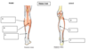
Biceps femoris: long head

3
Q

2

A

Semimembranous

4
Q

3

A

Semitendinosus

5
Q

4

A

Tibial nerve

6
Q

5

A

Popliteal artery

7
Q

6

A

Common fibular nerve

8
Q

What is the relation of the popliteal artery to the popliteal vein?

A

it lies medial and deep to it

9
Q

Which muscles form the superior boundaries of popliteal fossa?

A

Medial – Semimembranous and semitendinosus

Lateral – Biceps femoris (long head)

10
Q

Which muscle forms the inferior boundary of popliteal fossa?

A

Gastrocnemius

This important muscle is split and has a lateral and medial head, you will cover this in Part 2.

11
Q

what is the contents of the popliteal fossa?

A
  1. Fat
  2. Common fibular nerve
  3. Tibial nerve
  4. Sural nerve
  5. Posterior femoral cutaneous nerve
  6. Popliteal Lymph Nodes and Vessels
    (7. Termination of the lesser saphenous vein)
12
Q

Remember that the popliteal artery is the continuation of the femoral artery as it emerges from the _______ ______\_

A

adductor hiatus

13
Q

The posterior compartment of the leg is the _______ of the 3 compartments. The muscles in the posterior leg are divided into a superficial and a deep group

A

largest

14
Q

Gastrocnemius powerful, two-headed muscle which dorsiflexes the ankle and is essential for a walking gait.
Which nerve innervates gastrocnemius?

A

Tibial nerve (S1, S2)

15
Q

What is the function of the small muscle with its very long tendon, plantaris?

A

Plantar flex ankle joint and flex the knee joint, weakly assists in plantarflexion

16
Q

a

A

Medial head of gastrocnemius

17
Q

b

A

Lateral head of gastrocnemius

18
Q

c

A

Aponeurosis of gastrocnemius

19
Q

d

A

Soleus

20
Q

e

A

Plantaris

21
Q

f

A

Achilles Tendon

22
Q

The achillestendon is the strong terminal attachment of ___________, passing down to insert onto the posterior aspect of the _________ bone.

A

gastrocnemius

calcaneus

23
Q

_ muscles make up the deep compartmen

A

4

24
Q

1

A

popliteus

25
Q

2

A

Flexor hallucis longus

26
Q

3

A

Tibialis posterior

27
Q

4

A

Flexor digitorum longus

28
Q

where do the deep groups of muscles pass?

A

These muscles must pass to the sole of the foot, but do not travel around the calcaneus, where they would be compressed

Rather they pass medially to enter through the ‘doorto thefoot’, posterior to the medial malleolus, covered by the flexor retinaculum

29
Q

what is the function of the deep group of muscles?

A

These muscles flex the toes and/or the ankle.

30
Q

Which nerve and vessel accompany these 3 muscle tendons through the ‘door to the foot’?

A
  1. Tibial nerve
  2. Posterior tibial artery
31
Q

Which nerve innervates all the muscles of the posterior compartment of the leg?

A

tibial nerve

32
Q

Which branch of the popliteal artery supplies the posterior compartment of the leg?

A

Posterior tibial artery

33
Q

a

A

Tibial nerve

34
Q

b

A

Posterior tibial artery

35
Q

c

A

Common fibular nerve

36
Q

d

A

Popliteal artery

37
Q

lesson the posterior tibial artery is a pulse point in the lower limb but where?

A

behind medial malleolus

38
Q

As a rule, the main motor nerves of the foot follow the _______ supply, with some of the sensory nerves following the ________ supply

A

arterial

venous

39
Q

what is the role of the foot?

A

The foot functions to support the body weight and plays an important role in locomotion.

40
Q

1

A

Talus (articular surface)

41
Q

2

A

Calcaneus

42
Q

3

A

Navicular

43
Q

4

A

Cuboid

44
Q

5

A

Cuneiform

45
Q

6

A

Metatarsal

46
Q

As with the carpal bones in hand there are a number of _________ and ______ structures which reinforce this framework

A

ligaments

fascial

47
Q

The deep fascia of the foot is known as the _______ fascia

A

plantar

The plantar fascia is a thick, weblike ligament that connects your heel to the front of your foot. It acts as a shock absorber and supports the arch of your foot, helping you walk. Plantar fasciitis is one of the most common orthopedic complaints

48
Q

what is the role of plantar fascia?

A

This acts in a dual role by supporting the arches of the foot, and acting as a spring when pushing off in normal walking gait.

49
Q

Clinical – Plantar Fasciitis:

what is it? what causes it? what is the treatment?

A

Plantar fasciitis is a common presenting condition, often felt as heel pain and is generally brought on by overuse (running) or increase in weight and/ or age.

Treatment of this involves resting, strengthening exercises or invasive interventions such as steroid injection or as a last resort surgery.

50
Q

What movements of the ankle are achieved at the mortise joint of the ankle?

A

Dorsiflexion and plantarflexion

51
Q

The other movements of the ankle occur at the _______ (below the talus) joints – these are _______ and ________

A

subtalar

eversion

inversion

52
Q

what is 1 and its function?

A

The talonavicular joint (1), is a ball and socket joint which allows the foot to pivot from side to side

53
Q

what is 2 and its function?

A

The talocalcaneal joint (2), helps this process allowing for some lateral sliding movement

54
Q

The talonavicular joint (1), is a ball and socket joint which allows the foot to pivot from side to side.

The talocalcaneal joint (2), helps this process allowing for some lateral sliding movement

both of these are essential for what?

A

Both of these are synovial joints and are essential for weight distribution when walking on uneven ground and balancing on one leg.

55
Q

The foot bones are arranged in longitudinal and transverse arche

what is their function?

A

These arches distribute body weight on to the main fat pads and the curvature acts as a shock absorber

56
Q

In the view of the foot below, see how the body weight acting vertically downwards on the talus will tend to force the _______ and _________ bones apart.

This force is resisted by many of the muscle tendons which cross these joints but also by some important ligaments. This is the plantar ______________ or ‘spring’ ligament. It also helps to maintain the ______ longitudinal arch and contributes to the subtalar joints.

A

navicular

calcaneus

calcaneo-navicular

medial

57
Q

The integrity of the arch is maintained by both passive and dynamic factors.

Passive factors include what?

A

Shape of the united bones

Plantar calcaneonavicular ligament or spring ligament

Long plantar ligament

Short plantar ligament

58
Q

The integrity of the arch is maintained by both passive and dynamic factors.

Dynamic factors include what?

A

Intrinsic muscles of the foot

Long flexor tendons

Tendon of tibialis anterior and fibularis longus

59
Q

Clinical – Arch Problems:

A number of conditions can contribute to changes in the arches of the foot

what are some examples?

A

1) High arches (pes cavus) are often due to congenital abnormalities (cerebral palsy), causing fixed contractions of the flexor muscles.
2) Flat feet (pes planus) is a common problem as you get older, due to the dysfunction of one or more of the above dynamic or passive factors.

Both can impact on the normal walking gait of a person, this with or without pain.

60
Q

The muscles of the foot are arranged in _ layers – you are expected to study and know the first 2 layers of the foot, along with the major nerves and vessels. A basic understanding of the deeper layers is all that is required

A

4

61
Q

a

A

Abductor digiti minimi

62
Q

b

A

Abductor hallucis

63
Q

c

A

Flexor digitorum brevis

64
Q

d

A

Flexor Hallicus Longus

65
Q

e

A

Flexor Digitorum Accessoris

66
Q

f

A

Flexor digitorum longus

67
Q

g

A

Medial plantar nerve and artery

68
Q

h

A

Lateral plantar nerve and artery

69
Q

Once the plantar fascia is removed the layers of the foot become apparent. The first layer of muscles do what?

A

flexes the toes; initiating the first part of the gait cycle (the ground phase)

70
Q

Deeper to this in layer two, what is found here and what is the function?

A

the two terminal branches of the tibial artery and nerve and artery are found, with the muscles which flex the toes

71
Q

What are the two terminal branches of the tibial nerve which innervates the intrinsic muscles of the foot?

A

The medial plantar and lateral plantar nerves

72
Q

Which are the two terminal branches of the posterior tibial artery which supply the foot region?

A

Medial plantar artery and lateral plantar artery

73
Q

what does layer 3 contain?

A

Layer 3 contains the adductors and short flexors of the toes.

74
Q

what does layer 4 contain and their function?

A

Layer 4 contains the interossei – these are subdivided into plantar interossei which adducts the digits and dorsal interossei which abducts the digits.