Muscle pathologies Flashcards
(37 cards)
What are the 3 main types of human muscle tissue ?
- Skeletal
- Smooth
- Cardiac
What are the 4 characteristics of skeletal muscle tissue ?
They are voluntary, striated (due to sarcomeres), not branched, and multinucleated.
What are the characteristics of cardiac muscle
- Involuntary
- Striated - due to sarcomeres
- uninucleated
- Have intercalated discs
- Gap junctions between cells
What are the characteristic features of smooth muscle ?
- Cells not striated - No sarcomeres
- Single central nucleus
- Involuntary
Describe the organization of skeletal muscle
Muscle fibres are the smallest contractile units , containing nucleii, mitochondria, sarcomeres
- Each muscle fibre is covered by endomysium
- 20-80 muscle fibres each surrounded by endomysium group together to form a facile which is encapsulated by perimysium
- A large number of fasicles each encapsulated by perimysium group together and are covered by epimysium to form a muscle

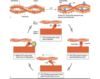
Describe the steps of excitation contraction coupling in skeletal muscle
- Acetylcholine relased at neuromusclar junction
- AP generated in response which goes down T-tubules of muscle cells
- AP in T-tubules causes relase of Ca2+ from the sarcoplasmic recticulum
- Ca2+ binds to tropnin on actin filaments
- Tropomyosin is therefore physically moved aside to uncover cross-binding bridges on actin filament
- Myosin cross-bridges attach to actin and bend pulling actin filaments towards the centre of sarcomere (contraction)
- When no longer AP Ca2+ is taken back up in the sarcoplasmic recticulum
- Ca2+ no longer bound to troponin, tropomyosin slips back to original postition over binding sites on actin, contraction ends, actin slides back to original resting place
Describe the different areas of the sarcomere
Note sarcomere is the smallest functional unit of skeletal muscle
Sarcomere is located between the Z bands
Think IAHM

Define what a fasciulation is and what they might occur in
- Visible, fast, fine , spontaneous twitch
- May occur in healthy muscle – precitated by stress, caffeine, fatigue
- Occur in denervated muscle which become hyperexcitable
- Usually a sign of disease in the motor neurone , not the muscle
What is myotonia ?
Is a symptom of a small handful of certain neuromuscular disorders characterized by inability to relax voluntary muscle after vigorous effort.
What are the general symptoms/signs suggestive of muscle disease specifically ?
- Myalgia (muscle pain)
- Muscle weakness – often specific patterns of weakness depending on cause eg proximal limbs in polymyositis
- Wasting
- Hyporeflexia
What is polymyositis and how does it typically present ?
An idiopathic inflammatory myopathy that causes symmetrical, proximal muscle weakness.
Typically presents with:
- Usually present with symmetrical, progressive (over weeks to months) proximal muscle weakness in the upper and lower extremities
- Commonly noticed as difficulty with particular activities e.g. climbing stairs
- Some have myalgia (muscle pain) or arthralgia (pain in joints)
- Dysphagia secondary to oropharyngeal and esophageal involvement occurs in about one third of patients with polymyositis
- Can also develop Interstitial lung disease
What is dermatomyositis and how does it usually present ?
It is clinicaly similar to polymyositis so presents with the same symptoms but also has typical cutaneous manifestations
Typical cutaneous manifestations are:
- Heliotrope rash
- Gottons papules
- V-shaped rash over chest
What age group does polymyositis and dermatomyositis most commonly affect ?
Usually above 20, esp aged 45-60
Describe the pathogenesis of polymyositis (think this is the same for dermatomyositis also)
- It is a T-cell–mediated cytotoxic process directed against unidentified muscle antigens
- CD8 T cells, along with macrophages, initially surround healthy nonnecrotic muscle fibers and eventually invade and destroy them.
What investigations are done to help diagnose polymyositis and dermatomyositis ?
- Inflammatory markers are often raised.
- Serum creatine kinase (CK) level is usually raised, often more than 10 times the normal level.
- Anti-Jo-1 and anti-SRP (these are the antibodies more specific to myositis)
- MRI may be useful to localize the extent of muscle involvement.
- Electromyographic (EMG) (abnormal in 90% of patients with it)
- Muscle biopsy is crucial in helping to diagnose polymyositis and in excluding other rare muscle diseases (believe this sounds the gold standard)
What is their an increased risk of in patients with polymyositis and dermatomyositis ?
- There is an associated risk of malignancy. This is found in around 25% in patients and is greatest in the 5 years following diagnosis. Common cancers include breast, ovarian, lung, colon, oesophagus and bladder.
- Malignancy should be screened for at the time of diagnosis.
What is the treatment of polymyositis?
Prednisolone (initially around 40mg) combined with immunosuppressive drugs such as methotrexate or azathioprine.
What is inclusion body myositis ?
A degenerative (because little response to steroids) muscle disease characterised by slowly progressive weakness in 6th decade of life with characteristic thumb sparing
Describe the typical presenting features of inclusion body myositis
IBM causes progressive weakness of the muscles of the wrists and fingers, the muscles of the front of the thigh, and the muscles that lift the front of the foot. Unlike in other inflammatory myopathies, the heart and lungs are not affected in IBM

What is myotonic dystrophy ?
It is part of a group of inherited disorders called muscular dystrophies. It is the most common form of muscular dystrophy that begins in adulthood.
Describe the typical features suggestive of myotonic dystrophy
- Characterized by progressive muscle wasting and weakness - typically distal-onset with weakness (hand/foot drop), weak sternomastoids, facial weakness and muscle wasting
- People with this disorder often have prolonged muscle contractions (myotonia) and are not able to relax certain muscles after use e.g. a person may have difficulty releasing their grip on a doorknob or handle
- Also, affected people may have slurred speech or temporary locking of their jaw.
Other signs include:
- cataracts
- Ptosis (drooping of eyelid)
- frontal balding in men
- cardiac defects
What age does myotnic dystrophy usually present and state its inheritance pattern
- Autosomal dominant - Trinucleotide repeat disorder affecting Cl- channels, with anticipation (affects people younger each generation)
- Usually affects people in their 20s/30s
What are duchenne and bekers muscular dustrophies ?
They are part of a group of genetic conditions characterized by progressive muscle weakness and wasting
What mutation causes duchennes and what causes bekers muscular dytrophy
Both are X-linked recessive conditions
- In duchennes the mutation results in dystrophin produced
- In bekers the mutation produces partially functioning dystrophin (hence symptoms are milder)
Both diseases caused by mutation in dystrophin protein – large deletion
Dystrophin allows connection between actin and myosin thus allowing muscle cells to renew and replace themselves