Muscles Flashcards Preview

Year 1 Histology > Muscles > Flashcards

Flashcards in Muscles Deck (80)
Loading flashcards...
1
Q

Muscle fibres comprise of several hundred ….

A

Myofibrils

2
Q

Define syncytium

A

a single cell or cytoplasmic mass containing several nuclei, formed by fusion of cells or by division of nuclei

4
Q

Features of a muscle fibres

A

Lipid droplets - fuel for respiration
Nuclei found in periphery of fibre
Lots of mito - high rate of resp

4
Q

Compare cardiac muscle to skeletal muscle

A

Cardiomyocytes form shorter fibres than skeletal muscle fibres
Cardiac muscle not built of syncytial fibres; they consist of single cells
Cardiac muscle contains single mono/dinucleated cells with limb-like extensions that allow the cells to connect to each other via intercalated discs
Centrally located nuclei in cardiac muscle; peripheral nuclei in muscle fibres
More mitochondria/capillaries in cardiomyocytes

5
Q

Describe the length-tension relationship briefly

A

Amount of force exerted by a sarcomere depends on the degree of overlap between the thick and thin filaments
At full stretch , few myosin heads can attach to actin so the force exerted is weak
Beyond full contraction, the ends of the thin filament get in the way of each other and thick filaments are forced against the Z lines so force is reduced
At optimal length, all myosin head have access to actin so force is maximal

6
Q

Structure of the fibres in heart walls

A

Muscle fibres arranged in several directions to achieve spiral like formation which allows the ventricles to contract in a wringing motion

7
Q

What are intercalated discs?

A

Connections between cardiomyocytes
Appear as dark jagged lines due to desmosomes

8
Q

Skeletal muscle fibres have gap junctions
True or false

A

False

9
Q

Features of intercalated discs

A

Desmosomes allow longitudinal force transfer due to their strong adherence
Low resistance gap functions allow fast transfer of APs

strong adherence between fibres

10
Q

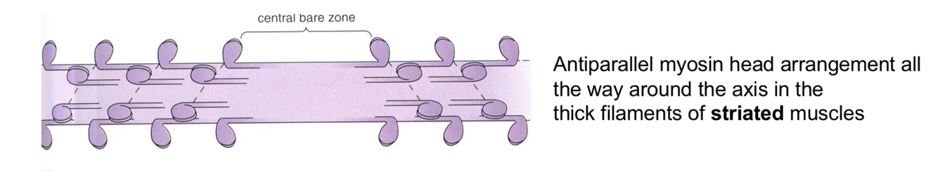
Arrangement of myosin heads in skeletal/cardiac muscle

A

Antiparallel all the way around the axis

11
Q

Arrangement of myosin heads in smooth muscle

A

Antiparallel at opposite sides

12
Q

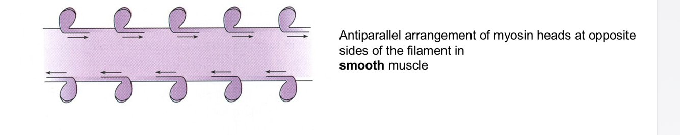
Why is the arrangement of myosin heads in smooth muscles advantageous ?

A

Allows longer sliding distances compared to skeletal muscle

higher amount of contraction can occur

13
Q

Structure of smooth muscle

A

Contraction shortens the smooth muscle cell to a fraction of its length while making it thicker

3D network of myosin, actin and intermediate filaments (not sarcomeres)

actin filaments held together by cytoskeletal proteins called dense bodies

14
Q

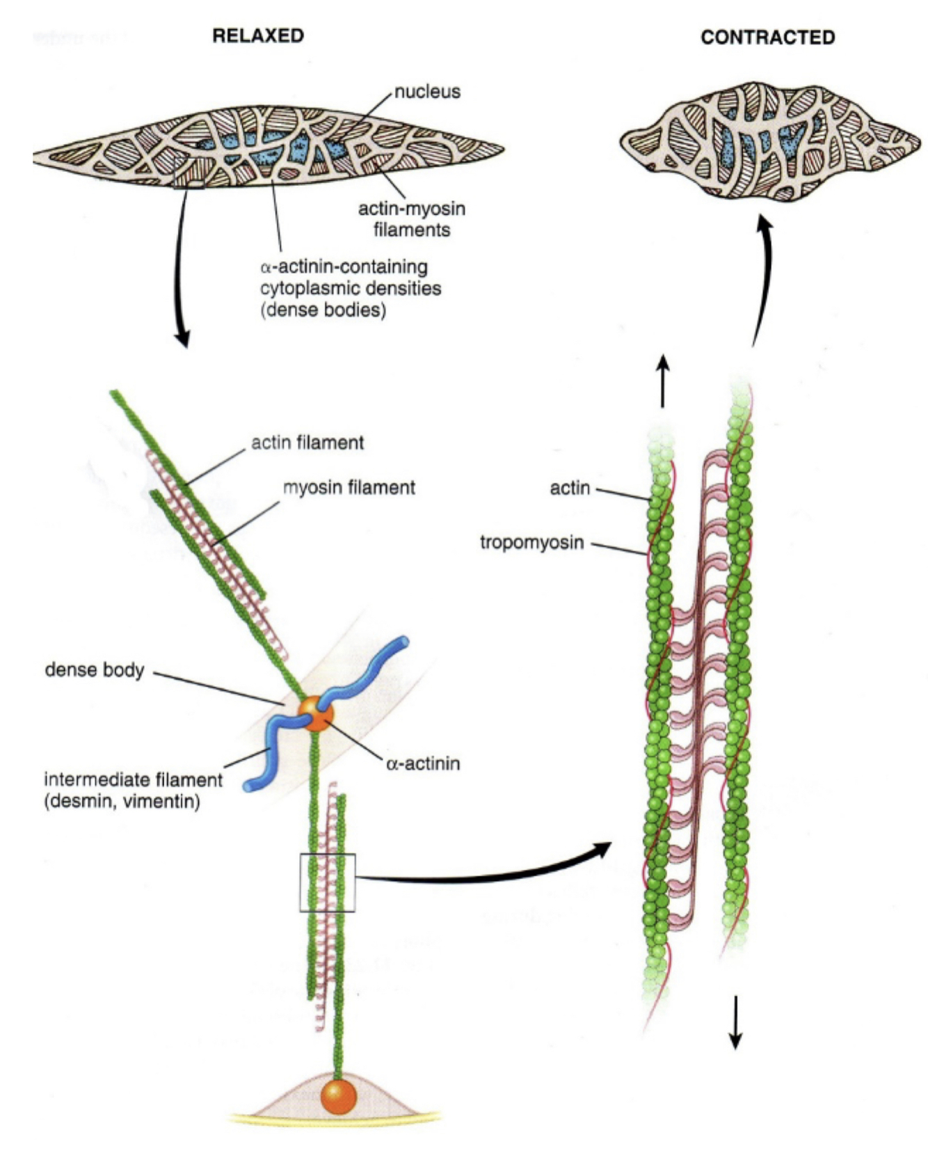
Smooth muscle cell features

A

Mononucleated

spindle shape

( dense network of filaments with one nucleus in the middle )

15
Q

Compare the three muscle types

A
16
Q

How does striation of skeletal muscle occur

A

Intermediate filaments such as desmin link Z lines laterally and myofibrils longitudinally

17
Q

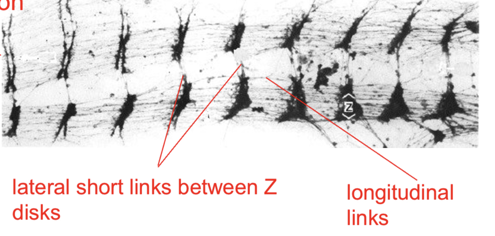
Z lines are laterally linked by

A

Desmin intermediate filaments

18
Q

What proteins is mainly responsible for preventing overstretch in muscles ?

A

Titin

19
Q

Role of myofibril cytoskeleton

A

Composed of mainly desmin fibres

Links myofibrils to each other

links synctycia to the basement membrane and into the surrounding connective tissue at the periphery

this allows force of contraction ti be transmitted both longitudinally and laterally

20
Q

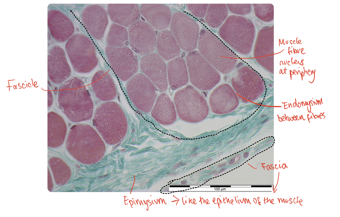
What is the endomysium ?

A

Connects to BM

Loose connective tissue with mixture of delicate/strong fibres that surrounds each muscle fibre

21
Q

What is the perimysium ?

A

Mixed connective tissue , some dense, some loose, that groups muscle fibres into fascicles

where most nerves and blood vessels are found

22
Q

What is the fascia ?

A

Dense layer of connective tissue that covers the muscle

23
Q

What is the epimysium

A

Relatively loose connective tissue between the fascia and muscle

24
Q

Identify the endomysium, a muscle fibre, the perimysium, the epimysium, a fascicle and the fascia

A

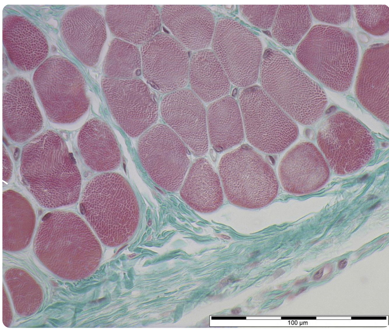
perimysium surrounds the fascicle

25
Q

Complete the sentences:

Within muscle fibres, sarcomeres are arranged into a striated pattern by 1 _____ _____ which also transfer some of the force onto the 2 ________ , which surround the muscle fibres.

muscle fibres are bundled into 3______ which are surrounded by 4_______

the muscles are packed by 5 _____

A

1 intermediate filaments

2 endomysium

3 fascicles

4 perimysia

5 fascia

26
Q

Which structures within a muscle are visible by naked eye ?

A

Fascicles can be seen as stripes

27
Q

What are motor end plates

A

Where a motor neurone synapses with a muscle fibre

1 synapse per muscle fibre

28
Q

Can AP be generated between muscle fibres?

A

No, AP can only be generated by motor end plates ; there are no gap junctions between muscle fibres

29
Q

What is proprioception?

A

Sesnors in muscle fibres for stretch and speed of movement work in association with sensory neurones to allow your body to sense movement, action, and location

30
Q

What is a motor unit ?

A

Refers to the motor neurone and the several hundred muscle fibres it attaches to

each motor neurone synapses with the same muscle fibre type

31
Q

What is the size principle

A

The idea that type 1 fibre motor units are recruited first

if more force is required , type 2 motor units recruited

32
Q

What is the difference between different fibre types ?

A

The isoform of myosin present

33
Q

Describe structure of NMJ

A

Only present in skeletal muscles not cardiac muscle

34
Q

State events that occur during chemical transition at a NMJ

A
  1. AP arrive at presynaptic bouton
  2. depolarisation of presynaptic membrane = voltage gated Ca2+ channels open
  3. calcium-mediated exocytosis of ACh into synaptic cleft
  4. ACh interacts with nAChR on sarcolemma
  5. Conformational change of nAChR ; influx of Na+ into muscle cell ; effluent of K+ Via nAChR ; Receptor Generated Potentials (RGPs) formed
  6. If RGPs exceed threshold , AP is generated; this AP is called an End Plate Potential (EPP)
  7. EPP propagated down muscle ; release of Ca2+ Into cytosol from SR ; muscle contracts
35
Q

Adaptations of muscles that require fine muscle control

A

1:1 ratio of motor neurone to muscle fibre

36
Q

Explain Ca2+ regulated release of ACh

A

AP arrives at axon terminal

terminal membrane depolarises

voltage gated Ca2+ open

Ca2+ diffuse into presynaptic bouton down conc grad

this causes exocytosis of secretory vesicles of ACh

37
Q

How is the capacity for detection of ACh release at the motor end plate enhanced?

A

Many more nAChR proteins present in sarcolemma than ACh molecules release by a single AP

38
Q

What type of receptor are nAChR receptors ?

A

Ionotropic

39
Q

How many NMJ does each myocyte have ?

A

One

one presynaptic AP leads to one post-synaptic AP

40
Q

describe how the neurones recover after an AP is initiated

A
  • enzymes found in the synaptic cleft break down the neurotransmitter
    • acetyl cholinesterase AChE hydrolyses ACh
    • ACh→ acetic acid + choline
    • these products diffuse across synaptic cleft back to presynaptic knob where they are recombined to from the neurotransmitter again
    • this requires ATP
  • this causes the neurotransmitters to no longer be complementary to receptor sites so the neurotransmitter is removed from nAChR in postsynaptic membrane
  • NAChR therefore close
  • Na+/K+ pumps work to restore resting potential
41
Q

What is myasthenia gravis

A

Autoimmune disease of NMJ

autoantibodies against nicotine AChR = trouble with initiating muscle contractions

42
Q

How is MG treated

A

inhibition of AChE via drugs ; prolong time ACh is present in cleft ; increase chances of binding to receptors and causing AP

43
Q

What is quantal exocytosis

A

Small quanta (packets ) of ACh are released at rest as voltage-gated Ca2+ channels are inefficient

this causes a small membrane depolarisation called miniature EPP (mepp)

44
Q

What happens inside a myocyte after muscle contraction occurs ?

A

Ca2+ are removed from cytosol and returned to the SR by ATP-Ca2+ pumps

45
Q

How does an ACh binding to receptors of the postsynaptic membrane cause muscle contraction

A

ACh binds to nAChR = epp = volatge gated Na+ channels = depolarisation to threshold = AP generated

AP travels down T-tubules to SR

AP communicates ryanodie receptor

Ca2+ released by SR via voltage gated Ca2+ channels

muscle contraction

46
Q

compare type 1 and type 2 fibres in terms of :

contraction time

size of motor unit

resistance to fatigue

max duration of use

force produced

mitochondrial density

capillary density

glycolytic capacity

myosin heavy chain human gene

A
47
Q

Function of muscle spindles

A

Innervated by γ motor neurons ; provide info about length changes of muscle

48
Q

Function of golgi tendon organs

A

Provide information about the strength of a muscles contraction

49
Q

What are satellite cells?

A

Myoblasts that remain in muscle fibres to act asa stem cell reserve in case damage to myonuclei/muscle fibre occurs

50
Q

Adaptation of skeletal muscles to exercise

A

Endurance training = Increased mito

Resistance training = muscle hypertrophy = more myofibrils within fibres

51
Q

How do satellite cells replace fibres ?

A

Cell fusion to syncytium , myofibril synthesis and maturation

52
Q

How much blood is expelled from heart every heart beat

A

Around 70% in a healthy person

53
Q

Two main features of heart

A

Constant contraction

Synchronous muscle activation in ventricles

54
Q

Name the 2 ways to increase function of heart

A

Inotripy - increased force of contraction = higher SV

chonotropy = increased frequency of contraction = increased HR

55
Q

Difference between AP in skeletal muscle and cardiac muscle

A

Duration of cardiac AP is much longer

shorter AP in skeletal muscle allows more control over its contraction

56
Q

How is cardiac muscle adapted to achieve synchronous contractions of the heart

A

longer AP to avoid tetanus in cardiac muscle

(tetanus is prolonged contraction of a muscle caused by rapidly repeated stimuli - you want cardiac muscle to contract with same short duration and power each time)

Longer AP achieved by plateau in AP due to influx of L-type Ca2+ channels (L for long-opening) to delay repolarisation

this allows twitch contraction of the muscle

57
Q

muscle force in a single myocyte regulated is regulated by …

A

Frequency of action potentials

summation - no time for muscle to relax before new contraction so prolonged muscle contraction

58
Q

Muscle paralysis occurs when ….

A

No action potentials are initiated in the muscle

59
Q

How is neuromuscular blockade useful?

A

Prevent muscle contractions

Maintain muscle relaxation/paralysis without the need of deep general anaesthesia

surgery (inc. cosmetic )

60
Q

What are the 4 ways to prevent neuromuscular transmission ?

A
  1. Prevent ACh synthesis by inhibiting choline uptake - no clinical use , slow effect and slow to reverse
  2. prevent exocytosis of ACh e.g botulinum toxin prevents vesicles from fusing with membrane
  3. block receptors e.g nicotinic receptor antagonists
  4. increase reuptake/inactivation of AChE
61
Q

Botox - clinical uses

A
  • Treat cervical dystopia (neuromuscular movement disorder involving head and neck)
  • treat blepharospasm (involuntary eye muscle contractions )
  • treat primary auxiliary hyperhidrosis (excessive sweating )
62
Q

Explain how non-depolarising agents work

include details about :

Onset ; Duration ; mechanism of action ; is block preceded by stimulation of muscles?

A

Non-depolarising agents work by competing for the receptor binding sites on nAChR; this is called competitive antagonism

longer duration ~ 30 mins

relatively fast onset of effect ~ 3-5 mins

Block is not preceded by stimulation of muscles

During block high freq stimulation causes tetanus with slightly onset duration of twitch (called tetanic fade)

63
Q

Agonism vs antagonism

A

Agonism = activates receptor

Antagonism = deactivates receptor

64
Q

Anticholinesterases can be used to ….

A

Reverse neuromuscular blockade due to action of antagonists

65
Q

Explain how depolarising agents work

include details of:

Mechanism of action : onset ; duration ; use ; is its blocking effect preceded by muscle stimulation?

A

work by leaving the nAChR permanently open ; they mimic shape of ACh or nicotine (which both fit receptor sites on nAChR)

new APs cannot be formed as overwhelms voltage gated Na+ channels ;

rapid onset <1min ;

short duration ~ 5 min

ideal for quick procedures e.g intubation , ECT(electro convulsive therapy) and dislocation

Block preceded by muscle twitches

66
Q

What happens upon high freq stimulation of non-depolarising block

A

Tetanus with duration only slightly longer than a twitch

called tetanus fade

67
Q

What happens upon high freq stimulation of depolarising block

A
68
Q

How can you antagonise a non-depolarising block ?

A

Using agents that depolarise the sarcolemma

drugs that increase ACh release e.g adrenaline / 4-aminopyridine

anticholinestersases (AChE inhibitors )

69
Q

name 3 common nACh-R antagonist/non-depolarising blockers

A

atracurium

vecuronium

gallamine

70
Q

name 2 nACh-R agonist/depolarising blockers

A

suxamethonium

dexamethonium

71
Q

name 2 common ACh-esterase inhibitor (“anticholinesterase”)

A

physiostigmine

neostigmine

72
Q

What is Ca induced Ca release (CICR) ?

A

L-type Ca2+ channels are very close to ryanodine receptors on SR

Ca2+ influx during cardiac AP results in Ca2+ release by SR

small increase in [Ca2+]i

Ca2+ then re-sequestered by SR via Ca2+ATPase pump and then expelled from cell by Na+/Ca2+ exchange

73
Q

difference between calcium handling apparatus in cardiac vs skeletal muscle

A

skeletal - thin t-tubules, thick SR, triads ( each t-tubule connects with 2 SR membranes) ; cardiac - thick t-tubules, thin SR, diads ( each t-tubule connects with 1 SR membrane)

74
Q

What is the effect of increasing the diastolic length of the cardiac myocyte

A

Increases sensitivity of myocyte to Ca2+ = more powerful heart contraction

75
Q

What does inotrope mean

A

Agent that alters the force of muscle contraction

76
Q

How do positive cardiac inotropes work

A

E.g caffeine

cause more Ca2+ release from channels of the SR

77
Q

How do negative inotropes work?

how do they help reduce angina/blood pressure

A

Block L-type Ca2+ channels

Reduced contraction = lower energy demand and blood ejected at lower force

78
Q

Cardiac muscle needs ATP for

A

The myosin heads to detach and reset it forward

79
Q

Energy sources of cardiac muscle

A

Mostly fat then glucose then lactate,glycolysis and ketones

80
Q

Describe cardiac muscle mitochondria

A

They have continuous reticulum

2 distinct population :

  • interfibrillar - provide ATP for muscle contraction
  • subsarcolemmal - provide ATP for ATP-dependant processes in the AP