Myelodysplastic syndrome and Myeloproliferative neoplasm Flashcards
(30 cards)
- List the two main features that characterize myelodysplastic syndrome (MDS).
MDS is a group of conditions where the marrow is replaced by a malignant clone, derived from a transformed _stem cel_l or progenitor cell, characterized by:
1) Ineffective hematopoiesis: the clone is not able to make normal functioning blood cells. In fact, they are often die before leaving the marrow, and usually look abnormal (dysplastic).
2) - Increased risk of transformation to acute leukemia: MDS is often regarded as a precursor to acute myeloid leukemia (AML).
- Describe the typical clinical presentation of patient with MDS
If MDS has a persistent cytopenia it is almost always______.

A diagnosis is usually considered in the setting of a persistent (at least several months) peripheral cytopenia in one or more lineages that cannot be otherwise explained.
If MDS has a persistent cytopenia in only one lineage, it is almost always persistent anemia.
-Isolated persistent neutropenia and isolated persistent thrombocytopenia are usually not due to MDS (but can be in a minority of cases).
Persistent cytopenia in 2 or more lineages in a patient of advanced age is suspicious for MDS.
- List three different types of tests that could be performed to make a diagnosis of MDS.
IN THE SETTING OF A PERSISTENT CYTOPENIA, THE DIAGNOSIS OF MDS CAN BE ESTABLISHED USING ONE OR MORE MODALITIES:
1) Morphologic evidence of dysplasia
2) Increased myeloblasts in blood or marrow, but less than 20% of blood and marrow cells (5-19% of marrow cells, or _2-19% of peripheral blood white blood cell_s)
3) Presence of a clonal cytogenetic abnormality, especially of an abnormality known to be associated with MDS
What is the morphological evidence of dysplasia?
What are the three classes of dyshematopoiesis?
When more than 10% of the cells in one lineage (erythroid cells, granulocytes, megakaryocytes) appear dysplastic, that qualifies as dyshematopoiesis.
Dyshematopoiesis can be subclassified as:
- Dyserythropoiesis
- Dysgranulopoiesis
- Dysmegakaryopoiesis
Dyserythropoiesis

Presence of dyserythropoiesis often results in the red blood cells in the peripheral blood exhibiting:
- Macrocytosis (CBC with increased MCV)*
- Anisocytosis (variation in cell size) and poikilocytosis (variation in cell shape); these result in increased in CBC with increased RDW
*A concurrent iron deficiency anemia, which is not uncommon in elderly patients, may mask the macrocytosis, and result in normal or even decreased MCV.
Dyserythropoiesis
Molecular correlation
Which gene mutation is associated with ring sideroblasts?
Good or bad prognosis?

The presence of increased ring sideroblasts is now known to be frequently associated with (and probably due to) mutation of SF3B1, with such mutations found in around 80% of such cases.
The presence of this mutation is associated with a relatively favorable course and prognosis*
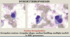
Dysgranulopoiesis

Nuclear hypolobation of mature neutrophils, including neutrophils with bilobed nuclei called pseudo‐Pelger‐Huet cells, also cytoplasmic hypogranularity of neutrophils.

Dysmegakaryopoiesis

Megakaryocytes with hypolobated or non‐lobated nuclei, often
hyperchromatic nuclei, megakaryocytes often of small size.
What is some cytogenetic evidence of MDS?
The presence of a clonal cytogenetic abnormality in the marrow of a persons with persistent cytopenia(s) is strong evidence for MDS.
The presence of one of the following MDS-related abnormalities within the clone in such a patient is diagnostic of MDS:
- complex karyotype with whole or partial deletions of chromosomes 5 and/or 7
- isolated deletion 5q
- trisomy 8
- List four possible causes of secondary myelodysplasia that might mimic MDS
A patient with suspected MDS, but negative for elevated myeloblasts and negative for a clonal cytogenetic abnormality, and only having morphologic evidence of dysplasia, be sure to exclude potential causes of secondary myelodysplasia. These include:
- VITAMIN DEFICIENCY (B12, folate, etc.)
- TOXIN EXPOSURE (e.g. heavy metals)
- EXPOSURE TO CERTAIN DRUGS
- VIRAL INFECTIONS
- Contrast low grade MDS and high grade MDS with regards to diagnostic criteria and prognosis.
____1______: Myeloblasts are not increased in frequency (myeloblasts account for <5% of marrow cells and <2% of peripheral blood WBCs)
___2_____: Myeloblasts are increased in frequency, but less than 20% (myeloblasts account for 5-19% of marrow cells and/or 2-19% of peripheral blood WBCs)
1- Low grade MDS
2- High grade MDS
Types of low grade MDS

Types of high grade MDS:


MYELOPROLIFERATIVE NEOPLASMS
- Compare and contrast MDS and myeloproliferative neoplasms (MPNs) in regards to usual number and appearance/functionality of cells in the blood and marrow.

-Clonal hematopoietic neoplasm arising from a transformed hematopoietic stem cell
-The neoplastic clone usually partially or entirely replaces the normal marrow cells in multiple lineages.
The neoplastic clone usually gives rise to increased numbers of normal (not dysplastic as in MDS) blood cells in one or more lineages.
Usually occurs in middle-aged to elderly adults, rare in children
Incidence of 6-10 cases per 100K persons per year
What are some common themes in MPNs?
What organs are usually enlarged?
1) Early disease characterized by increase in one or more blood cell types, with corresponding increase in marrow cellularity
2) Patients often have splenomegaly and/or hepatomegaly.
3) Usually have insidious onset
4) Without treatment, progress to….
- excessive marrow fibrosis with resultant bone marrow failure
- transformation to acute leukemia (much less common for MPNs than for MDS)

- Compare and contrast the four MPNs covered in the notes with regard to blood cell counts, marrow findings, and usual cytogenetic and molecular abnormalities.
Chronic Myelogenous Leukemia (CML)
CML is an MPN that manifests primarily as a persistent ____1_____, and is associated with the presence of a BCR-ABL1 gene fusion.
In what phase is usually CML diagnoses at?
What cells other than neutrophils are increased in the blood?

1- Neutrophilic leukocytosis

Majority of patients present with non-specific signs/symptoms, possibly including night sweats, weight loss, splenomegaly, and anemia
Significant minority of patients are asymptomatic at diagnosis (often incidental diagnosis due to abnormal CBC results).
Incidence of 1-2 cases per 100K persons per year.
Typical age at diagnosis: 40-60, rare in children and young adults
Chronic Myelogenous Leukemia (CML)


Chronic Myelogenous Leukemia (CML)
What is the blast phase?
What cells predominate in 70% of cases? In 30% of cases?

-If left untreated, CML often progresses to a blast phase, defined by 20% or more blasts in the marrow or blood (essentially, CML-blast phase is CML that has transformed to acute leukemia).
-In 70% of cases of CML-blast phase, the blasts are myeloblasts; in 30% of cases, the blasts are lymphoblasts.
CML - GENETICS
CML is defined by the presence of a BCR-ABL1 gene fusion, resulting in a BCR-ABL fusion tyrosine kinase protein.
This gene fusion is usually attained through a translocation t(9;22), with the derivative chromosome 22 containing the BCR-ABL fusion gene; this der(22)t(9;22) is called the Philadelphia chromosome
The translocation usually involves a breakpoint in the major breakpoint cluster region of BCR (M-BCR), giving rise to a 210kd fusion protein (p210)
In contrast, in Ph+ ALL (ALL with a Philadelphia chromosome), the BCR breakpoint is usually in the minor breakpoint cluster region (m-BCR), giving rise to a 190kd fusion protein (p190)
CML - DIAGNOSIS
STEPS IN DIAGNOSIS OF CML INCLUDE:
- Documenting the typical findings in blood and marrow, as discussed above.
- Documenting the presence of BCR-ABL1 fusion gene. This can be accomplished by:
- Conventional cytogenetics (karyotype)
- FISH (BCR-ABL1 fusion probes)
- RT-PCR (BCR-ABL mRNA transcripts)

- Explain why there is a need for a second and third generation of protein tyrosine kinase inhibitors (PTKIs).

Polycythemia Vera (PV)
Characterized by an increase in ________.
All cases of PV contain an activating mutation of _____,
PV is an MPN chiefly characterized by an increase in RBC mass (erythrocytosis)
Usually, also see increased neutrophils and platelets in the blood, and trilineage hyperplasia in the marrow.
Bone marrow biopsy often shows clusters of *bizarre megakaryocytes
Essentially all cases of PV contain an activating mutation of JAK2, usually a V617F point mutation.

Polycythemia Vera (PV)
If a patient has a persistent erythrocytosis in the absence of a demonstrable JAK2 mutation, consider the possibility of a secondary (reactive) erythrocytosis. Secondary erythrocytosis may be seen in:
- Smokers (due to carboxyhemoglobin)
- Chronic hypoxia
- Certain hemoglobin disorders
- Other conditions
Polycythemia Vera (PV)
-PV usually is diagnosed in a _________, with increased blood cell counts.
Over time, PV may progress to a ________, also known as post-PV myelofibrosis. Spent phase is characterized by extensive marrow fibrosis with a corresponding fall in blood cell counts.
Initial presenting symptoms in PV patients include:
- headaches - dizziness - plethora (dusky, reddish skin) - pruritus (itching) - paresthesias (“pins and needles” sensations) - splenomegaly (70%); hepatomegaly (40%)
1- polycythemic phase
2- Spent phase


