Myocarditis & Cardiomyopathy Flashcards
(36 cards)
The provided image is an examle of what?
Label the indicated features

healthy heart
P: pericardium
M: myocardium
E: epicardium
PM: papillary muscle

The provided image is an example of what?

Healthy pericardium & myocardium
F: fibrous pericardium
M: myocardium
CA: coronary artery

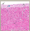
The provided image shows what histology?

normal myocardium
The provided image shows what histology?
normal endocardium (lighter on top of image)
normal myocardium (pinker bottom of image)
What is the definition of cardiomyopathy?
What are the three main types?
dysfunction of the myocardium, not secondary to ischemia, valvular disease, or hypertension
- Dilated CM: Large, flabby heart with ventricular dilation, +/- ventricular thickening; systolic dysfunction
- Hypertrophic CM: thickened, stiff left ventricle and septum; no ventricular dilation; diastolic dysfunction
- Restrictive CM: Rare. Rigid, but not necessarily thickened ventricles, diastolic dysfunction
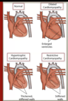
The heart on the left shows what pathology as compared to the normal heart on the right?
What are the primary causes of this pathology?

Dilated Cardiomyopathy, 4 chamber dilation
- Primary causes
- idiopathic – up to 50%
- familial – 30-50% (many mutations
- mostly proteins of cytoskeleton, could be sarcolemma or nuclear envelope proteins
What is the most common type of cardiomyopathy?
dilated cardiomyopathy
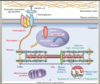
Identify the indicated proteins & the type of cardiomyopathy that would result from a mutation in said gene.
** unclear how important it is to memorize this
-
Red: dilated cardiomyopathy
- delta-sarcoglycan
- dystrophin
- desmin
- mitochondrial proteins
- titin
- lamin A/C
-
Blue: hypertrophic cardiomyopathy
- myosin binding protein C
- myosin light chains
-
Green: either (but different mutation)
- troponin I/T
- alpha-tropomyosin
- actin
- beta-myosin heavy chains
What are the secondary causes of dilated cardiomyopathy? They account for what overall percent of dilated cardiomyopathy?
-
Secondary Causes – 50% all cases
-
inflammatory
- post-infectious (especially viral)
- non-infectious
- autoimmune diseases (ie. lupus)
- peripartum cardiomyopathy (late pregnancy/postpartum)
- sarcoidosis
-
neuromuscular
- Muscular or myotonic dystrophy
-
toxic
- chronic ETOH toxicity
- heay metals, iron overload
- chemotherapeutic agents (doxorubicin, tratuzumab)
-
metabolic disorders
- hypothyroidism (esp. older individuals)
- chronic hypocalcemia or hypophosphatemia
-
inflammatory
What are the clinical features of dilated cardiomyopathy?
-
Clinical features
-
heart failure
- pulmonary congestion (LV dysfunction)
- dyspnea, exercise intolerance, orthopnea
- chronic systemic venous congestion (RH/combon LV & RH failure)
- jugular venous distension, ascites, pedal edema
- pulmonary congestion (LV dysfunction)
- arrhythemia
- thromboembolic complications
-
heart failure
-
clinical course is unpredictable
- depends on person & type of dilated cardiomyopathy
- some cases may recover, some may not
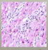
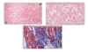
The provided image is an example of what pathology?
What histological features do you notice?

- Dilated Cardiomyopathy
- Pretty nonspecific
- myocyte hypertrophy (red)
- interstitial fibrosis (blue = collagen)
- can end up with conduction disturbances
- may also see some thrombus due to stasis
- white arrow in gross image

Describe how myocyte injury can lead to pulmonary congestion, systemic congestion & mitral regurgitation.
What clinical presentation would you expect with these conditions?

How does the kidney respond to a decrease in cardiac outlfow? This ultimately has what effect on the heart?
- When cardiac outflow is declining, blood flow to kidney will also decrease
- This prompts kidney to secrete renin –> activating the renin, angiotensin, aldosterone system forcing the body to retain more salt & with that fluid to try to increase the intervascular volume
- this worsens the condition, because the heart is already not working properly & increasing intervascular volume increases the workload on the heart
The provided image is an example of what pathology?
What are the caracteristics of this condition?

Hypertrophic Cardiomyopathy
heart is heavy, muscular, hypercontractile & stiff with poor diastolic relaxation. Asymmetrical septal hypertrophy
ventricular outflow obstruction in 1/3 of cases (anterior leaflet may be flapping up against the endocardium causing thickening and perhaps plaque formation)
Mutations in what three genes account for 70-80% of all hypertrophic cardiomyopathy?
- beta-myosin heavy chain
- myosin binding protein C
- troponin T
What is the most common cause of hyertrophic cardiomyopathy?
What demographics are most effected?
100% inherited
Incidence 1/500
all age groups
no populatin predilection
What is the most common cause of sudden cardiac death in young athletes?
hypertophic cardiomyopathy
What are the clinical features of hypertrophic cardiomyopathy?
- often asymptomatic
- syncope
- palpitations
- exertional dyspnea
- chest pain
- atrial fibrillation
- sudden cardiac death
Describe how myocyte hypertrophy & dynamic left ventricle outflow obstruction can lead to angina, dyspnea, syncope and sudden death.
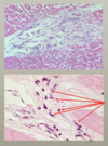
The provided image is an example of what pathology?
What are the key histological fearures?
hypertrophic cardiomyopathy
myocyte disarray
interstitial fibrossis (pink in between the myocytes in image C)
Trichrome stain (D): stains collagen blue, you can see the large amounts of collagen
What are the common clinical features of dilated cardiomyopathy and hypertrophic cardiomyopathy?
heart failure
sudden death
atrial fibrilation
stroke
The provided image is an example of what type of pathology?
Prominent features?

Restrictive Cardiomyopathy
- Left: prominent biatrial dilation
- Right: prominent interstitial fibrosis
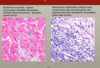
The provided images are an example of what type of pathology?

Amyloidosis – can cause restrictive cardiomyopathy
- H&E- pale pink, smudgy material
- Congo Red, polarized Amyloid (upper half) is green
What is the most common cause of restrictive cariomyopathy in non-tropical countries? Most affected demographic?
What is the most common cause of restrictive cardiomyopathy in tropical countries? Most affected demographics?
- non-tropical
- Amyloidosis
- middle aged & older adults
- tropical - nutritional deficiencies and worm infections (hypereosinophilia)
- endomyocardial fibrosis
- children & young adults