Nephrology Flashcards
(35 cards)
ACUTE NEPHRITIS: Outline the aetiology, clinical features and management.
Aetiology: Accumulation of immune complexes at the GBM causes inflammation of the membrane and endothelial proliferation. In short, this leads to reduced glomerular perfusion/filtration.
There are autoimmune, infectious and hereditary causes.
Common causes: Post-streptococcal infection, vasculitis (HSP, granulomatosis with polyangiitis, SLE, microscopic polyangitis), IgA nephropathy a.k.a. Berger’s, Goodpasture’s disease
Clinical features:
- Hypertension (→ seizures)
- Oedema (periorbital)
- Haematuria and proteinuria
- Renal dysfunction. leading to:
- Oliguria
- Volume overload ( = oedema)
Management:
Supportive care as resolution is often spontaneous
- Fluid restriction with use of diuretics when necessary, prophylactic antibiotics
- Monitoring urine output
Monitoring of renal function to check for damage
ACUTE NEPHRITIS: Be aware of the long term complications of HSP
-
Renal impairment
- Follow-up appointment after HSP diagnosis to check blood pressure, urine dip ( check for proteinuria) and for any signs of renal impairment.
- Follow-up with the GP at regular intervals to complete the above (timing dependent upon +/- proteinuria at time of presentation)
- Severe proteinuria may lead to nephrotic syndrome
- Progressive chronic kidney disease: Hypertension, heavy proteinuria, oedema and declining renal function are all seen
-
Intussception
- ! WARN about red currant jelly stools
- Ileus
- Orchitis
- Testicular torsion/haemorrhage
Intussception, ileus and orchitis are rare complications
ACUTE NEPHRITIS: Outline the management plan of patients with HSP including follow-up for detection of HSP nephritis.
*Consider the triad of symptoms*
TRIAD OF SYMPTOMS: Arthalgia (knees, ankles), purpuric rash, colicky abdominal pain (+/- diarrhoea, haematemesis, obstruction)
Classically affects children between the ages of 3 - 10 years
Child is usually clinically well - unlike meningococcal disease
Preceeded by an illness or vaccination
Management
Arthalgia: NSAIDs
CAUTION If there is renal involvement then be wary of NSAID use
Rehydration with fluid balance monitoring
?Low dose corticosteroids - no evidence of benefit in terms of reducing risk of renal complications
Year long follow-up to check blood pressure and urinalysis
- Renal involvement is often delayed, with renal detriment seen after resolution of the rash (4/52)
- Hypertension and proteinuria indicate renal pathology and need for specilaist referral
ACUTE NEPHRITIS: Name the most common cause of acute glomerulonephritis in childhood.
- Post-infectious
- Group A Streptococcus
- Vasculitis
- HSP (IgA vasculitis - IgA and IgG accumulate at the GBM, causing complement activation. Precipitates an inflammatory response with vasculitis)
- SLE
- Granulomatosis with polyangiitis
- IgA nephropathy (Berger’s)
- Mesangiocapillary glomerulonephritis
- Goodpasture syndrome
ACUTE NEPHRITIS: List the initial investigations in patients presenting with acute Glomerulonephritis
ACUTE NEPHRITIS: Be aware that IgA nephropathy shares the histopathological features of HSP nephritis
Histopathological features:
- IgA deposition
- Endothelial proliferation at the glomerulus
Differentiating factors
- Systemic involvement in HSP (joints, GI, skin), only renal in IgA nephropathy
- HSP is more commonly seen in younger children (3-10 years), IgA nephropathy is more common between the ages of 15 - 30
NEPHROTIC SYNDROME: Know the aetiology, incidence and presenting features of childhood nephrotic syndrome.
Aetiology: Dysfunction of the GBM leads to heavy proteinuria
- Secondary to systemic disease
- HSP, SLE, malaria, bee sting
- Minimal change disease
- Focal segmental glomerulosclerosis
- Membranous nephropathy
- Membranoproliferative glomerulonephritis
Incidence:
Usually seen between 2-6 years
Presenting features:
- Oedema
- Proteinuria (> 1g/m2/day or 3+ of protein on dipstick)
- Hypoalbuminaemia
- Hyperlipidaemia
Clinical features
- Oedema: Most often periorbital up on waking, swollen lips
- Oedema of the scrotum/vulva, ankles and legs
- Infections (due to loss of immunoglobulins in the urine)
- Ascites
- Foamy urine
- Discomfort relating to swelling or skin breakdown
- Weight gain
- Abdominal distension
- Tiredness
NEPHROTIC SYNDROME: Name the most common type in childhood
- 80% of cases are due to minimal change disease
- Focal segmental glomerulosclerosis
- Membranous nephropathy
NEPHROTIC SYNDROME: Outline the initial management of children who present with nephrotic syndrome.
High dose corticosteroids for 4/52, with dose weaning
- MONITOR blood pressure - hypertension is a recognised complication
Dietary sodium restriction: Helps to reduce fluid retention
Prophylactic antibiotics: To reduce infection risk (2º to loss of immunoglobulins)
Fluid management: Diuretic therapy and IV albumin may be required
Vaccination: Pneumococcal vaccination and varicella immunisation should be considered
NEPHROTIC SYNDROME: Be aware of the atypical features which would prompt consideration of second line treatment and/or a renal biopsy.
Nephrotic syndrome unresponsive to steroid treatment or frequent relapse indicates the need for a renal biopsy, prior to commencing immunomodulatory agents.
Atypical features that suggest that the aetiology is unlikely to be minimal change disease (hence likely to be steroid unresponsive)
- Age < 1 or > 12 years
- Gradual onset of oedema
- Macroscopic haematuria
- Rash/features of systemic disease
- Hypocomplementaemia
- Renal dysfunction
- Steroid resistance with failure to respond to steroids by 4 weeks
- Significant hypertension
- Confirmation of calcineurin nephrotoxicity
UTI: Know the incidence and common organisms which cause childhood UTI.
Incidence
- 1/10 girls, 1/30 boys have a UTI before the age of 16
Causative microorganisms
- E.Coli
- Proteus
- Klebsiella
- Enterococcus
- Pseudomonas: May indicate a structural abnormality
UTI: Reach a differential diagnosis for children presenting with haematuria.
UTI
Nephritis – nephritic syndrome
Urinary system trauma
Renal calculi
Congenital abnormality e.g. stricture
Renal malignancy
Bleeding disorders
Hypercalciuria

UTI: List the presenting features of UTI in infants, preverbal children and verbal children.

UTI: Know methods of collecting urine i.e. clean catch urine, bag urine, catheter specimen and suprapubic aspirate and be aware of some of the advantages and disadvantages of each meth

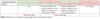
UTI: List the criteria for diagnosis of UTI based on urine dipstick and urine culture
Diagnosis from urine culture
- > 105 organisms/mL in pure growth and clinical suspicion
- Any growth from suprapubic aspirate
Diagnosis from urine dipstick
(see image)
UTI: Know the definition of atypical UTI and recurrent UTI as stated in the NICE guideline CG54 (Childhood UTI) and outline the investigation schedule based on these definitions.
Atypical UTI:
- Seriously ill
- Poor urine output (? congenital abnormality)
- Abdomial/renal mass
- Raised creatinine
- Sepitcaemia
- Failure to respond to antibiotic therapy within 48 hours
- Causative agent not E.Coli
Recurrent UTI:
- 2 or more upper UTIs OR
- 1 upper and 1 lower UTI OR
- 3 lower UTIs
Investigations:
- Age dependent
- Renal USS is indicated for children < 6 months presenting with first UTI, children > 6 months with features of an atypical UTI and children with recurrent UTIs
- Atypical UTI:
- USS during acute infection for all age groups
- DMSA and KUB scan after infection for those < 6 months old
- Recurrent UTI
- USS during acute infection for those < 6 months
- DMSA (4/6 months) after the infection
- Assess for the presence of renal scarring
- MCUG for those < 6 months
- May allow for posterior urethral valves (in males) or VUR to be detected
UTI: Know the incidence of Vesicoureteric Reflux (VUR) in the general population and in children who present with a UTI.
Incidence
General population - 1%
Children presenting with UTI - 30-45% of < 5s
UTI: Outline the diagnostic tests for VUR
Antenatal scans: Small, smooth kidneys
MCUG: Catheterisation to allow introduction of a contrast dye into the bladder. The flow of dye in the urine is checked using radiography (X-ray). Dye will travel contralaterally in reflux
Indirect cystogram: Injection of radioactive isotope, which is then taken up by the kidneys. The flow is then analysed by asking the child to urinate in front of a detector. Only suitable for children who are able to mictuate on demand
UTI: Define pyelonephritis and cystitis as stated in the NICE guideline CG54 (Childhood UTI)
*think of definitive signs/symptoms*
Pyelonephritis:
- Bacteriuria
- Fever (> 38ºC)
OR
- Fever < 38ºC with loin pain/tenderness
?Elevated CRP
Cystitis:
- Bacteriuria WITHOUT systemic signs/symptoms
UTI: Be aware of the treatment of Pyelonephritis.
- Immediate referral to paediatrics for infants less than 3 months of age
- Analgesia
- Ensure adequate hydration
- Antibiotic therapy
- PO: Cefalexin, coamoxiclav
- IV: Coamoxiclav
UTI: Understand that Vulvo-vaginitis is common in young girls and the initial steps in management.
Vulvo-vaginitis: Inflammation of the vagina and vulva
Signs/symptoms: Erythema, pruritis, pain, discharge (yellow/green)
Pathophysiology: The lower levels of sex hormones in younger females mean the genital skin is thinner and less acidic, creating a predisposition to infection.
Initial steps in management:
- Further investigations, namely swabs, may be indicated if the conditon is causing distress
- Personal hygiene adjustments: Wipe front to back, wash the genitals after urination/opening the bowels
- Clothing: Avoid tight fitting trousers and wearing underwear at night
- Acitivities: Avoid direct pressure on the vulva, avoid chlorinated water
- Relief from soreness: Apply a cold compress, application of sudocrem/bepanthen
AKI: Know the presenting features of acute kidney injury (AKI) in childhood.
- Reduced urine output (< 0.5 ml/kg/hr for 8 hours)
- Increased serum creatinine
- Increase from baseline or value greater x1.5 the upper limit for the age group
- Systemically unwell child (PEWS)
AKI: Name the most common causative organism of childhood diarrhoea associated Haemolytic Uraemic Syndrome (HUS)
Verocytotoxin-producting E.coli O157:H7 (VTEC)
- Acquired through contact with farm animals or eating uncooked beef
Less commonly, HUS may be seen as a consequence of Shigella infection
AKI: List the triad of abnormalities which define HUS
- Acute kidney injury
- Haemolytic anaemia
- Thrombocytopenia

The E.coli preferentially localises to the kidney and causes intravascular thrombogenesis, leading to AKI. This depletes platelets, leading to thrombocytopenia, and RBC become damaged as they circulate through the occluded vessels, promopting their lysis.

