Nephrotic Syndrome I and II Flashcards
(53 cards)
Glomerulus
•Anastomosing capillaries lined by fenestrated endothelium, supported by mesangium, surrounded by Bowman’s capsule, with two layers of epithelium (visceral and parietal).

Glomerular Capillary Wall
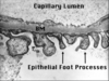
- Thin layer of fenestrated endothelium.
- Glomerular basement membrane (lamina densa, lamina rara interna and lamina rara externa), composed mostly of Type IV collagen.
- Visceral epithelial cells (podocytes) – Interdigitating “foot” processes, separated by slit diaphragm (filtration slit).

Mesangium
- Supports the capillary loops.
- Consists of cells and matrix

Bowman’s Capsule
•Lined by parietal epithelial cells.
Nephrotic Syndrome
Clinical condition related to dysfunction of glomerular podocyte. Four key components:
- Proteinuria > 3.5 gm/24 hours.
- Hypoalbuminemia
- Hyperlipidemia and lipiduria
- Edema
Acute Nephritic Syndrome
- Clinical condition associated with glomerular capillary dysfunction/inflammation (active glomerulonephritis).
- Main clinical features: Hematuria, proteinuria, increased blood pressure, edema, increased serum creatinine, and active urinary sediment.
- Active urinary sediment – Red blood cells and casts (made of red blood cells, white blood cells and/or epithelial cells) detected in the urine; indicates active glomerulonephritis
•Rapidly progressive glomerulonephritis - Severe form of acute nephritic syndrome with rapid rise in serum creatinine (renal emergency).
Persistent Asymptomatic Urine Abnormalities
•Usually subnephrotic range proteinuria (< 3.5 gm/24 hours) or persistent/recurrent microscopic hematuria.
Renal Failure
•Elevated serum creatinine.
- Acute – Glomerular, vascular or tubulointerstitial disease.
- Chronic – Advanced renal disease, usually irreversible, due to a variety of primary diseases.
Clinical Presentations Leading to Renal Biopsy
- Nephrotic Syndrome
- Acute Nephritic Syndrome
- Persistent asymptomatic urine abnormalities
- Renal Failure
Three Modalities of Renal Biopsy Examination
- Light Microscopy
- Immunofluorescence Microscopy
- Electron Microscopy
•All samples must have cortex with glomeruli
Light Microscopy
- Multiple level sections of formalin-fixed tissue cut and mounted on glass slides.
- Basic stain: Hematoxylin and Eosin (H&E)
•General stain, good for inflammatory cells
- Special stains:
•Periodic Acid Schiff (PAS)
- Mesangium, basement membranes
•trichrome
- Fibrosis, necrosis
•Jones silver stain
- Basement Membranes

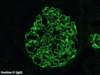
Immunofluorescence Microscopy
- Used to detect presence of immunoglobulin and complement proteins.
- Multiple level sections of frozen tissue cut and mounted on glass slides.
- Proteins detected: IgG, IgM, IgA, C3, C4, C1q, fibrinogen, albumin, kappa light chain, and lambda light chain.
• Immunofluorescence Microscopy (IF) Antibodies used:
– Immunoglobulins:
- Heavy chains: IgA, IgG, IgM
- Light chains: kappa, lambda
– Complement: C3, C4, C1q
– Fibrin/fibrinogen
• Marker of severe injury (necrosis, crescents)
– Albumin
• Good barometer for background staining - “Stickiness” factor
Electron Microscopy
- Transmission electron microscope is used to examine ultrastructure of renal tissue.
- Tissue fixed and embedded in hard epoxy resin; ultrathin sections cut using diamond blade; sections mounted on a grid and examined using electron microscope.


Three Proposed Mechanisms of Immune Complex Formation
•Some forms of renal disease are caused by antigen-antibody complexes.
- Antigen-antibody complexes form in the blood, circulate to the kidney and are then deposited into renal tissue.
- A circulating antigen is first deposited into the kidney, and the recognizing antibody then binds to the planted antigen.
- A protein normally present in renal tissue acts as an auto-antigen, and the recognizing antibody binds to this intrinsic renal protein.
Mechanisms of Renal Injury
- Antigen-antibody complexes activate the complement cascade.
- Some results of complement activation:
a. Elaboration of cytokines and chemokines.
b. Recruitment of inflammatory cells.
c. Damage to renal tissues from cell lysis, actions of inflammatory mediators, activation of digestive enzymes, etc. - Inflammatory type of injury more severe in diseases that cause acute nephritis vs nephrotic syndrome.
Pathophysiology of Nephrotic Syndrome
- Glomerular Proteinuria
- Hypoalbuminemia
- Edema
- Hyperlipidemia and Lipiduria
Glomerular Proteinuria
– Increased filtration of macromolecules across glomerular capillary
– Due to abnormalities in podocyte
– Albumin – Principal urinary protein
- Clotting inhibitors
- Transferrin
- Vitamin D binding protein

Hypoalbuminemia
– Consequent to urinary albumin loss
– Hepatic albumin synthesis increases but is unable to sufficiently replete serum levels
– Serum albumin levels are low
• Usually <2 g/dl (nl 3.5-5.5 g/dL)
Edema
– Hypoalbuminemia causes egress of fluid into interstitial space
• Due to decreased plasma oncotic pressure
– Stimulation of the Renin-Angiotensin system
- aldosterone release causing marked sodium retention
- sympathetic stimulation
- reduced natiuretic peptide release
– Soft dependent, pitting edema
Hyperlipidemia and Lipiduria
• Hyperlipidemia
– Decreased oncotic pressure stimulates hepatic lipoprotein synthesis
- Manifests as hypercholesterolemia, hypertrigliceridemia
- Lipiduria
– Lipid in urine becomes entrapped within protein material in renal tubules “lipid casts”
– Enclosed by cytoplasmic membrane of degenerated cells = “oval fat body”
• Cholesterol in oval fat bodies appear as maltese crosses under polarized light

Complications of Nephrotic Syndrome
A. Altered coagulation
- Thromboembolism - Typically seen with >10 grams of proteinuria.
a. Increased hepatic production of procoagulation factors.
b. Anticoagulant factors lost through glomerular capillaries (anti-thrombin III).
c. Concomitant volume depletion from diuretic therapy + reduced oncotic pressure in the vasculature leads to hemoconcentration and increased platelet aggregation.
d. Thrombus formation
i. Predilection for renal vein thrombosis (likely due to loss of fluid across the glomerulus and ensuing hemoconcentration in the postglomerular circulation).
B. Infection
- Due to immunoglobulin loss in the urine.
- Common infections: Staphylococcus and Streptococcus pneumoniae
Generalized Treatment of Nephrotic Syndrome (Non specific Therapy)
A. Renin-angiotensin system: All should be treated with ACE inhibitors (ACEi) and or angiotensin receptor blockers (ARB) to reduce intraglomerular capillary pressure and reduce proteinuria.
B. Diuresis: Loop diuretics for edema, low sodium diet (<2g/day).
C. Strict blood pressure control: Goal <130/80 mm/Hg.
D. Statin: Treatment of hyperlipidemia (Statin = HMG-CoA reductase inhibitor).
Causes of Nephrotic Syndrome
- Primary Glomerular Diseases
- Membranous Nephropathy
- Minimal Change Disease
- Primary Focal Segmental Glomerulosclerosis
- Idiopathic/Autoimmune Membranoproliferative Glomerulonephritis - Secondary Systemic Diseases
- Diabetic Nephropathy
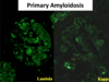
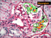
- Amyloidosis
- Systemic Lupus Erythematosus