Neuraxial Anesthesia Flashcards
(40 cards)
How many vertebrae in the spine? Categories of vertebrae?
Features?
- The building blocks of the spine are the individual bones called vertebrae.
- Cervical (7)
- Thoracic (12)
- Lumbar (5)
- Sacral (5) fused
- Coccygeal (4)fused
-
all vertebrae have a vertebral body (Except C1)
- vertebral body increases in size as you go down the spinal column
-
have 2 pedicles from the vertebral body which join together with lamina
- transverse process for muscle attachmenet
- spinous process also for muscle attachment
-
pedicle with lamina and vertebral body make the vertebral foramen, which houses the spinal cord
- superior and inferior articulating processes/facets
- where adjoining vertebrae articulate
- when 2 vertebral come together, intervertebral foramen is created
- this is where spinal nerves exit the vertebral column
- superior and inferior articulating processes/facets
-
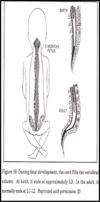
spinous process shape changes down vertebral column
- cervical region- spinous process short and bifid until C7 (vetebral prominent)
- thoracic- spinous process elongated and inferior/posterior direction
- spinous process overlay inferior body of next vertebrae
- lumbar- spinous process is short and hatchet shaped

What are the vertebral curves in the supine position?
- High
- C5
- L3
- Low
- T5
- S2

What are the ligaments of the spinal column? Purpose?
- Purpose of ligaments (5 ligaments)
- to Stabilize Vertebral body
- Supraspinous- runs C5-sacrum
-
Interspinous- entire length
- in between each spinous process
- Ligamentum flavum
- Posterior longitudinal ligament- posterior surface of vertebral bodies (C2-sacrum)
- Anterior longitudinal ligament- anterior surface of vertebral bodies (C1-sacrum)

What are the spinal meninges?
Protective membranes continuous with cranial meninges:
-
Dura mater
- thickest meningeal tissue
- Begins at foramen magnum and ends caudally at S2/Dural sac (posterior superior iliac spine); S3 in infants
- Abuts the arachnoid mater (subdural space)
-
Arachnoid mater
- Principal physiologic barrier for drugs moving between the epidural space and the spinal cord
- Pressure of CSF pushes arachnoid against Dura Mater
- underneath arachnoid mater is subarachnoid space
- Gives rise to the Subarachnoid space
- house CSF
- Ends at S2; delicate and nonvascular
- Pia mater
- Adheres to the spinal cord

Where does the dural sac end?
What composes the cauda equina?
What is the filum terminal internum? filum terminal externum?
S2
- Superficial landmark is posterior superior iliac spine (PSIS)
- cauda equina is ventral and dorsal roots of lumbar and sacral region of spinal cord
- filum terminal internum (pia mater) piereces dura sac and picks up arachnoid and dura mater which becomes flium terminal externum
- anchors SC to coccyx

Anatomy of spinal cord?
- Foramen magnum to conus medullaris (termination of the spinal cord)- L1-L2
- Spinal cord ends at L1 in adults, L3 in pediatrics
- Gives rise to 31 pairs of spinal nerves
- Each with an anterior root (motor) and posterior root (sensory)
- Roots are in turn composed of rootlets.
-
Conus medullaris ends L1 in adult
- has cauda equina in area
- Cauda equina (ventral/dorsal roots of lumbar and sacral region)
- has filum terminal internum
- comprised of pia mater
- as pierces dural sac–> picks up arachnoid and dura mater form filum terminal externum (anchors SC to coccyx)
- comprised of pia mater
- has cauda equina in area
- Dural sac- ends at S2 (PSIS)
What is a dermatome?
Segment?
Which dermatomes do we need to memorize?
- The portion of the spinal cord that gives rise to all the rootlets of a single spinal nerve is called a segment
- Dermatome is the skin area innervated by a spinal nerve and its segment
- Cutaneous distribution of spinal nerves
- C6 (thumb)
- C7 (2nd and 3rd finger)
- C8 (4th and 5th finger)
- T4 (nipple)
- T6 (xiphoid)
- T8 (last rib)
- T10 (umbilicus).
Goal of neuraxial anesthesia?
- Goal: Blockade of nociceptive impulses
- Nociceptive impulse is a stimulus that causes pain or injury
- Blocks all impulses regardless of fiber type (also order of blockade. Return of sensation happens in REVERSE order)
- Autonomic
- Sensory
- Proprioception
- Motor
-
Autonomic and motor function are also blocked !!
- blocking both dorsal and ventral root
-
Order of fibers blocked (this was not covered adv prin, but covered in previous classes? don’t know if we need this level of detail…)
- B fibers – lightly myelinated, pre-ganglionic ANS fibers
- C fibers – sympathetic, non-myelinated post ganglionic ANS fibers
- C fibers – dorsal root, non-myelinated slow pain fibers – slow pain/ temperature / touch
- A delta – medium myelination, fast pain, temperature touch
- A gamma – medium myelinated, skeletal muscle tone
- A beta – heavy myelinated touch/pressure
- A alpha – heavy myelinated, skeletal muscle, motor, proprioception.
What is a differential blockade?
Different nerve types have different sensitivities to local anesthetic (LA)
- Autonomic nerves highly sensitive with rapid onset of blockade
- will see decrease in BP and hypotension before loss of sensory/motor
- if blockade high enough, can see bradycardia
- Sensory nerve intermediate sensitivity
- next modality blocked
- Motor nerves more resistant to LA and have slower onset of blockade
- last modality to be blocked
- patient may not be able to sense leg, but will be able to move it until motor is blocked
How does the autonomic and motor blockade differ between SAB and Epidural blockade?
- Spinal (SAB) Blockade:
- Autonomic blockade 2-6 levels above sensory blockade
- Motor blockade 2 below sensory blockade
- Epidural Blockade:
-
Autonomic blockade same level as sensory blockade
- because of diffusion required through dura/arachnoid mater
-
Motor blockade 2-4 levels below sensory blockade
- iif patient says no feeling at nipple line (T4), autonomic blocked at same T4, cardiac accelerators still intact. not as much bradycardia
-
motor blockade at T6-T8
- not as much of a respiratory blockade c/t spinal
-
Autonomic blockade same level as sensory blockade
Example
-
if patient has SAB and says they can’t feel anything at nipple line, sensory blockade is T4
-
autonomic blockade will be 2-6 levels above that. T1-T4 is where cardiac accelerators are
- so patient will lose ability to regulate heart rate in response to vasodilation that occurs with spinal, causing bradycardia/hypotension
-
autonomic blockade will be 2-6 levels above that. T1-T4 is where cardiac accelerators are
-
if same patient has sensory at T4, motor blockade is T6–> impairs accessory respiratory muscles
- may not matter in young, healthy individual but may matter in pt with resp decline (copd etc)
Advantages/disadvantages to neuraxial anesthesia
Advantages
- Decreased incidence of DVT, cardiac morbidity and death
- Decreased lower extremity vascular graft occlusion, due to vasodilationà increased tissue blood flow below level of blockade
- Decreased incidence of pneumonia
- minimal pain, get up, move, cough, prevent PNA
- Decreased stress response
- decreased catechol release
- beneficial for pt with CAD
- Avoids airway manipulation
- caution neuraxial technique in those with known difficult airway
-
safest option for known difficult airway is to have control over airway throughout the procedure
- if blockade goes high and limits respiratory drive, then have to deal with difficult airway in middle of sx.
- Decreased incidence of PONV
- Intra and postoperative pain relief
Disadvantages
- Hypotension
- If pt can’t tolerate big drop in BP, may want GA
-
ex- pt withs severe aortic stenosis, CAD
- epidural may be better option because hypotension is not as profoudn
- may decide GA is safer
- Delayed case start
- Failure rate depends on experience
- Not a benign anesthetic
CV effects of neuraxial block
- Loss of sympathetic activity results in vasodilation below level of blockade, decreasing SVR (15-20%)à decreased preload therefore CO (decrease 10-15%).
- Venous dilation > arterial dilation
- If blockade is at or cephalad to T1-T4 the cardiac accelerators are blocked resulting in bradycardia.
- Results in profound hypotension
- Treatment includes: vasopressors, volume load (15ml/kg), +/- vagolytic drug to treat bradycardia
Pulmonary effect of neuraxial blockade
- Low levels of blockade- minimal effect on MV/TV/RR/dead space
- As block ascends, accessory muscle paralysis occurs, a perception of ineffective breathing and decrease ability to cough develops/protect airway (T4-T6 rea)
- even young healthy patients will not be able to feel themselves breath. need to reassure everything is ok
- if history of asthma, may want to get lower blockade if possible
- No direct respiratory effects except those related to positioning unless high block (C3-5 phrenic nerve) …
- With profound hypotension, may see ischemia of the central respiratory centers causes respiratory arrest
- will need respiratory support. have back up supplies ready!
GI/Renal effects of neuraxial anesthesia?
-
Nausea & vomiting (20%)
- r/t hypoTN
- OB complaining → give phenylephrine
-
Hyperperistalsis d/t unopposed parasympathetic activity (cranial component & vagus nerve)
- Vagus- innervation of abdomen to left colonic flexure → makes unopposed vagus = hyperparastalsis
- Flow to liver- BP dependent
- Maintenance of MAP – NO untoward liver effects
- Renal blood flow is autoregulated therefore minimal effect
- Bladder dysfunction - Urinary retention
- (sacral component of blocked PSNS)
- If no catheter is present- avoid excessive IV fluids
Metabolic and Endocrine effects of neuraxial anesthesia?
- Blocks the stress response to surgery (decrease catecholamine release) – bc nerves at surgical site are anesthetized
- Catecholamine release may be blocked from the adrenal medulla
- good for pts with CAD as long as you maintain perfusion (DBP)
- Cortisol secretion is delayed
-
Shivering – altered thermoregulation w/vasodilation
- Don’t have thermoregulation after spinal (sometimes epidural) and they vasodilate (loss of autonomics) → unable to shiver!
- Keep warm after receive spinal/epidural.
- Don’t have thermoregulation after spinal (sometimes epidural) and they vasodilate (loss of autonomics) → unable to shiver!
Neurological effects of neuraxial anesthesia?
- CBF maintained unless MAP < 60 mmHg
-
S/S Low BP:
- *N/V
- Eventually leading to → apnea and hypoxia
-
S/S Low BP:
- Decreased signals to Reticular Activating System (RAS)à drowsiness (normal)
- Why need to know DAW
Considerations for choosing neuraxial technique?
- Anatomy
- Age
- young female in 20s will be easier to place epidural/spinal c/t elder back with arthritic changes/osteoporosis
- Pregnancy
- Pathophysiology/Comorbidities
- mild/mod aortic stenosis should do epidural instead of spinal (more controlled onset of autonomic blockade with epidural)
- severe aortic stenosis- may not want neuraxial technique at all
- Sensory level required vs adverse physiological effects
- if need sensory up to T4, then you’re going to get autnomic blockade that blocks cardiac accelerators. can your pt tolerate that?
- may want to do epidural instead of spinal, because autnomic blockade will also only be at T4 with epidural, leaving cardiac accelerators intact.
- Length of procedure
-
a good spinal anesthetic lasts about 2 hours (can prolong)
- if sx is longer, may need epidural with catheter so you can redose
-
a good spinal anesthetic lasts about 2 hours (can prolong)
- Post-op analgesic needs
- need one shot and done, or will you need the added analgesia postop?
Contraindications to neuraxial anesthesia
- Patient Refusal<< only absolute C/I
- Infection at injection site
- may need to go to diff level
- Increased ICP
- Clotting defects/anticoagulant therapy
- know pt PLT level
- need >100 k PLT
- know site protocol
- Severe hemorrhage or hypovolemia
- CNS disease/meningitis
- MS or meningitis for example
- Hysteria/inability to remain still for block placement
- Bacteremia
- Septicemia
- Valvular lesions with fixed stroke volume
- severe AS/MS - maybe just use epidural
- hypertrophic cardiomyopathy
- Difficult airway
- Full stomach- relative
- Peripheral neuropathies- relative. need thorough documentation of baseline abnormalities
Pre-procedure preparaiton for SAB/Epidural insertion?
- Appropriate monitors
- Suction (@HOB)
- Oxygen delivery (nasal cannula/face mask)
- Fluid Bolus
-
*15mL/kg or 500-1000 ml – if pt can tolerate.
- 15-20 min before block** (don’t wait too long)
- Prevent hypoTN (esp SA)
-
*15mL/kg or 500-1000 ml – if pt can tolerate.
- Equipment for airway management and resuscitation are available
- Emergency drugs drawn and available
- ~Lipids
- Consider sedation prior to procedure
- Identify landmarks – before prep or drape.
-
if obese, fill where hips are and ask the patient if your hands are on their hip
- ask patient if you’re in the midline of their back
- can be difficult to find spinous processes
- feel where back is located, feel spinous processes
-
if obese, fill where hips are and ask the patient if your hands are on their hip
What are common landmarks for an epidural?
- C7 – vertebra prominens
- T3- scapular spine
- T7 - inferior angle of scapula
- T12- last rib
-
L4- Intercristal line or Tuffiers line = iliac crest
- Lumbar epidural/SA block
- Stay below level of conus medullaris (L1) → SC ends at L1
- S2- posterior superior iliac spine- for caudal epidural

Landmarks for SAB?
- Below level of spinal cord – conus medullaris @L1-L2 (L2-L5 interspaces)
- Spinal cord typically ends at L1 (in adults)
Describe the median approach for SAB/Epidural?
- The most common approach
- the needle or introducer is placed midline, perpendicular to two spinous processes, aiming slightly cephalad.
- Landmark: Spinous process
- the needle or introducer is placed midline, perpendicular to two spinous processes, aiming slightly cephalad.
-
With median/midline approach will go through: “triple S I LoveEpidurals”
- Skin
- Subq tissue
- Supraspinous
- Interspinous
- Ligamentum flavum
- Epidural space
Describe the paramedian approach?
- Indicated in patients who cannot adequately flex because of pain or whose ligaments are ossified
- Spinal needle placed 1.5 cm laterally and with a slight cephalad direction to the center of the selected interspace (toward midline)
- Initially angle needle towards midline, but then place it 1-2 cm off midline and then go in a cephalad direction, find the transverse process, adjust angle more cephalad and work more midline.
- Landmark: Lamina (gives good direction on where need to be)
-
Good USES:
- Older- interspaces small
- arthritis – cant push out back
-
Thoracic blocks
- Thoracic spinous processes – go in inferior/posterior direction (in the way of midline approach)
-
With paramediam approach will go through:
- Skin
- subQ
- LIGAMENTUM FLAVUM
- Designed to spread the dural fibers → help reduce the occurrence of post dural puncture headache
- Do not cut dura fibers (move dural fibers out of way)
- Yields a distinct "pop"
- as the pencil point penetrates the ligamentum flavum & additional pop when going through dura matter
- Offers increased "tip strength" to minimize bending or breakage
- Precision-formed side hole enables directional flow of anesthetic and reduces the possibility
of straddling the dura
- Tracks straight when advancing through ligaments toward the dura
- Less likely to injure cauda equina
- Does require more force

