Neuro Flashcards

1
Q

What are the 3 types of ataxia?

A

Vestibular = inner ear, CNVIII periheral (often CN 7 and horners), vestibular nuclei rostral medullar oblongata - central (often CP def, islat CN5&7 def)

proprioceptive (lesion at or caudal to midbrain, usually spinal)

cerebellar = hypermetria

2
Q

What is decerbrate rigidity?

A

opisthotonus + rigidity extension 4 limbs =midbrain/rostarl cerebellar lesion

3
Q

What is decebellate ridgitiy?

A

opisthotonus + rigidity extension 4 limbs with hip flexed = cerebellum

4
Q

What is pleurothotonus?

A

head and neck deviated to one side - mid rostral brainstem, cerebral lesions

5
Q

What spinal tracts are associated with the general proprioceptive pathway and UMN pathway?

A

General: spinocerebellar tracts (unconcisous proprioception), fasciculatus gracillus and cuneatus (CP HL and FL)

UMN pathway: reticulospinal and rubrospinal

6
Q

What is the modified Frankel score?

A
7
Q

What spinal segments are associated with the femoral n?

A

L4-L6

8
Q

How do you grade the patellar reflex?

A

0 = absent

1= hypo

2= normal

3= hyper

4= clonic

9
Q

What nerves to the biceps and triceps reflex test?

A

Biceps = musculocutaneous (C6-C8)

Triceps = radial = (C7-T2)

10
Q

What nerves are responisble for withdrawal in the forelimb and hind limb?

A

Forelimb = dorsal thoracic, axillary, musculocutaneous, median, ulnar and radial (C6-T2)

HL = sciatic (L6-S1)

11
Q

Where is the cutaneious trunci reflex relayed to?

A

C8-T1

12
Q

What does the cross extensor test indicate?

A

UMN lesion on the opposite side tested

Opposite side extends during withdrawl

13
Q

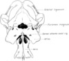
What are the cranial n?

A

1= oflactory 2 = optic, 3 = occulomotor 4= trochlear 5=trigeminal 6= abducens 7= fascial 8= auditory/vestibulocochlear 9= glossophyngeal 10= vagus 11= spinal/accesory 12= hypoglossal

on old olympus towering top a finn and German veiwed some hops

Some say marry money but my brother says big busniess make money

14
Q

What CN do menace, pupillary light and palpebral test?

A

menace = CN 2 and 7

pupillary reflex = CN 2 and 3 (also palpebral fissure)

palpebral = CN 5 and 7

15
Q

What is the pathway of the menace reflex?

A

CN2, optic tract, lateral genicular nucleus (thalmus/diencephalon), occiptal lobe cerebrum, cerebeullum and facial.

16
Q

When can evidence of Horner’s syndrome be seen (aka what are possible areas of damage to the sympathetic tracts)?

A

T1-T3

C1-C5

bracial plexsus

otitis media/interna - sympathetic fibers in petrous portion of the temporal bone

17
Q

What are signs of CN 3 dysfunction?

A

Ventrolateral strabismus; ptosis; dilated pupils; diminished to absent PLRs

Normally: Motor to extraocular muscles; parasympathetic to pupil

Test with physiologic nystagmus and PLR

18
Q

What are signs of dysfucntion of CN 4?

A

Dorsomedial strabismus (cat); lateral deviation of retinal vein (dog)

Normal: Motor to dorsal oblique muscle

Test: Resting eyeball position (cat); fundic examination (dog)

19
Q

What are signs of CN 5 dysfunction?

A

Masticatory muscle atrophy; dropped jaw if bilateral; decreased or absent facial/nasal sensation

Normal: Motor to muscles of mastication (mandibular); sensory to face (ophthalmic, maxillary, mandibular)

Test: Jaw tone; muscle bulk; sensation to face, cornea, and nasal mucosa

20
Q

What are signs of CN 6 dysfunction?

A

Medial strabismus

Normal = Motor to lateral rectus and retractor bulbi

Test = Physiologic nystagmus; resting eyeball position

21
Q

What is CN 7 responisble for besides facial movement?

A

Motor to muscles of facial expression; parasympathetic to lacrimal glands; sensory (taste) to rostral tongue

Test: Menace response; palpebral reflex; lip retraction; ear movement; Schirmer tear test

22
Q

What does CN 10 do?

A

Sensory and motor to pharynx, larynx, and viscera

Test with Gag reflex or oculocardiac reflex

Diminished gag reflex; dysphagia; laryngeal paralysis; megaesophagus

CN 9 is also associated with gag reflex and dysphagia

23
Q

What does the accessory n do?

A

Motor to trapezius

Test: Evaluation of muscle mass

Dysfunctional = Atrophy of trapezius

24
Q

Describe the pathway for vestibular nystagmus?

A

CN8 → brainstem → vestibular nuclei → medial longitudinal fasciculus → abducens and oculomotor

25
Q

What nerves are associatied with the palpebral reflex?

A

CN5 - opthalmic br (medial palpebral), maxiallary branch lateral palpebral

CN7 (palpberal br)

To evaluate mandibular br = evaluate symmetry of masseter and temporalis mm.

26
Q

What does testing nasal sensation evaluate?

A

ipsilateral opthalmic and maxillary n innervate masal mucosa = ipsilateral trigeminal lesion

nociceptive pathway = contralateral prosencephalic lesion

27
Q

Signs assoicated with proencephalon disease

A

Seizures; abnormal behavior; propulsive activity; depression to coma

Head turn, normal gait; propulsive circling (usually ipsilateral to lesion) or pacing; aimless wandering; head pressing; movement disorders (rare)

Postural Reactions = Contralateral deficits

Cutaneous Sensation = Contralateral (often facial/nasal) hypalgesia

Cranial Nerves = Contralateral menace deficits with normal (optic radiation and occipital cortex) or abnormal (optic chiasm, optic tracts) PLRs; facial; tongue or pharyngeal weakness (rare)

Other = Abnormalities in thirst, appetite, thermoregulation, endocrine dysfunction

28
Q

Neurologic Signs That May Be Associated With Mid to Caudal Brainstem Disease

A
29
Q

Table • 26-4 Neurologic Signs That May Be Associated With Cerebellar Disease

A
30
Q

How does Shiff fherrington posture occur?

A

Disrtuption asending inhibitory axons arising from interneurons (border cells) in the dorsal border of the grey colunm of spinal seg L1 - L4

31
Q

What is spinal shock?

A

T3-L3 lesion with LMN signs to hind limbs

32
Q

What are signs associated with LMN disease?

A
33
Q

What comprizes the lower motor neuron unit?

A

nerve cell body in ventral grey matter CNS

ventral nerve root

peripheral n

muscle/disease effecting neuromuscular transmission mediated by acetylcholine

34
Q

What tests can be used to differentiate between junctionpathies, neuropathies and mypoathies?

A

creatinine kinase, aminotrasferase (AST), tensilon test, Ach receptor antibody titer, EMG, and nerve/muscle biopsies

35
Q

What are differentials for diffuse/multifocal neurologic disease?

A

disorders myelin formation

diffuse meningitis (shaker syndrome, GME, infectious)

metabolic

dengenerative disease (lysosomal storage disease)

intoxications (mold, algae, ethylene glycol)

36
Q

What are the essential components of a motor unit?

A
  1. motoneuron = cell body (located within the central nervous system, either in the cranial nerve nuclei of the brainstem or in the ventral horns of gray matter in the spinal cord) and its peripheral axon, supported by Schwann cells.
  2. Neuromuscular junctions.
  3. Myofibers innervated by the motoneuron.
37
Q

What are 3 syndromes associated with neuromuscular disorders and their CS?

A
  1. Myopathic syndrome = weakness, stiff gait, tremors, atrophy or hypertrophy, muscle pain
  2. Motor neuropathic syndrome (classic LMN)= flaccid paralysis/esis, neurogenic atrophy, decreased reflexes, fasiculations
  3. Sensory neuropathic syndrome = hypalgesia/hypesthesia (decreased pain and sensation), CP deficits, decreased reflexes without muscle atrophy
38
Q

List tests to diagnosis neuromuscular disorders

A

blood: CK, lytes, lactate, myoglobinuria (aka ammonium sulfate precipitation test), thyroid acthylcholine recptor antibody

Electomyography = peripheral n. conduction

Muscle biopsy

Nerve biopsy

39
Q

Diagram of the major components of the motor unit (1 through 7) and reflex arc. A, Sensory nerve fiber. B, Dorsal root ganglion. C, Dorsal nerve root. 1, Motor neuron. 2, Ventral nerve root. 3, Spinal nerve. 4, Plexus. 5, Motor nerve fiber. 6, Neuromuscular junctions. 7, Muscle fiber

A
40
Q

Table • 27-1 Pathoanatomic Classification of Neuromuscular Disorders

A
41
Q

What are types of electrodiagnostic testing?

A

Electromyography

Nerve conduction velocity testing (motor and sensory)

Somatosensory evoked potentials (cord dorsum potentials)

Late wave analysis (F-wave and H-reflex)

Repetitive nerve stimulation

Single-fiber electromyography

Muscle and nerve biopsy

42
Q

How do you diagnose masticatory myopathy?

A

can be made with a positive 2M-antibody titer

43
Q

Can electromyography ID disorders restricted to sensory neurons?

A

No

44
Q

What are normal and abnormal spontaneous activty on EMG?

A

Normal = insertional activity, miniature end-plate potential (needle positioned in proximity to NM junction), End plate spikes (complete depolarization, motor unit action potential (normal m. not completely at rest).

Abnormal = Fibs and sharps (firing single myofibers due to destablization sarcolemmal membrane), complex repetitive discharges (group of fibers firing in synchrony due to denervation), myotonic potential (congenital myotonia)

45
Q

Figure 27-2 Electromyography. A, Normal baseline (“electrical silence”). B, Miniature end-plate potentials with a single end-plate spike. C, Fibrillation potentials. D, Positive sharp waves. E, Positive sharp waves and fibrillation potentials. F, Complex repetitive discharges. (Calibration: Amplitude is represented by the vertical line: 100 µV in D, and 50 µV in all other tracings. Time base is represented by the horizontal line: 10 msec in all tracings.)

A
46
Q

Peroneal motor nerve conduction study in a normal dog (B) and a dog with a sensorimotor polyneuropathy (C), following stimulation of the peroneal nerve at the hock, stifle, and hip. Compound motor unit action potentials (M-waves) were recorded from the extensor digitalis brevis lateralis muscle (recording electrode). A, Diagram of electrode placement. B, Normal canine peroneal motor nerve conduction study. Note that latency of the M-wave increases with greater distance between stimulating and recording electrodes. Motor nerve conduction velocities calculated in this 1-year-old dog were 69 m/sec (hip to stifle) and 57 m/sec (stifle to hock). C, Abnormal canine peroneal motor nerve conduction study. Note the polyphasia, temporal dispersion, and decrease in amplitude of the M-wave when compared with B. Motor nerve conduction velocities calculated in this 6-year-old dog were slowed: 39 m/sec (hip to stifle) and 32 m/sec (stifle to hock). These abnormalities are consistent with demyelination.

A
47
Q

Somatosensory evoked potential study in a normal dog (B) and a dog with a sensorimotor polyneuropathy (C), following stimulation of the peroneal nerve at the level of the fourth distal metatarsus (stimulus site). Somatosensory evoked potentials were recorded from the hock, stifle, hip, and L4-5 (cord dorsum potential [CDP]). Latency measurements for sensory conduction velocity determinations are represented by T1, T2, T3, and T4. A, Diagram of electrode placement. B, Normal canine somatosensory evoked potential study. Although the somatosensory evoked potentials appear to be of similar amplitude at each recording site, the sensitivity increases from hock to L4-5. Note that 1000 individual potentials (N = 1000 to right of figure) were averaged to produce these tracings. C, Abnormal canine somatosensory evoked potential study. All sensory nerve action potentials are severely reduced in amplitude, and sensory nerve conduction velocities are slowed. Dispersion of the potentials is difficult to evaluate, as potentials are barely distinguishable from background noise. Note that compared with (B), 4000 individual potentials (N = 4000) were averaged in an attempt to minimize random background noise from the time-locked electrical signal. These abnormalities are consistent with a severe sensory neuropathy.

A
48
Q

Diagrams showing the generation of late waves after stimulation of a mixed nerve and recording from the motor point of a muscle. A, M-wave. B, F-wave. C, H-reflex.

A
49
Q

How do you do an EMG?

A

most frequently used technique for electromyography involves the use of a concentric needle electrode. The central wire of the electrode (active or exploring electrode) is insulated from the surrounding cannula (reference electrode) so that the actual area being tested (between the two) is very small. A ground electrode is used to minimize extraneous “noise.”

50
Q

Normal canine M-waves (left) and F-waves (right) recorded following peroneal nerve stimulation at the hock (32 superimposed individual tracings). B, Normal canine M-waves (left) and F-waves (right) recorded following peroneal nerve stimulation at the hip (32 superimposed individual tracings). Note the longer latency of the M-waves (left) and the reduced latency of the F-waves (right) when compared with (A). C, Normal canine H-waves recorded following peroneal nerve stimulation at the hip, using a low stimulus intensity (0.6 mA). Note that M-waves are not present (left). D, The same stimulation and recording sites as used in (C), after a slight increase in stimulus intensity (to 1.0 mA). M-waves now are present (left).

A
51
Q

Repetitive nerve stimulation. A, Normal canine repetitive nerve stimulation following 1 Hz stimulation of the peroneal nerve at the hock. Note in the data table that M-wave amplitude (Peak Amp mV), area under the M-wave (Area mVms), and percentage amplitude decrement (Amp Decr %) remain constant with consecutive stimuli. B, Decremental response in a cat following 3 Hz peroneal nerve stimulation at the hock. Note that amplitude and area under the M-wave decrease with consecutive stimuli, which is measured as a percentage amplitude decrement of 17% to 44%. The cat had muscular dystrophy. C, Facilitation in a dog following 30 Hz peroneal nerve stimulation at the hock. Note that both area and amplitude of the M-wave are increased (i.e., the M-wave becomes taller and is not narrower). The increase in amplitude is represented by a negative Amp Decr %. A cause could not be determined in this dog presenting with exercise-induced weakness that occurred in warm weather. D, Pseudofacilitation in a normal dog following 30 Hz peroneal nerve stimulation at the hock. Note the increase in amplitude with no change in the area under the curve (i.e., the M-wave becomes taller and narrower). As in (C), the increase in amplitude is represented by a negative Amp Decr %.

A
52
Q

What are the physiologic tests to evaluate the NM junction?

A
  1. Repetative nerve stimulation - repeated supramaximal while recording M waves
  2. Single-fiber electromyography - end plate in NM junction = v. sens for aquired myasthenia gravis
53
Q

What are criteria for selection for muscle biopsy?

A
  1. Affected
  2. minimal approch/morbidity
  3. Ideally fibers oriented in 1 direction
  4. Previously described characteristics: latear head triceps (distal 1/3), vastus lateralus (distal 1/3), cr. tibial (prox 1/3), temporalis
  5. Away from tendon of insertion or aponeuroses
  6. Free of artifact
54
Q

What are criteria for selection of nerves for biopsy?

A
  1. affected by disease, if diffuse established normal and innervates routine m. biopsy
    aka: common peroneal, tibial, ulnar = all contain motor, sensory and autonomic
  2. For sensory = cutaneous sensory n (cd cutaneous antebrachial or cd cutaneous sural)
  3. Nerve root biopsy
  4. CN biopsy - facial or trigeminal
55
Q

What are the techniques for nerve biopsy?

A

Fascicular

full thickness

56
Q

How does tissue contrast differ between T1 and T2 weighted images (short vs. long)?

A

T1 = short T1 relaxtion (hyperintense) - fat, long T1 relaxation (hypointense) = CSF

T2 = short T2 relaxation (hypointese) - muscle, long T2 relaxation (hyperintense) = CSF

57
Q

Schematic illustrating the two basic pulse sequences, spin echo and gradient echo, and their variations. BOLD, Blood oxygenation level dependent; FIESTA, fast imaging employing steady state acquisition; FLAIR, fluid attenuation inversion recovery; FLASH, fast low-angle shot; fMRI, function magnetic resonance imaging; GRE, gradient echo; HASTE, half Fourier acquisition single-shot turbo spin echo; PDW, proton density weighted; SE, spin echo; SPGR, spoiled gradient recalled; SSFP, steady state free precession; SSFSE, single-shot fast spin echo; STIR, short-tau inversion recovery; T1W, T1-weighted; T2W, T2-weighted; true FISP, true fast imaging with steady state precession.

A
58
Q

Substances With High Signal Intensity on T1W and T2W Images

A
59
Q

What is the dose of water soluable iodinated contrast and gadolinium?

A

Water-soluble iodinated contrast medium is usually administered intravenously at a dosage of 400 to 800 mg iodine/kg

Gadolinium: intravenously at a dosage of 0.1 mmol/kg body weight = paramagnetic (enhances/strengthens the magentic field)

60
Q

What is the Larmor frequency?

A

Spin at frequrecy porportional to the strength of the magnetic enviroment

61
Q

Hounsfield Unit Measurements for Various Substances

A
62
Q

What is the dose of contrast agents for the spine on CT?

A

Nonionic contrast media of choice are iohexol (Omnipaque, 240 mgI/mL) and iopamidol (Isovue, 200 mgI/mL), which are administered at dosage of 0.45 mL/kg (full spine) and 0.3 mL/kg (regional): using a cervical or lumbar puncture technique

63
Q

What is the path of flow of spinal fluid?

A

CSF (blood ultrafiltrate) formed in Choroid plexus (lateral ventricles) → foramina 3rd ventricle → mesenchepahlic aquaduct → 4th ventricle → lateral apertures from 4th ventricle → subarachnoid space → central canal of spinal cord

64
Q

What are the 3 meniginges?

A

pia mater

arachnoid mater

dura mater

65
Q
A
66
Q

Anatomy of spinal cord

A

3 meniges: dura mater (outide side), arachnoid mater, pia matter (inside)

2 spaces = subarachnoid = CSF, subdural = vessels

epidural = between bone and dura mater = fused with periosteum skull

67
Q

What is in the grey mater and white matter?

A

Grey = neuronal cell bodies

White = axons and glial cells

Spinal cord grey on inside (outside in brain and majority of brainstem) = divides white matter into compartments (funiculi)

68
Q

What is the falx cerebri?

A

falx cerebri = separates 2 hemispheres (connective tissue)

tentorium cerebelli = separates cerebrum from cerebellum (connective tissue)

69
Q

What is the resting cell membrane potential for neurons?

A

Negative 80mV

Action potential = rapid depolarization by influx of Na+ through voltage gated ion channels

Iniated at axon hillock (junction between axon and neuronal cell body

all or nothing

Repolarizes by clsoure of Na channels and eflux of K through open potassium channels

Active extrusion Na in exchange for K and by K uptake by astrocytes

70
Q

What is produced by oligodendrocytes?

A

Myelin

71
Q

What is pressure autoregulation?

A

pressure sensative smooth m. cells maintain constant P in CNS as long as systemic BP between 50-160mmHg

Affected by intracranial disease

72
Q

Graph of autoregulatory control of cerebral blood flow (CBF) and vascular diameter, in response to changes in mean arterial pressure (MAP), arterial partial pressure of oxygen (PO2), and arterial partial pressure of carbon dioxide (PCO2).

Profusion highly responsive to arterial paCO2 (as CO2 increase, profusion increase)

1mmHg change PaC02 = 5% change profusion

Intercranial disease = hypoventilation = vasodilation = increase ICP = herniation

Markedly increased profusion if PaO2 <50mmHg

A
73
Q

What is the definition of cerebral perfusion pressure?

A

mean arterial pressure - intracranial pressure

74
Q

How does low BP or high ICP affect cerebral profusion?

A

Both cause decreased cerebral perfusion pressure → ischemia in medulla → increased vasomotor tone → increase MAP → increased cerebral profusion

Hypertenion systemic → barorecptors → bradycardia

75
Q

What is brain - heart syndrome?

A

If cerebral pressure continues to drop → increase catecholamines → myocardial ischemia → arrythmias

76
Q

What is normal ICP?

A

8-15mmHg

ICP >15-20mmHg → abnormal, should be treated

>30mmHg significant decrease in perfusion

Durotomy decrease ICP 65% vs craniotomy alone = 15%

77
Q

List drugs based on if they are good or poor at crossing the blood brain barrer.

A

Good = 3rd gen cephal, fluroquin, metronidazole, sulfas, chloramphenicol, trimethoprim

Mod = tetracycline, erythomycin, penicilins, rifampin

Poor = 1st and 2nd gen cepha, aminoglycocides, clindamycin, vancomyacin

78
Q

What potion of the CNS is not immunologically privlaged?

A

meninges and choroid plexus = no BBB = normal response to inflammation (ie increased neutrophils and mononuclear cells)

79
Q

What are the 2 areas in the brain where constitutive neurogenesis occurs in the adult nervous system (ie stem cells)?

A

subventricular zone/olfactory system

dentate gyrus of the hippocampus

80
Q

What are the primary and secondary mechanisms of injury related to contusion?

A

1 = mechanical - swelling, hypoxia, axonal injury

2 = trauma and ischemic

  • hemorrhage → decreased perfusion/energy → increase cell permability to Na, Cl, Ca
  • damage → increased glutamate →increased Ca and Na uptake (via NMDA, AMPA< kainate receptors)
  • increase Ca = apoptosis and activates phopholipase = increased inflammation
  • increased mitochondrial permiability →free radicals → vascular damage, edema and vasospasm via increased Trpm4 and parenchyma pressure
81
Q

What do micoglial (inflammatory cells) release in response to inflammation?

A

IL-1, TNF alpa, hydrogen peroxide, NO, proteinases

82
Q

What does compression induce in the brain?

A

demyelination, edema, axonal degeneration and neuronal necrosis

  • via physical deformation myelin (reversible) , vasogenic edema, increase glutamate = demylination via oligodendrocyte damage
83
Q

Increased intracellular calcium concentration does the following: (3)

A
  1. Activates intracellular proteases such as calpains and caspase, which destroy the cytoskeleton and chromosomes and initiate programmed cell death
  2. Activates phospholipase A2, thereby producing eicosanoids and initiating an inflammatory response
  3. Binds intracellular phosphates, further depleting the cell of energy sources
84
Q

What is the max amount of CSF that can be collected?

A

1ml per 5kg

85
Q

What are the 2 locations for a CSF tap and what are the landmarks?

A
  1. Cerebellomedullary cistern: full flex skull to 90 degree with cervical spine. Intersection lines between occiptal protuberence - C2 and line along cr aspect of aspect of the wings of C2. Keep needle perpendicular to dorsal lamina.
  2. Lumbar cistern: Legs flexed, L5-L6 dogs, L6-L7 cats
86
Q

What is considered pleocytosis for CSF (cloudy CSF)? What is a normal cell count CSF?

A

>500 x 10^6 WBC/L

normal = 0-5 x 10^6 WBC/L

87
Q

What is a normal protein count for CSF?

A

<250mg/L (25mg/dL) for cerebromedullary cistern

<450mg/L (45mg/dL) for lumbar cistern

88
Q

What are common serologic tests for neuro disease?

A

Toxo, neospora, ehrlichia, rickettsia, coccidiodies

Can do IgG index = CSF IgG/serum IgG, if low it has migrated across BBB,high then CSF is source

89
Q

What is the histopathologic characteristic of DM

A

Necrosis primarily in lateral and ventral funiculi of TL spine

Breeds: GSD, Welsh Corgi, Boxer, Rhodesian Ridgeback

Genetic risk factor = mutation in SOD1 gene

90
Q

Where do the inflammatory lesions occur for steroid responsive meninditits-artertitis (SRMA)?

A

Leptomeninges and associated a. - etiology unknown

Breeds: Beagle, Boxer, Berner, Weimies, NS duck trolling retrievers

Usually 6-18m range upto 7yr

91
Q

What are the 3 forms of GME?

A

disseminated - most common

Focal - slowly progressive, suggestive of mass lesion

Ocular - pupils often fixed or dilated, optice n. swelling, chorioretinitis

92
Q

What is the treatment for toxoplasma?

A

Clindamycin 10-25mg/kg BID 3-4 weeks

or

TMS 15mg/kg BID with pyrimethamine 1mg/kg SID

93
Q

For discospondylitis, what percent are diagnosed on UA with blood culture and on direct percutaneous aspiration of the IVD?

A

Urine and blood culture = 40%

Percutaneous disc = 60%

94
Q

What spinal segments are commonly diagnosed with FCE?

A

L4-L3 = 44-50%

C6-T2 = 37-42%

95
Q

Where is FCE usually diagnosed?

A

L4-S3 = 50%

C6-T2 = 40%

96
Q

What is the appearence of FCE on MRI and what percent have not lesion?

A

Appearence = hyperintensity on T2

21% have no lesion

97
Q

What can potentially indicate sucessful recovery on MRI for FCE?

A

MRI lesion:length ratio >2 = 40% sucess

<2 = 100% sucess

98
Q

What is a 2 engine gait and what is it associated with?

A

C6-T2 lesion

short strided front limb, long stride hind limbs

99
Q

What spinal segment is associtated with phrenic n?

A

C5-C7

100
Q

Schematic representation of the cutaneous sensory innervation of the thoracic limb.

A
101
Q

What are the 2 approaches to the AA joint?

A

1. Ventral approach

  • midline incision cd1/3 mandibule to 5cm caudal to larynx
  • blunt dissection right sternothyroideus and sternocephalicus to expose longus coli
  • ventral prominence C1

2. right parasagittal approach

  • improved exposure, avoid dissection right thyroid, trachea and recurrent laryngeal
102
Q

What is the lateral approach to cervical spine?

A
  1. lateral skin incision C2-scapula at level zygapophyseal joint
  2. Platysma
  3. C3-C6: trapezius and grid brachicephalicus = serratus ventralis and splenius
    - dissect serratus ventraus = longissmus system. C5-C7: retract brachepahlicus cr-lat and trapeziums cd-dor-lat = serratus and splenius (ligate superficial cervical a.). C6-C7: abduct and caudal retract scapula
  4. Dissect longissiumus capitis (disect from transverse pro. and retract ventral) and complexus mm.
  5. Incise attachments of compleux and mulitifidus from articular process (retract dorsal)
103
Q

gain access to the caudal cervical vertebra, the splenius muscle needs to be exposed by separating the brachiocephalicus and trapezius muscles from each other, as indicated in this illustration

A
104
Q

Describe dorsal dissection for AA joint

A
  1. skin incision = dorsal midline, occipital protuberance to C3 or C4.
  2. expose the occipitalis, cervicoscutularis, and cervicoauricularis superficialis muscles. Caudal and lateral to these muscles are the thin fibers of the platysma muscle. These muscles are incised on the midline fibrous raphe
  3. Separation of the paired bellies of the biventer cervicis superficially and the deeper rectus capitis attached to the dorsal spine of C2. The insertion of the rectus capitis muscle is incised along the lateral border of the spine of C2 to allow its elevation from the bone by combined sharp and blunt dissection.

D. Avoid the vertebral artery, which courses through the muscles slightly ventrolateral to the articular processes. The interarcuate (yellow) ligament covering the foramina between C1 and C2 is carefully incised to expose the spinal cord and the root of spinal nerve C1. The dorsal atlanto-occipital membrane of the foramen magnum may also be incised to expose the cranial rim of the dorsal arch of C1.

105
Q

Describe dosal cervical approch for C2-C7

A
  1. skin incision occipital protuberance to the first thoracic vertebra.
  2. Almost transparent fibrous aponeurosis of the platysma muscle comes into view. Incision through the median fibrous raphe. This incision is deepened until the nuchal ligament (missing in the cat) is exposed.
  3. The dorsolateral cervical muscles (cleidocephalicus, semispinalis capitus, biventer complesus) separated by this incision are retracted laterally to expose the nuchal ligament.
  4. An incision is made in the rectus capitis, spinalis et semispinalis cervicus, and multifidus muscles along one side of the nuchal ligament. The incision is deepened along the lateral side of the spinous processes to the vertebral laminae.
  5. Elevation with a periosteal elevator and retraction of the muscles from the vertebrae are done first on the side that was incised. The insertion of the nuchal ligament is now elevated from the spinous process of the axis, and the ligament is retracted with the muscles on the side opposite the incision. The ligament remains firmly attached to the muscles of one side and cranially to the axis.

Lateral elevation of muscles from the laminae should be limited to the lateral aspect of the articular processes to avoid branches of the vertebral artery coursing ventrolaterally to the processes.

106
Q

Lateral approach C3-C6:

  1. skin and underlying platysma- cranial border of the scapula to the wing of the atlas.
  2. The incised wound margins are retracted to expose the brachiocephalicus and trapezius muscles. Separate between these muscles in a dorsal to ventral, oblique direction. The superficial cervical artery and vein, which emerge from between these two muscles, are retracted caudally and ligated. Underlying these vessels are the superficial cervical lymph nodes. The brachiocephalicus muscle is transected at the level of the articular facets.
  3. Retraction of the transected brachiocephalicus muscle exposes the splenius muscle dorsocranially and also the serratus ventralis muscle that runs obliquely from under the cranial border of the scapula in a cranioventral direction. Cranial nerve XI (accessory nerve) crosses the serratus ventralis muscle to innervate the trapezius muscle.

— As an alternative, exposure of the C5-C7 vertebral segments can be achieved without transection of the brachiocephalicus muscle, by further extension of the separation between the brachiocephalicus and trapezius muscles in an oblique direction. The brachiocephalicus muscle is retracted cranioventrally, rather than being transected. Exposure of C6-C7 is increased further by simultaneous abduction and caudal retraction of the scapula, without the need to incise the muscular attachments on the cranial border of the scapula.

In the C3-C5 region, the brachiocephalicus muscle is incised longitudinally in the direction of the muscle fibers, instead of being transected. Using a grid technique, the divided brachiocephalicus muscle is separated to expose the underlying splenius and serratus ventralis muscles.

E. The dorsal border of the serratus ventralis muscle is elevated all the way caudally to its origin on the scapula. It may then be reflected ventrally to fully expose the underlying splenius muscle. Vascular bundles between the deep surface of the serratus ventralis and splenius muscles are ligated and divided.

F. Reflection of the serratus ventralis continues to its insertions on the lateral processes of the vertebrae, exposing the longissimus cervicis and longissimus capitis muscles. These muscles are elevated and retracted ventrally. For orientation, the prominent transverse process of C6 is located by palpation.

G, H. The splenius muscle is retracted dorsally to expose the underlying semispinalis complexus muscle. The articular facets of C3-C4, C4-C5, and C5-C6 can be palpated at the junction between the semispinalis muscle and the two longissimus muscles. The tendinous origins of the semispinalis complexus and multifidus cervicis muscles are incised from the articular facets with scissors or electrocautery. These two muscles are then elevated from the dorsal lamina of the cervical vertebrae to the base of the spinous process using a periosteal elevator.

A
107
Q

Describe lateral approach to the brachial plexus (C5-T1)

A
  1. long curvilinear incision 3-4 cm cranially from the midpoint of the cranial border of the scapula to a point located slightly distal to the greater tubercle of the humerus.
  2. The platysma and fascia cervicalis are incised, exposing the cleidocervicalis, omotransversarius, and trapezius muscles (Figure 31-7, A). The superficial cervical artery and vein, which emerge between the cleidocervicalis and trapezius muscles, should be ligated.
  3. The omotransversarius muscle incised near spine of the scapula, retracted cranially.
  4. The dissection is continued medially through the deep fascia along the dorsal border of the cleidocervicalis muscle, which is withdrawn ventrally. The scapula is withdrawn caudally.
  5. (C5-T1) are now exposed by transecting the superficial and deep portions of the scalenius muscle
108
Q

What pieces do the altas and axis develop from?

A

atlas = three bony elements (a pair of neural arches that become the vertebral arch and a body) - fusion by 115d

the axis develops from seven bony elements (a pair of neural arches, three parts of the vertebral body, the dens, and the apical element on the dens) -fusion from 30-396d

109
Q

Atlas anatomy

A

Transverse foramen

Lateral foramen

Fovea of the dens

glenoid cavities - articulate with axis - no joint

modeified articular process articular with occipital condyles - no joint

110
Q

Atlas anatomy

A

dorsal spinous process

cr vertebral notches = large intervertebral foramina - 2nd cervical n and intervertebral vessels

dens (odontoid process)

transverse lig

111
Q

AA lig

A

Transverse lig

Alar ligaments

Apical lig of dens

Dorsal atlanto-axial lig

Lateral atlanto-occipital lig

112
Q

Nerves of the brachial plexus and associated spinal n?

A

SSMAR M U

Suprascapular (C6-C7)

Subscapular (C6-C7)

Musculocutaneous (C6-C8)

Axillary (C6-C8)

Radial (C6-T2)

Median (C7-T1)

Ulnar (C8-T2)

113
Q

Schematic representation of the spinal root origins of the major nerves of the lumbosacral plexus. (Based on the work of Fletcher, T. F. 1970. Lumbosacral plexus and pelvic limb myotomes in the dog. Am. J. Vet. Res. 31:35-41; and Bailey, C. S., R. L. Kitchell, S. S. Haghighi, and R. D. Johnson. 1984. Spinal root origins of the cutaneous nerves of the canine pelvic limb. Am. J. Vet. Res. 45:1689-1698.)

A
114
Q

What are congential and developmental abnormalities associated with AA lux?

A

34% dysplasia

46% aplasia

dorsal angulation

separation of the dens

absence of the transevere lig

incomplete ossification fo the atlas (4/5 with concurrent subluxation)

block vertebrea (fulcrum effect)

Up tp 24% normal dens

115
Q

What radiographic changes are noted with AA lux?

A

increased space between the dorsal lamina of atlas and dorsal process of axis

malalignment of atlas and axis bodies

Angle between arlas and axis less than 162 degrees more predicitive of instability than decrease in AA overlap

VD to evaluate dens

116
Q

What are dorsal and ventral stabilization techniques for AA lux and what are the disadvantages of dorsal?

A

Dorsal = wires, wire and PMMA, braided suture, portion of nucal lig, metallic retractor and pins

CONTAINDICATED: dorsal deviation of dens. Also no fusion = so fail cause recurrence

More biomech sound and prefered if <2kg

Ventral fixation = plate, pins. pins ans PMMA, transarticular lag screw, screws and pins with PMMA

117
Q

Dorsal atlantoaxial wiring technique. A–D illustrate the loop of wire being passed cranially under the arch of C1, then folded back and cut with the ends twisted together through the two predrilled holes in the spine of C2.

Variation = loop used to pass suture through holes

A
118
Q

What is nuhal ligament technique?

A

split lig longitudionally

transect at T1

passed like wire procedure

Tie to self through hole in spinous process or after placing through notch

119
Q

Atlantoaxial (AA) joint dorsal cross-pinning; the Kirschner wires are directed into the caudal half of the wings of the atlas.

A
120
Q

Kishigami AA tension band

A

Less risk of damage to the spinal cord

121
Q

crews placed into the caudoventral and sometimes the cranioventral part of the body of C2 can be used as anchors to elevate this vertebra into apposition with C1 using cerclage wire.

A
122
Q

Transarticular screws can be inserted in lag fashion across the atlantoaxial (AA) joint, toward the medial aspect of the alar notch.

1.5 mm hole for a 1.5 mm cortical lag

approximately 30 degrees from midline

similar technique using Kirschner wires or pins (0.045 inch or 0.062 inch) has been described

A
123
Q

Cortical bone screws can be placed into the medial aspect of each wing of the atlas caudal to the transverse foramen in a craniolateral direction

A third screw can sometimes be placed in the middle of the ventral body of C1, depending on the size of the patient; this screw can be inserted at 90 degrees to the bone surface

A

Two ventral C2 screw patterns have been described.

he first pattern, four cortical screws are placed into the body; one screw is placed in the middle of the caudal aspect of each of the cranial articular surfaces of C2 at the insertion point of the longus colli muscle. The screws are directed craniolaterally at 30 to 40 degrees. The second pair of screws is placed at the base of the transverse processes of C2 or C3 and is directed laterally at 30 to 40 degrees to the midline.

A second pattern uses just two cortical screws, one of which can be the screw that facilitates secure realignment. second screw can be placed in the cranial half of the C2 vertebral body.165 This latter technique additionally differs from the first screw and PMMA technique in that transarticular 0.035 to 0.062 inch threaded or nonthreaded Kirschner wires are used to secure the atlantoaxial joint, subsequently incorporating all of the screws and the wires into the PMMA

124
Q

What is the complication rate of stabilizaiton for AA lux?

A

dorsal = 71%, failure of fixation 25-48%

ventral = 53%, failure of fixation 18-44%

125
Q

What is the prognosis for long term outcome for conservative vs ventral vs dorsal AA stabilization?

A

Conservative 38% longterm success (better if CS <30d)

Periop mortaliity = 10-30%

dorsal approach = 61% (~50%dorsal loop/suture, ~75% nucal lig and Kishigami)

Ventral = 47-92% pinning alon 47%, transarticular screws 40-90%

Overall better px sucess 1st sx if <24m or long term sucess if mild CS and <10m

126
Q

Nerve roots lack epineurium = susceptible to avulsion

A
127
Q

Describe the structure of nerves related to the layers surrounding the nerve.

A

Endoneurium surrounds each axon, which is composed of collagen fibrils, fibroblasts, and vascular tissue, and groups of axons in turn are surrounded by perineurium, which is composed of collagen and elastic fibers; the connective tissue around the entire nerve, called the epineurium, is composed of areolar connective tissue and collagen fibrils

128
Q

What are the nerves of brachial plexus - all not just SSMARMU

A

The nerves that are branches of the brachial plexus or are direct continuations of the formative ventral branches include the brachiocephalic, suprascapular, subscapular, axillary, musculocutaneous, radial, median, ulnar, dorsal thoracic, lateral thoracic, long thoracic, pectoral, and muscular branches

129
Q

Anatomy nerves

A
130
Q

Six different classifications have been assigned for the types of injury that can occur after peripheral nerve trauma

A

Class 1: neuopraxia = interrupt function/conduction w/o structural changes (hours-6w)

Class 2: axonotmesis = Wallerian degeneration of distal stump following severe trauma (endoneurium and Schwann sheath preserved)

Class 3: neurotmesis = disrupt axons and endoneurium, perineurium intact

Class 4: neurotmesis = Class 3 + perineurium disrupted

Class 5: neurotmesis = severed

131
Q

What is the widest and narrowest cervical joint space?

A

wide C4-5 and 5-6

narrow C2-3

132
Q

Describe fibrous arrangement of disc

A

lattice like pattern (paralelle fibers running obliquely), more than 8 layers ventrally

ventral 1.5 - 3x thicker than dorsal

more cartilagenous close to nucleus

133
Q

What are ligaments associated with spinal canal

A

dorsal longitudonal (thickest in cervical)

Ventral longitudonal lig

interspinous lig

yellow lig (or interarcuate)

ligament head of rib & costotransverse lig

intercapital lig - absent 1st, 11th, 12th, and 13th rib +/- 10th

134
Q

What percent of IVDD is cervical?

A

25%

135
Q

What are the most common cervical IVDD spaces - extrusion?

A

C2-4 80%

C2-3 44-59%

136
Q

Where is the most common protrusion/large breed disc?

A

C6-7

137
Q

What percent are associated with various neuro deficits for cervical IVDD?

A

up to 60% pain only, 40% amb tetra, 10-20% non-amb tetra, 2-7% plegic

Deficits More common with C4-5 and C6-7

36% C1-C5 will present like C6-T2

138
Q

What are the landmarks for a slanted V-slot?

A

20% width, 20-25% length on the cranial aspect of the disc

139
Q

What % mortality for Cervical spinal surgery is realted to respiratory compromise, what % require PPV for hypoventalation and what increases risk for hypoventilation?

A

Overall 8% mortalitiy for cervical sx, 1.8-3.5% due to respiratory failure

5% require PPV, increased risk if dorsal lam or C2-C4 lesions

140
Q

Why are intrap cardiac dysrythmais common with V-slot?

A

Retraction carotid and vagosympathetic trunck

disrupt sympathetic tectotegmental tract

141
Q

What is the risk of recurrence for cervical disc disease: sx vs conservative?

A

Conservative = 36%

Sx = 5-10% - meantime 3m

142
Q

Why are corticosteroids potentially beneficial for spinal disease?

A

Decrease vasogenic edema

protection glutamate toxcity

decreased apoptosis

143
Q

What is the difference between ossesous associated and disc associated cervical spondylomyelopathy?

A

Disc associated (dobe): middle aged 6.5-8yr vs young for osseous 3.5yr (giant breed)

Disc: canal stenosis + cervical torsion (3x more axial rotation in cd vs cr) + large relative volume of disc = IVDD C5-7

Ossesous: congenital, stensis (proliferation vertebral arch, articular processes or process and pedicles), veterbral malformations and OA at zygaphyseal joint, lig. flavum hypertrophy uncommon

large breed: 50/50 1 site vs more sites, giant 20% one site

144
Q

Which sites are more commonly affected Dobie vs Giant breed?

A

Dobie = C6-C7 > C5-C6, combined 90%

Giant = C6-C7 > C5-C6 >C5-C4, combined 80% one of these, 20% C2-3 and C3-4

Recently T1-T2 14% giant breed, C7-T1 22% all dogs

145
Q

What is the outcome of conservative management vs surgery for CSM?

A

Conservative (aka minimize free activity +/- drugs) = 54% improve

Surgery = 81% improve

146
Q

What are direct decompression techniques for CSM?

A

Dorsal lam

dorsal laminoplasty

ventral slot

inverted cone slot

hemilam

147
Q

Indirect decompressive techniques?

A

aka distraction and stablization: bone graft, pins or screws with PMMA, interbody screws, washers, metallic spacers, metallic plates, platic plates, k-wire spacers, Harrinton rods, interbody PMMA plugs, fusion cages (combined with discectomy or partial or complete V-slot)

  • IVD fenestration in motion-presevering techniques: disc arthroplasty or artificial disc replacement (recently proposed)
148
Q

What techniques are discussed for ventral disc associated CSM?

A

Ventral static compression = V-slot or inverted cone slot

Dynamic (distraction/stab) = PMMA plug and pins/screw PMMA bar. Continous dorsal laminectomy if multiple sites

149
Q

What sx techniques for osseous CSM?

A

Static = dorsal of hemi lam

distraction/stabilzation may be used but doesn’t treat compression

150
Q

Disc-associated cervical spondylomyelopathy (CSM). Top image shows ventral spinal cord compression and nerve root compression at C5-C6 caused by intervertebral disc protrusion. Dorsally, hypertrophy of the ligamentum flavum (yellow) causes mild spinal cord compression. Bottom images: A, Transverse section at the level of the C4-C5 disc region showing normal spinal cord and vertebral canal. B, Ventral compression at C5-C6 region caused by intervertebral disc protrusion and hypertrophy of the dorsal longitudinal ligament (yellow) and ligamentum flavum (causing mild dorsal compression). C, Asymmetric intervertebral disc protrusion at C6-C7, causing spinal cord and nerve root compressions.

A
151
Q

Osseous-associated cervical spondylomyelopathy (CSM). A, Severe dorsolateral spinal cord compression at C2-C3 caused by osseous malformation and osteoarthritic changes. B, Normal C3-C4 disc region. C, Bilateral compression at C4-C5 caused by osteoarthritic changes and medial proliferation of the articular process, resulting in absolute vertebral canal stenosis and foraminal stenosis leading to spinal cord and nerve root compressions, respectively. Bottom image shows dorsal spinal cord compression at C3-C4 caused by lamina malformation and hypertrophy of the ligamentum flavum. Osteoarthritic changes are also shown at C2-C3.

A
152
Q

Inverted cone slot. This is a modification of the traditional ventral slot that minimizes the risk of hemorrhage and subluxation.

A

width of the ventral decompression window limited to 20% of the cranial vertebral body.

This technique allows more complete removal of the protruded disc from the vertebral canal, with less risk of hemorrhage, compared with the traditional ventral slot. The inverted cone technique also minimizes the risk of collapse of the disc space, which could lead to postoperative nerve root entrapment and lameness as seen with the traditional slot technique

153
Q

Dorsal laminectomy. Representation of a dorsal laminectomy from C4 to C7

Continous = C4-T1

A

high postoperative morbidity rate (70%), which can be challenging to manage in large- and giant-breed dogs.

Most dogs will improve over time (mean time to optimum recovery, ≈3.6 months), and the success rate for dorsal laminectomy ranges from 79 to 95%

30% of dogs can have signs of recurrence

154
Q

What is this?

A

Caspar distractor

allows for controlled distraction, another option is modefied Gelpi retractor

155
Q

Polymethylmethacrylate (PMMA) plug distraction technique. Representation of the PMMA plug is in place (gray). Two anchor holes in the cranial and caudal end plates prevent plug displacement. The dorsal annulus is left intact. Multiple small holes should be drilled in the ventral aspect of the vertebral bodies to promote incorporation of the cancellous bone graft (dark red) and fusion

A
156
Q

Representation of the technique of pins and polymethylmethacrylate combined with a partial slot. A, Transverse view. Note the location of pin insertion avoiding the transverse foramina and vertebral canal. B, Lateral view, showing the position of the pins

A

Angle 30-35 degree, 45 degrees C7

distract

graft in slot then PMMA

73% sucess

1 site prefered can potentially do 2 sites but increases risk

157
Q

What are the complication rates associated with CSM?

A

V-solt = 14.9% - resp, cardiac, sublux, hemorr, seroma

Mortality = 3% (0-6%)

Increased risk DObies due to Dobies disease

70% worse post-op w dorsal, 42% worse V-slot - 2.5m to ambulate w/o assistence

Other sx comp: a_djacent segment syndrom (20% distact/stable), Laminectomy membrane, implant failure (7.5-30% implant extrusion/intrusion, innerspace collapse, fx), collapse foramen (post-Vslot), insufficent decompression._

158
Q

For bicortical pin implantation what percent have canal or transeverse foramen pentration?

A

25-57%

159
Q

What is px for CSM?

A

80% improve with sx, 50% conservaitve, 33% fenestration (not recommend)

24% recurrence (direct or indirect)

160
Q

What are Extradural synovial cysts?

A

Extradural synovial cysts (intraspinal synovial cysts, juxtafacet cysts, and ganglion cysts

These cysts originate from the zygapophyseal joint of the spine and are located extradurally = associated with instability and OA

Histologically, the cysts can be divided into synovial cysts, which are those that have a synovium-like lining of epithelial cells, and ganglion cysts, which do not have this lining and are thought to result from mucinous degeneration of the articular cartilage

Tx conservatuve (rest, steroids, percutaneous injection steroid), no improvment sx (remove cyst and articular surface - 70-80% excellent outcome). Generally neurosigns but not painful.

161
Q

Types of sacral fx

A

Type 1: Alar

Type 2: Foraminal

Type 3: Transverse

Type 4: Avulsion

Type 5: Comminuted

162
Q

What are the biomechanics of the vertebra, artcular process and IVD?

A

body = buttress to resist bending and axial loading

Articular processes = resist all forces

IVD = resist rotation and lateral bending

163
Q

Dorsolateral approach TL spine

A

The skin incision slightly to side midline.

Subcutaneous fat is elevated from the deep fascia 1.5 cm lateral to the dorsal spinous

Both layers of the fascia are incised 5 to 10 mm from the dorsal spines

The multifidus and longissimus muscles are separated by blunt dissection of the intermuscular septum.

The tendons are attachments of the longissimus muscle to the accessory processes of the vertebrae. The dorsal branches of the spinal nerves emerge just ventral to these tendinons and the intervertebral disk space is located caudoventral to the insertions.

An elevator is used to remove muscle and fascia overlying each disk. The insertions of the longissimus tendons are used as landmarks for locating the disk spaces. A nerve root retractor allows the spinal nerves to be retracted cranially and protected from the elevation and fenestrating instruments.

T10-T11 is the most cranial disk accessible and L5-L6 the most caudal (tobias says T9-L7)

164
Q

What is the primary type collagen in the NP?

A

nucleus pulposus originates from remnant notochord and chondrocyte-like cells. It is normally well hydrated, contains glycosaminoglycans such as chondroitin sulfate and keratan sulfate, and has a dispersed matrix of type IV collagen

165
Q

The thoracic spine is supplied by spinal branches (A) from the intercostal arteries (B), which enter the vertebral canal via the intervertebral foramina. The internal vertebral venous plexus drains into the major veins of the dorsal thorax (C), mainly in the azygous vein.

A
166
Q

Dorsal view of the muscles encountered during the dorsal approach to the cranial thoracic spine.

A
167
Q

Approach to lateral TL spine

A

The deep thoracolumbar fascia is incised to reveal a second layer of fat and the underlying muscles. The lateral processes can now be palpated quite easily. To approach the thoracic spaces, it is necessary to transect some bundles of the serratus dorsalis muscle. D. The thirteenth rib is a good place to start because it allows counting cranially and caudally to identify the other vertebrae. Blunt dissection and separation of the fibers of the iliocostalis lumborum muscle allow the proximal end of the thirteenth rib to be exposed. A periosteal elevator is used to clear soft tissues from the cranial border of the rib and the lateral surface of the disk. Staying close to the cranial edge of the rib and strong cranial retraction of the muscle will protect the spinal nerve and vessel (see Plate 20D inset drawing). Care must be taken to prevent penetrating the thoracic pleura when clearing the disk surface. The lumbar disks are exposed by blunt separation of muscle tissue over the end of the appropriate lateral process. Confining the dissection and elevation to the dorsal surface of the process will protect vessels running along the cranial and caudal borders of the tip of the process. The elevation of muscle tissue is continued medially until the disk space is exposed. As the vertebral body and disk are approached, all elevation and retraction should be from a caudal-to-cranial direction to protect the spinal vessel and nerve. A blood vessel will be seen crossing the surface of the disk in the lower lumbar spaces, and it is usually lacerated during the fenestration process.

168
Q

What are the different Funquist laminectomies

A

During Funkquist type A dorsal laminectomy, remove zygapophyseal joints and pedicles to expose the dorsal half of the spinal cord. B, During Funkquest type B dorsal laminectomy, remove the dorsal lamina, taking care to preserve the cranial and caudal portions of the zygapophyseal joint. C, When performing deep dorsal laminectomy, remove the dorsal lamina, the entire zygapophyseal joint, and pedicles to the level of the ventral aspect of the vertebral canal.

169
Q

What is the accuracy of radiographs to determine sight disc extrusion?

A

51-61%

170
Q

What is the accuracy of myleography vs CT?

A

Myelography 85-97%

CT 95-100%

171
Q

What is the prognostic value of an intramedually pattern with myelography?

A

intramedullary/L2 >5 = 26% recovery rate vs. 61% <5

172
Q

What is the freq of seizures with myelogram?

A

21%

173
Q

How often is FCE recognizable on MRI?

A

78.8%

174
Q

What are prognostic indicators on MRI for TL disc disease?

A

No Tw2 signal change = better px voluntary ambulation

Hyperintesity >3x L2 poorer px ambulation, odds recovery decreased 1.9 fold per unit hyperintesity

Cross sectional area: Intramedullay T2 hyperintesity >90% = 86% sens and 96% spec poor long term function outcome

175
Q

What percent of TL disc disease has elevated protein and pleocytosis on CSF?

A

66% and 61%

176
Q

What values are associatd with failure to ambulate on CSF anaylsis?

A

high CK, pleocytosis, increased protein = failure to ambulate on long term follow up

177
Q

What is the px and what complications are associated with percutaneous discectomy?

A

88% deep pain (+) sucess

32% deep pain (-)

Complications = pneumothorax, discospondylitis

178
Q

What CS or progression of disease before and after surgery for IVDD are associated with px?

A

Protrusion poorer px

Fast non-ambulatory status

not regaining deep pain status by 2 weeks post surgery

179
Q

What is the recurrence rate associated with TL IVDD?

A

15-20% within 3y if surgery

40% within 4y if not surgery

Dachsunds higher risk

Non-dachsund dogs with 5-6 opacified discs at the time of surgery had 50% recurrence

180
Q

What are alternatives to fenestration for prophylaxis?

A

Laser disc abalation, chemonucleosis (chondroitinas ABC, callagenase, chymopapain)

181
Q

Where do cats most commonly get disc herniation?

A

caudal lumbar spine (2/3 cases)

182
Q

Cerebrospinal Fluid Characteristics in Dogs With Thoracolumbar Disc Herniation

A
183
Q

Chronic disc hernias can produce a “wrapping” effect: The cord spreads out on either side of the vertebral canal floor around the margins of the protruded material. B, Craniolateral view of thoracolumbar corpectomy: A lateral slot has been drilled under the prolapsed material through the intervertebral disc and the vertebral epiphysis of two adjacent vertebral bodies. Excision of the protruded disc has been achieved when the spinal cord has returned to its normal position

A

his surgery involves the creation of a slot in the vertebral body, ventral to the intervertebral foramen. The slot is the length of each vertebra, centered over the disc. The pedicle and lamina remain intact

184
Q

What are advanatages and disadvantages of lateral corpectomy?

A

advantages of lateral corpectomy include enhanced removal of herniated disc material with minimal spinal cord manipulation; disadvantages include possible vertebral column instability and increased risk for fracture/subluxation.

185
Q

The hypogastric nerve is derived from the first through fourth lumbar spinal cord segments and provides adrenergic input to the internal urethral sphincter, the pelvic (parasympathetic) ganglia, and the detrusor muscle. Sympathetic input to the pelvic ganglia inhibits parasympathetic activity during urine storage. The hypogastric nerve contains sensory fibers from the bladder wall and is involved in bladder nociception.

A

To empty the urinary bladder, adrenergic input to the bladder is decreased, allowing increased detrusor muscle and decreased internal urethral sphincter tone. Cholinergic stimulation (pelvic and pudendal nerves) results in detrusor muscle contraction and external urethral sphincter relaxation.

alpha-adrenergic antagonists, such as phenoxybenzamine (0.25 to 0.5 mg/kg PO every 12 to 24 hours) or prazosin (1 mg/15 kg PO every 8 to 24 hours)

186
Q

What are risk factors for UTI with spinal cord injurgy?

A

gender

incomplete voiding

elevated intravesical paressure

prior dexamethazone administration

mucosal trauma due to catheteriztion

contamination during catheterization

normal to alkaline urine

low urine osmolality

prexisiting medical conditions

187
Q

What are hemivertbra, butterfly vertebra and block vertebra?

A

Hemivertebrae are incompletely formed and tend to have a wedge-like shape (kyphosis, lordosis, or scoliosis). Screw-tailed breeds of dogs (English Bulldogs, French Bulldogs, Boston Terriers, and Pugs) are most commonly diagnosed. An inherited form of thoracic hemivertebra has been diagnosed in German Shorthaired Pointers. Hemivertebra results from failure of one or more sclerotomes to form during embryogenensis.

Block vertebrae result from failure of vertebral segmentation during early embryonic development. The disc space normally adjoining adjacent vertebrae does not develop, and adjacent vertebrae are fused. The spinal canal diameter may be altered by compression of the spinal cord or nerve roots.

Butterfly vertebrae are characterized by a sagittal cleft within the affected vertebral body. This cleft may be visible on a dorsoventral radiographic view, creating a “butterfly” appearance of the affected vertebra.

188
Q

What congential and developmenta malformations of the TL spine?

A

Abnormal vertebra

Carilagenous exostoses - benging proliferation physis long bones, ribs, back - sx excise

Pilonidal (dermoid sinus)

Epidermoid cyst - ectodermal cells in CNS, often intracranial

Spinal arachnoid cyst

spinal dysraphism - Weimie, abnormal fusion, non-progress/pain (1-2m age)

Tumoral calcionsosi - soft tissue mineralization - sx excise

Leukodystrophy - abnormal mylein, poor px (<1y age often)

189
Q

Where does the canine spinal cord end and become cauda equine?

A

Large = L4

<15kg = L6

toy breeds and cats = L7

dural sac extends 1-2cm beyound termination

190
Q

What is pathophysiology LS diease

A

Hansen type II IVD, congenital osseous stenosis vertebral canal/foramina, sacral OCD, proliferation joint cap/lig, osteophtosis of articular process, epidural fibrosis, instability or malalignment of L7-S1

191
Q

Methods used to dx LS disease?

A

Electormyography - F waves (ventral n. root) and somatosensory evoked potentials

Linear tomography - blurring anatomic structures in ROI

Myelography

Discography - abnormal if >0.3ml

Epidurography (S3-Cd1 injection) = accuracy 50-93%

CT - degree compression correlates poorly with CS

MRI

192
Q

What is the outcome/px for LS disease?

A

dorsal laminectomy and discectomy - 77-73% good to excellent

79% to normal function, 93% improved.

Recurrence = 18% between 3-36m

Medical management = 50% improve (2-4m activity restriction)

193
Q

What is the probablity of poor outcome with LS disease with urinary incontinence greater than 1 month?

A

5.8x higher for poor outcome

194
Q

Schematic relationship between vertebral bodies and spinal cord segments in the lumbosacral spine.

A
195
Q

Pathologic changes in degenerative lumbosacral stenosis contributing to compression of nerve roots include the following: (A) bulging of the disc or annulus fibrosus; (B) thickening of the dorsal annulus fibrosus; (C) mechanical instability with subluxation of L7-S1; (D) osteophyte formation within spinal canal and around the articular process joints; (E) thickening of the joint capsule of the articular process joints; and (F) thickening of the ligamentum flavum (interarcuate ligament).

A
196
Q

Sagittal computed tomography (CT) scan demonstrating osteochondrosis lesion of the dorsal aspect of the S1 end plate (arrow). This is the most common area for osteochondrosis lesions to be seen in the lumbosacral region.

A
197
Q

Flexion (top) and extension (bottom) sagittal computed tomography (CT) images demonstrating exacerbation of L7-S1 disc protrusion (arrow) and nerve root compression in the extended position.

A
198
Q

Postoperative lateral (A) and ventrodorsal (B) radiographs showing positive-profile end-threaded pins driven into the body of L7 and S1 and polymethylmethacrylate used to stabilize the lumbosacral region.

A
199
Q

Postoperative lateral (A) and ventrodorsal (B) radiographs showing positioning of the SpondyloFitz bolt with dorsal augmentation with screws, pins, and methylmethacrylate.

A
200
Q

Sensativity radiographs for spinal fx?

A

48-72%

201
Q

What are the 3 compartments of the spinal colum?

A

Dorsal = spinous process, veterbral lamina, articular process, vertebral pedicles, dorsal ligamentous complex (ligaments and capsule)

Middle = dorsal longitudinal lig, dorsal annulus, dorsal vertebral body

Ventral = remainer vertebral body and AF, NP, ventral longtudinal lig.

Instabilty greatet to least = Failure of the body > disc > articular facet

If greater than 1 compartment compromised = instability

202
Q

What are good candidates for medical management?

A

Minimal neuro deficits

intact ventral buttress

lack concurrnet abnormalities

Not recommended if non-complicant owner/patient (aka splint can do more harm than good if displaced)

203
Q

What are landmarks for placement of spinal pins?

A

Entry points:

thoracic = accessory process or tubercle of rib

lumbar = btwn base transverse process and accessory process

Aim: medial lateral and either cr-ventral or cd ventral towards endplate

Insertion angles = T10-T13 (30-35, 60, 22-44), L1-L6 (30-25, 60, 60)

204
Q

What is the sensativity of radiographs and CT for vetebral canal perforation?

A

Radiographs = 50%

CT = 100%

205
Q

What are possible configurations for spinal Ex-fx?

A

Can be done open or closed (fluro guided = decreased liklihood compromising thoracic pleura or vasculature)

Type 1a, 1b or 1b with spiral arch

Strength: 1b with arch = 8pin PMMA > 1b or 4pin PMMA

mean time removal = 105d

206
Q

What are other techniques for spinal stablization besides PMMA/pins, Ex-Fix and SOP plate? These techniques are not recommended

A

Spinous process plating - (bilatearl metal: Auburn spinal plate, Lubra plate (propensity for failure and granuloma formation). Plates connect through (metal) or between (plastic) spinous processes.

Modified segmental fixation: multiple pins in dorsal lamina with encirlaing ortho wire

Spinal stapling: Holes at base spinous process with steel pins (bent in staple shape)

Complications - ishchemic necrosis spinous process, SP fracture, device pull out

207
Q

What area of the cervical spine is most frequently effected by fractures?

A

Body and dens of C2 most frequently effected

208
Q

What is the rate of mortality with cervical fractures?

A

36% (cardiopulmonary arrest)

Respiratory dysfunction = intercostal innervation (T4 nerve roots), phrenic n (C3-C5)

209
Q

What is this?

A

Scoville-Haverfield Laminectomy retractor

for cervical fracture distraction

210
Q

What is the appropriate insertion angle and location for pins for cervical spinal fractures?

A

Insert = midline on caudoventral aspect body

Angles: average ideal (34.2-37.5)

30 degrees = 58 and 92% canal violation

32.5 degrees = 41%

35 = 41%

40 = 33%

Safe corridor = 1mm 4kg dog and 4.5mm in 50kg dog

C7 = 47.5 degrees bc no transverse foramen

211
Q

What are other cervical stabilization techniques for fx?

A

locking plates

screws in transverse process joined by curved steel bar and encased in PMMA = equal stiffness to pins/PMMA

212
Q

Cross-sections of the L4 vertebra illustrating the basivertebral vein (arrow), which anastomoses with the internal vertebral venous plexus. This vessel can be a source of hemorrhage during pin placement.

A
213
Q

Cross-section of C5 vertebra, illustrating implant corridors for bicortical implants (green) and monocortical implants (purple), with proximity to the transverse foramen (A), vertebral artery (B), vertebral vein (C), and vertebral canal (D

A
214
Q

Ventrodorsal (A) and lateral (B) radiographs of UniLock System stabilization of a cervical lesion at C6-C7, using monocortical screws

A

Ventrodorsal (A) and lateral (B) radiographs of a C2 fracture-luxation stabilized with bone screws and polymethylmethacrylate (PMMA)

215
Q

How to fix

A

Lateral (A) and dorsoventral (B) radiographs of pin/polymethylmethacrylate (PMMA) stabilization of an L7-S1 fracture-luxation. Note the air pockets in the dorsal aspect of the PMMA. This weakens the pin/PMMA construct. Vacuum mixing of the cement and application while still somewhat viscous will help to minimize air pocket formation.

216
Q

LS safe corredors for pins?

A

L7 vertebra illustrating the wide pedicle available for implant purchase

Cranial implants = intact pedicles L7: pins just cd to base of cr. articular processes (directed ventrally or sl. laterally 0-5 degrees)

Cd implants = sacrum: same L7 or shaft ilium (enter sacrum cd/lat to cd articular process of L7, directed caudal-ventrally across SI joint (averge angle 29 saggital, 20 transverse)

217
Q

Fractures of the body of the axis

A

Fractures of the body of the axis are best stabilized through the ventral approach. The thin central body of C2 provides little purchase for implants. Pins or screws must be inserted into the cranial or caudal aspect of the body. Construct stability should be increased by crossing the atlantoaxial joint with implants (Figure 34-11). The cranial pins are directed in a craniolateral direction from C2 to C1. They should be placed at approximately a 30 to 35 degree angle in the sagittal plane and at a 40 to 45 degree angle in the transverse plane. To avoid the transverse foramen and vertebral artery, a point just medial to the alar notch of C1 can be used as a target during placement. Caudal C2 pins can be inserted in a transverse plane, directed laterally at a 30 to 50 degree angle

218
Q

The illustration depicts the anatomic locations where intracranial hemorrhage can occur secondary to traumatic brain injury. Differentiation between hemorrhages in the anatomic locations subdural (hemorrhage within the dura mater) and epidural (superficial to the dura mater) is challenging based on imaging alone. Intraventricular and subarachnoid (within the subarachnoid space) hemorrhages are not depicted.

A
219
Q

Modified Glasgow Coma Scale

A

Motor

Brainstem reflexes

Level of conscisous

Graded 1-6

220
Q

What is the dose of hypertonic saline for hypovolemic shock?

A

Saline 4-5ml/kg in 5-10 minutes dog

Cats 2ml/kg

Sodium doesn’t cross BBB - decrease edema by osmotic pull

positive inotroppic, immunomofulsyoty, decrease endothelial swelling

221
Q

What is the dose of pRBC and what percent dose it increase PCV?

A

1ml/kg pRBC or 2ml/kg whole blood = 1% increase PCV

dose = 10-15ml/kg over 4 hours

222
Q

What is the goal for oxygen and CO2 levels with brain injury?

A

Hyperoxygenation = nasal or transtracheal O2 = 40%, flow = 100ml/kg/min or 50ml/kg/min respectively

Monitor PaCO2 with arterial blood gas (EtCO2 underestimates),

PvCO2 usually 5mmHg less than PaCO2 (but PaCO2 can be much higher with Vq mismatch)

10-20 breathes/min = PaCO2 25-35mmHg if not lung parenchymal disease

223
Q

Why is CT prefered to MRI for brain injurgy?

A

Fast, easier to monitor

better to see acute hemorrhage and bone

224
Q

What is the MOA and dose of Mannitol?

A

MOA = reflex vasoconstriction ( better CPP at lower brain blood volume)- first few minutes, then osmotic effect after 15-30min

Dose = 0.5-1.5g/kg IV warmed, through filter over 20-30min

Do not give more than 3 bolus in 24hr or monitor osmolality & electrolytes (osmolality needs to measured (unmeasured osmole)

225
Q

What drugs are recommended for medical management of hydrocephalus?

A

Prednisone

Furosemide- loop diuretic

Acetalzolamide - carboxyanhydrase inhibitor

Omeprazole - decrease CSF by 26%

226
Q

schematic illustration of a typical ventriculoperitoneal shunting device

A
227
Q

Facts related to intracranial archnoid cyst

A

Congenital split in arachnoid membrane = CSF accumulation

Another name = Quadrigmal cyst

In neural tube, surround by perimedullary mesh that forms pia and arachnoid mater (pulsatile CSF flow separated layer)

aberrent flow forces separation in forming arachnoid = cyst

1/3 incidental, associated with caudal fossa and quadrigeminal cistern

Tx = fenestration or cytoperiotoneal shunt

228
Q

What is the approach for a cystoperitoneal shunt?

A

caudolateral craniotomy = rostrotentorial and subocciptial skull defets (occudle transverse sinus with bone wax)

high sucess rate with surgery

229
Q

Describe a chiari-like malformation

A

supraoccipital bone causes indentation of the caudal cerebellum (cranial cavity too small for both brainstem and cerebellum)

230
Q

Theories for the cause of syringomelia secondary to Chiari-like malformation

A

Theories that assume that the fluid in the syrinx is cerebrospinal fluid include the water hammer theory, the piston theory, and the suck theory (all prob incorrect).

water hammer theory, the systolic pulse pressure wave forces cerebrospinal fluid into the syrinx, and this fluid has difficulty returning to the intracranial compartment during diastole (because of the constriction).

piston theory is similar, but assumes that the herniated cerebellar tissue acts like a piston during systole by moving farther caudally and returning to a more intracranial position during diastole.

The suck theory suggests that cerebrospinal fluid is sucked into the syrinx cavity, especially during maneuvers that may increase intrathoracic or intra-abdominal pressure (Valsalva maneuvers).

Intramedullary pulse pressure theory = water hammer theory but ESF not CSF pushed out

vascular theory proposes that hydrostatically mediated damage to the spinal cord vasculature leads to leakage of fluid into the substance of the spinal cord

231
Q

What is the Venturi effect related to syrinx development?

A

The Venturi effect is based on the phenomenon of a jet of cerebrospinal fluid flowing from higher to lower velocity. Low pressure outside the spinal cord (cerebrospinal fluid) combines with high pressure inside the spinal cord, leading to the spinal cord substance being pulled in an outward direction, facilitating the accumulation of fluid in the syrinx cavity

232
Q

What percent of Cavies are asymptomatic for Chiari-like malformation?

A

40%

233
Q

What is the recommended surgical treatment for Chiari-like malformation?

A

Foramen magnum decompression with titanium mesh PMMA cranioplasty

  • suboccitpital craniotomy and CI laminectomy + titanium mesh/PMMA plate on titanium screw anchor posts inserted around the circumference of the occipital bone.
234
Q

What is the px for sx for foramum magnum decompression and cranioplasty

A

80% good short term outcome

Inverse relationship between length of CS and extent post-op improvement

25-47% relapse with decompression alone (scar), 7% with cranioplasty

Medical management = 36-50% euthanasia in 1.7-2yr due to progression

Sratching often persists

235
Q

for foramen magnum decompression in dogs with Chiari-like malformation. A, Occipitalis muscle; B, median raphe; C, cervicoscutularis and cervicoauricularis superficialis muscles.

A
236
Q

Illustration of deeper structures encountered during foramen magnum decompression in dogs with Chiari-like malformation A, Biventer cervicis muscle; B, rectus capitis dorsalis muscle; C, multifidus muscles; D, nuchal ligament.

A
237
Q

A) the region of the caudal occiput and the dorsal aspect of C1 vertebra removed for foramen magnum decompression, and (B) the extent of the foramen magnum decompression once completed.

A
238
Q

Figure 37-24 Sagittal T2-weighted MR image of a dog with atlanto-occipital overlapping. In addition to the overlap, atlantoaxial instability and occipital dysplasia are evident. A, Cranial-most aspect of the dorsal arch of C1; B, body of C1; C, dens; D, ventral-most aspect of the supraoccipital bone.

A
239
Q

What is the equation for cerebral blood flow?

A

CBF =CPP/cerebral vascular resistence

240
Q

What is normal cerebral blood flow?

A

75.9ml/min/100g

Declines linearly when MAP<60mmHg, increases linear when >180

241
Q

What is normal cerebral metabolic rate?

A

3.5ml/min/100g

20% O2 goes to brain (60% for ATP, 40% maintain cellular integrity)