Neuro WEEK 12 PT 1 (1-7) Flashcards

(157 cards)

1
Q

Striatum

A

Caudate nucleus & the Putamen

2
Q

Substantia Nigra is comprised of

A

cell rich Pars compacta & fiber rich Pars reticulate

3
Q

Basal Nuclei Includes

A

Caudate nucleus, Putamen, Globus Pallidus, Substantia Nigra & Subthalamus

4
Q

Basal Nuclei also includes the: PeNS 

A

Pedunculopontine nucleus Nucleus Accumbens Subthalamic nucleus

5
Q

Where is the Pedunculopontine nucleus located? 

A

in brainstem just caudal to substantia nigra.

6
Q

The Pedunculopontine N is part of the

A

Ascending reticular activating system (ARAS) and descending connections thru reticulospinal tracts to LMNs

7
Q

Nucleus Accumbens is also known as

A

ventral striatum

8
Q

Which system is the Nucleus Accumbens densely connected with?

A

limbic system.

9
Q

Nucleus Accumbens is a part of which pathway? MMR

A

mesolimbic motivation & reward pathway

10
Q

What runs through specific parts of the basal nuclei?

A

Distinct, parallel operating circuits or loops

11
Q

Naturally occurring disorders of the basal ganglia such as Parkinson’s disease (PD) or Huntington’s disease (HD) may affect

A

multiple loops

12
Q

Symptoms presented with basal ganglia disorders such as Parkinson’s disease (PD) or Huntington’s disease (HD) - MEC

A
  • Motor
  • Emotional
  • Cognitive,
13
Q

What is the key to basal nuclei function in motor activities?

A

Disinhibition.

14
Q

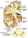
Thalamocortical (VA/VL to motor cortex) projections are

A

Excitatory (glutaminergic)

15
Q

In order for the thalamus to excite the cortex, basal nuclei must

A

Physically remove the inhibition of the motor nuclei of the thalamus (VA/VL).

16
Q

Excitatory (glutaminergic) motor nuclei are tonically inhibited (GABAnergic) by

A

Globus pallidus & substantia nigra pars reticulate

17
Q

Removal of inhibition of disinhibition is done by

A

The striatum via the GPi in the direct pathway

18
Q

Indirect pathway begins with excitatory glutaminergic pathway from

A

Cortex to striatum.

19
Q

In the indirect pathway striatal GABAnergic inhibitory neurons produces inhibition of inhibitory GABA output from

A

GPe to subthalamus.

20
Q

In the indirect pathway, the disinhibited subthalamus excites

A

GPi (again with glutamate).

21
Q

What happens when the GPi is excited in the indirect pathway?  .

A

The VA/VL thalamus is inhibited & cortex can no longer be excited - motor cortex will not produce movement

22
Q

what type of movement does the indirect pathway inhibit?

A

voluntary movement

23
Q

Balance of the direct & indirect pathways produces

A

movement or not.

24
Q

Pars Reticulata is very similar in structure and function to

A

Globus pallidus internus.

25
Pars Reticularis has input from which fibers?
Striatum – striatonigral fibers
26
Which part of the thalamus does Pars Reticularis inhibit?
VA/VL thalamus with GABAnergic inhibitory neurons
27
Neurotransmitter released by Pars Compacta Neurons
Dopamine (DA)
28
Dopamine has two actions on the striatum. 
1. Produces inhibition via D2 receptors on striatal neurons of the indirect pathway - ID2 2. Produces excitation via D1 receptors on striatal neurons of the direct pathway- ED1
29
Substantia Nigra Pars Compacta promotes
Thalamic excitation by activating the direct pathway and inhibiting the indirect pathway
30
Substantia Nigra Pars Compacta is inhibited by
Striatal GABAnergic input
31
Substantia Nigra Pars Compacta is excited by
Cholinergic input from pedunculo-pontine nucleus (PPN).
32
The pedunculo-pontine n is a brainstem n that contributes to the
Pontine reticulospinal tract and produces descending excitation of spinal LMN.
33
Motor loop is associated with the regulation of what type of movement?
Voluntary movement.
34
Where does the motor loop arise from? 
Supplementary motor cortex & other cortical areas
35
What are the functions of the motor loop? ![]()
Regulate VA/VL thalamic and motor cortex excitability via connections through the Putamen & GPi (direct pathway) and GPe and subthalamus (indirect pathway). * Preparation, organization and execution of action * Initiated by the intention to act * Endogenous generation of responses when environmental stimuli fail to provoke responses
36
The oculomotor channel (loop) is associated with regulation of
eye movements.
37
Where does the oculomotor loop begin?
In the SMA & Posterior parietal cortex
38
The oculomotor loop projects via- CGS
via caudate nucleus, GPi & SNpr to regulate excitability of the VA and MedioDorsal / dorsomedial thalamic nuclei.
39
Area of cortex where thalamic output of the oculomotor loop project to
Prefrontal areas - vicinity of frontal eye fields.
40
Functions of the oculomotor channel include: 
Higher-order control of eye movements -orientation of eyes towards specific objects in the environment and is implicated in visual exploration and visual learning
41
The prefrontal channel is associated with
Cognition.
42
Part of the prefrontal channel where the cortical input to the head of the caudate is primarily from?
The dorsolateral pre-frontal cortex.
43
In the prefrontal channel, the caudate influences the GPi & SNpr to regulate
Excitability of the VA and mediodorsal (MD) thalamic nuclei. These nuclei in turn project to the prefrontal cortex (PFC)
44
Functions of the prefrontal channel
* Processing information about fairness of decision-making, * Trusting in “fair” decisions & those who make them * ‘Altruistic punishment’ – the desire to punish violations of social norms even when we have not been personally wronged * Regulation of ‘worry’
45
The limbic channel is associated with regulation of
Emotions and motivational drives
46
The limbic channel Input from temporal cortex, hippocampus & amygdala into the basal nuclei is via the . NVC
* Nucleus accumbens * Ventral putamen & * Caudate.
47
Nucleus accumbens, ventral putamen & caudate connect to the .
GPi for output to the mediodorsal thalamic nucleus
48
The dorsomedial thelamic nucleus projects to the which cortex? AO
Anterior cingulate cortex & orbitofrontal cortex
49
Functions of the limbic channel
* Evaluation of personal actions and environmental resources * Social, behavioral and affective self-regulation of behaviors.
50
The limbic channel is the channel implicated in excessive gambling seen in which disorder?
Parkinson’s patients on L-Dopa & dopamine agonists
51
What is Parkinson’s Disease?
A progressive degenerative disease caused by death of dopaminergic (DA) neurons primarily in the SNpc.
52
What does the time course of Parkinson’s Disease begin with?
Loss of noradrenergic input into the dorsal motor nucleus of X & the noradrenergic neurons of the locus coeruleus.
53
What neurotransmitter is depleted / lost in Parkinson's disease?
Dopaminergic neurons seen in SNpc with normal aging (50% decrease ages 20 to 60)
54
True/false - aging may be a major factor in the accelerated development of PD
True
55
Patients with parkinson's disease shows progressive decline in DA in areas of the Mesolimbic system including projections from the
* Ventral tegmental area (VTA) to the amygdala * Nucleus accumbens * Prefrontal cortex * Hippocampus via medial forebrain bundle (MFB)
56
Progression of Parkinson’s Disease causes continued decline & loss of connections in which parts of the cortex?
* Prefrontal cortex / brain * Limbic cortex & * Hippocampus.
57
Continued development of Parkinson’s Disease produces loss in
* Cognitive skills, * Memory and * Higher associative cognitive functioning
58
Cardinal signs of Parkinson’s Disease includes: BRRP
* Bradykinsesia * Resting tremor  * Rigidity  * Postural instability.
59
Resting Tremor
4-6 Hz resting “pill-rolling” tremor. Begins in peripheral extremities but extends proximally as the disease progresses
60
Name the three components of Bradykinesia- HAB
* Hypokinesia-paucity of movement  * Akinesia – problem initiating movement * Bradykinesia – slowed movement 
61
Non-motor impairments include: CHAP2S
* Cognitive decline (up to 80%)  * Hallucinations  * Autonomic changes * Postural hypotension  * Pain * Sleepiness & Fatigue 
62
Etiology of Parkinson disease
* No known cause of degeneration / apoptosis of dopaminergic neurons * Some cases environmental or genetic causes are implicated - Combination of factors
63
What are some pharmacological approaches to the management of Parkinson’s patients: BRAVeD 
* Blocking the enzymatic breakdown of dopamine * Replacement therapy * Anticholinergic interventions * Various other approaches * Dopamine agonists
64
What is the medication used to supplement the loss of dopamine?
Levodopa (L-DOPA)
65
What is L Dopa co-administered with to minimize peripheral metabolism
Carbidopa
66
What does carbidopa do?
Blocks DOPA Decarboxylase and does not cross the BBB.
67
What is the combination of L-DOPA and carbidopa called?
SinemetTM
68
True/false- L-DOPA use requires intact dopaminergic neurons therefore as apoptosis of dopaminergic neurons continues, the use of L-DOPA is less effective with the progression of the disease
True
69
True/false- Patients on L-DOPA therapy may exhibit sudden ON-OFF shifts of symptoms late in therapy
True
70
Sudden ON-OFF shifts of symptoms late in therapy due to L-DOPA use may be due to
Apoptosis of dopaminergic neurons and the relatively short half-life of L-DOPA
71
How long does L-Dopa lasts?
 about 90 minutes
72
What helps to extend the action of L-DOPA?
Sustained release Sinemet-CR.
73
Patients on L-DOPA often suffer considerably from this disorder
Dyskinesias - may be an effect of dosing of the drug and its rapid metabolism.
74
True / false- there is some evidence that L-DOPA may accelerate DA neuron cell death secondary to increased free radicals or metabolic stress. So many neurologists suggest shifting to other medications early in the progression of the disease to minimize these effects.
true
75
Since dopamine cannot cross blood-brain barrier, what is the metabolic precursor that must be used to help it across?
Levorotatory 3, 4 dihydroxyphenylalanine (L-DOPA).
76
Why is L-DOPA rapidly metabolized peripherally?
Because large numbers of dopaminergic neurons are in the enteric nervous system (n surrounding the gut),
77
What is the newest approach to treatment of Parkinson’s patients? DoRA
Dopamine receptor agonists
78
What does dopamine receptor agonist do?
Mimics the effect of DA at DA receptors.
79
Advantages of dopamine receptor agonist over L-DOPA therapy include: **MoL2eS2**
* More selective action (D1 or D2 agonists) * Longer effect over the course of the disease * Less dyskinesias * Slower rate of metabolism so longer half-life of effectiveness * Slows SNpc cell death
80
Similar adverse reactions of dopamine agonists & L-Dopa?
Both increase the action on DA receptors either by: * Iincreasing DA or * Mimicking the effects of DA.
81
Adverse reactions of dopamine agonists & L-Dopa include: CHA2 D
* Constipation – due to action on DA neurons of enteric nervous system * Hallucinations * Addictive behaviors – excessive gambling * Abnormal thinking – hold onto false beliefs * Dyskinesias – choreoform movements
82
Inhibitors of dopamine metabolism:
Monoamine oxidase (MAO) inhibitors and Catechol-O-methyl transferase (COMT) inhibitors
83
Oldest therapies for Parkinson’s Disease before L-DOPA AA
Anticholinergic agents
84
Blocking \_\_\_\_\_\_\_\_\_\_\_\_receptors could reduce some of the symptoms of Parkinsons disease
Muscarinic receptors
85
Other drugs used to treat Parkinson’s patients include: **NA2**
**Amantadine** – Antiviral may also have effects on catecholamine pathways but has little efficacy **Anti**-**depressants** – other than MAO inhibitors probably have no direct effect per se but used for depression symptoms. **NMDA receptor antagonists** -Newest Tx, may slow cell degeneration (apoptosis).
86
Surgical Interventions for Parkinson's Disease: **PVD**
* Pallidotomy or lesion of the globus pallidus internus * Ventrolateral thalamotomy * Deep brain stimulation.
87
What is Pallidotomy or lesion of the globus pallidus internus?
Common approach involving use of a stereotaxic device to localize GP & stabilize patients. Pallidotomy reduces tremor & rigidity but not other symptoms.
88
What is Ventrolateral thalamotomy –
Lesioning of the VL nucleus- done with similar results to GPi lesions
89
Deep brain stimulation-
Implantation of leads to different brain regions along with an implanted pacemaker unit. Common targets for DBS are the thalamus, subthalamic nucleus, and GPi
90
True / False- Fetal nigral transplantation- has had Little efficacy despite positive MRI results. In addition Substantial adverse reactions noted in young patients
True
91
Huntington's Disease is also know as
Huntington’s Chorea
92
What is Huntington's disease?
An Inherited neurodegenerative disease.
93
T / F- Children of Huntington Disease patients have a 50% risk of developing the disease.
True
94
Test for Huntington disese
Genetic testing to identify gene or not.
95
Huntington’s Disease causes Gross atrophy of
The striatum (caudate and putamen)
96
What is Huntingtin?.
An autosomal dominant mutation in either of an individual's two copies of a gene
97
Characteristics of Huntington’s Disease CPM
* **Cognitive decline -**leading to dementia * **Psychiatric disorders**- paranoid and psychosis behaviors * **Motor changes** – choreoathetosis
98
Typical age of onset of Huntington disease?
40–50 years
99
Most Huntington patients progress to a vegetative state within
10–15 years and die
100
Medical Management of Huntington disease? FeD2S
* Fetal transplantation (unsuccessful) * Dopamine antagonists * Deep brain stimulation * Surgical ablation of the GPi all without much value
101
Hemiballismus
Wild, unpatterned, flinging movements of an entire extremity. 
102
Cause of Hemiballismus?
A discrete lesion of the subthalamic nucleus contralateral to symptoms which reduces activity of the indirect pathway and thus reduces the inhibition of movement.
103
Hemiballismus most commonly results from vascular disorder of which artery?
Penetrating branch of the posterior cerebral artery (PCA)
104
Three main Connections of the cerebellum to the Brainstem SIM
* Superior peduncle * Inferior peduncle * Middle peduncle *
105
Inferior cerebellar peduncle contains
Both cerebellar afferents and efferents
106
Inferior cerebellar afferents (cerebellopetal fibers) contains the foll. tracts? DROVe C
* Dorsal, Cuneo & Rostral spinocerebellar tracts * Olivocerebellar tract * Vestibulocerebellar tract
107
Nuclei of the Inferior cerebellar efferent (cerebellofugal fibers) contain? VR
* Vestibular nuclei * Reticular nuclei
108
Middle cerebellar peduncle tract and fibers
Pontocerebellar tract which are afferent or cerebellopetal fibers
109
Superior cerebellar peduncle contains
* Some affferent fibers - Ventral spinocerebellar tract, * Primarily efferent corticofugal fibers from cerebellum which terminate in the red nucleus and the VL motor nucleus of the thalamus
110
What fibers are the most numerous form of input into the cerebellum?
Mossy fibers
111
Each parallel mossy fiber excites
\>500 Purkinje cells and from 100-300,000 parallel fibers excite a single Purkinje cells (glutamate).
112
What type of synapses does Mossy fibers form?
Glutaminergic excitatory synapses with glutaminergic mediated EPSPs.
113
Where does EPSPs of Mossy fibers end?
* On granule cells (of the granule cell layer), whose axons form parallel fibers which ascend up into the molecular layer of the cerebellar cortex to synapse with glutaminergic excitatory endings on the dendrites of Purkinje cells.
114
Mossy fibers originate from- VSC
* Vestibular primary afferent fibers via inferior peduncle * Vestibular nuclei via inferior peduncle * Spinal cord (spinocerebellar tracts) via mostly inferior peduncle (ventral SCT via superior peduncle) * Cerebral cortex via pontine nuclei & pontocerebellar fibers (middle peduncle) * End also on Golgi cells – inhibitory interneurons
115
Most climbing fibers direct to- P
Purkinje cells
116
Climbing fibers originate from
Contralateral inferior olivary nucleus
117
Climbing fibers end with
Excitatory aspartate synapses directly on Purkinje cells
118
Climbing fibers produces
Large EPSPs which produce bursts of Purkinje cell action potention activity
119
Each Purkinje cell receives .
Single climbing fiber
120
Each climbing fiber innervates only
1-3 Purkinje cells
121
What type of signal does climbing fibers provide?
Feed forward signals
122
Feed forward signals from climbing fibers travel from the cerebral cortex to the cerebellum via COOC
Cortico-olivary and olivocerebellar fibers
123
Mossy fibers provide feedback signals from SVO
spinal, vestibular & other inputs
124
Cerebellar Interneurons are what type of cells? GBS
Golgi cells, Basket and Stellate cells
125
Golgi cells are innervated / excited by what fibers? MP
Mossy & parallel fibers
126
T/F- Golgi cells are inhibitory interneurons of the granule cell layer
True
127
Golgi cell Inhibitory action (GABA) on granule cell dendrites produces FF& FB
Both feed forward (via mossy fibers) and feedback (via parallel fibers) inhibition to granule cells
128
Basket & stellate cells produces GI
GABAminergic Inhibitory action on Purkinje cells
129
Basket and stellate cells produce a prolonged inhibition related to
Associative learning in cerebellum
130
Outflow from cerebellar cortex is via
Purkinje neurons
131
T / F- Output from cerebellar cortex is excitatory, 
False it is inhibitory (GABA)
132
Purkinje neurons project to which nuclei?
Deep cerebellar nuclei
133
Vestibulocerebellum Purkinje cells project directly to which nuclei?
vestibular nuclei
134
T / F- With just a few Purkinje cells leaving the cerebellum the principle outputs from cerebellum are from the deep cerebellar nuclei.
True
135
T / F- Mossy & climbing fibers create an inhibitory drive which is sculpted by the inhibitory influence of the Purkinje neurons
False it produces an excitatory drive
136
Where does vestibular afferents project to?
Cortex of vestibulocerebellum Fastigial nucleus through the inferior cerebellar peduncle
137
Vestibular afferents Influence TENT
* Tone in limbs * Extraocular eye muscles, * Neck * Trunk
138
Visual systems project indirectly to vestibulocerebellum through
Climbing fibers of the inferior olivary nucleus.
139
Input from which cerebellar peduncle assists in regulating the vestibular ocular reflex (VOR)
Inferior cerebellar peduncle
140
Vestibulocerebellum outputs go to. BVn
Brainstem Vestibular nuclei.
141
Vestibulocerebellum outputs to Projections influence
Extraocular motor neurons via the medial longitudinal fasciculus
142
Vestibulocerebellum outputs to the brainstem end in nuclei of the
Reticular formation which send both descending fibers forming the reticulospinal tracts and ascending fibers to extraocular motor nuclei
143
Role of vestibulocerebellum outputs to the vestibular nuclei
Influence body and limb tone and responses via the vestibulospinal tracts
144
Functions of the vestibulocerebellum (Flocculonodular lobe) include: 
* Balance in sitting, standing & gait * Plasticity of vestibulo-ocular reflex * Nodule function- sensitivity to motion sickness (ablated this makes subject immune to motion sickness)
145
Regions of the cerebellum.
* Pontocerebellum * Spinocerebellum & * vestibulocerebellum.
146
The Pontocerebellum / Neocerebellum belongs to which zone of the cerebellum?
Lateral zone- including the more lateral regions of anterior & posterior lobes
147
The Spinocerebellum / paleocerebellum belongs to which zone of the cerebellum?
Intermediate zone - includes medial aspects of both lobes, cerebellar tonsils and vermis
148
Vestibulocerebellum / archicerebellum
Most medial part of the cerebellum and Includes the flocculonodular lobe
149
4 pairs of deep nuclei in the cerebellum
* Fastigial * Globose & Emboliform or interposed nuclei * Dentate nucleus
150
The Fastigial nucleus
The most medial -associated with primarily the vestibulocerebellum but also relays projections from the spinocerebellum
151
Interposed Nuclei (Globose & Emboliform)
As their name implies are interposed between the fastigial and dentate nuclei - associated with spinocerebellum
152
The Dentate nucleus
Most lateral & largest of the deep cerebellar nuclei -associated with pontocerebellum
153
Three distinct layer of the cerebellar cortex:
* Molecular layer * Purkinje cell layer and * Granule cell layer.
154
The Molecular Layer
* Most Superficial layer. * Low neuron cell body density * Types of inhibitory neurons: stellate & basket cells. * Contain large dendritic arborizations of Purkinje cells- dendritic arborizations lie in a single plane which is perpendicular to long axis of folia, which are the gyri of the cerebellar cortex.
155
Two sets of afferent fibers in the Molecular Layer of the cerebellar cortex:
* Parallel fibers- Unmyelinated Granule cell axons, each runs 5 mm along (parallel to) long axis of folium activating hundreds of Purkinje cells * Climbing fibers – wraps around individual Purkinje cell dendritic trees
156
Purkinje Cell Layer
Middle layer of the cerebellar cortex - composed of cell bodies of Purkinje neurons. Neurons are Inhibitory (GABA mediated) to the deep nuclei and some axons will exit the cerebellum
157
Granular Layer
* Innermost layer of the cerebellar cortex- tightly packed with granule cells. * Many inhibitory interneurons in this layer called Golgi cells. * Axons of granule cells ascend into molecular layer to form parallel fibers. * Parallel fibers run about 5 mm along the cortex activating with excitatory (glutamate) endings with each parallel fiber ending on \>500 Purkinje cells