NeuroBiology66-100 Flashcards
(35 cards)
- Selectively activates Gs
A. Tetrabenazine
B. a-bungarotoxin
C. D-tubocurarine
D. Strychnine
E. Tetanus toxin
F. Cholera toxin
G. Barbiturates
H. Botulinus toxin
I. Pertussis toxin
J. LSD
K. Ondansetron
L. None of the above
F. Cholera toxin
Ion channels conduct ions at extremely high rates, are selective for specific ions, and are regulated or gated. Gated ion channels can be regulated by changes in voltage, chemical transmitters (ligands), and mechanical factors. Ligand-gated channels include glutamatergic channels, cholinergic channels, glycinergic channels, and GABAergic channels.
Acetylcholine-activated channels include nicotinic and
muscarinic receptors. Nicotinic cholinergic receptors are ionotropic channels that are permeable to Na+
and K+ and consist of five subunits: two a and the (3, y, and 8 subunits (a2py8). The a subunit binds a single molecule of ACh, thus requiring two ACh molecules to bind the receptor to elicit channel activation. The snake venom toxin cc-bungarotoxin binds the a subunit as well, effectively inhibiting channel function. Each subunit of the receptor contains four hydro-phobic a helices (Ml to M4) that traverse the plasma mem-brane. Opening of the nicotinic ACh channel results in the generation of a fast excitatory postsynaptic potential (EPSP).
Hexamethonium (ganglionic), succinylcholine (depolarizing), and D-tubocurarine (nondepolarizing) represent inhibitors of various nicotinic cholinergic receptors. The nicotinic receptor is found at the neuromuscular junction as well as preganglionic synapses of the autonomic nervous system.
The muscarinic cholinergic receptor is a metabotropic
receptor that is coupled to G proteins and consists of only two subunits (a and (3). This channel is a slow-activating K+ channel (M-type channel) that closes when stimulated and results in the generation of a slow EPSP. Muscarinic channels are found throughout the GNS (cerebellum, striatum, cortex,
Renshaw cells of the spinal cord) and in autonomic ganglia. These receptors are inhibited by atropine and scopolamine and stimulated by bethanecol (bladder), carbachol (Gl tract), pilocarpine (eye), and methacholine. Glycine is the neurotransmitter released by Renshaw cells (inhibitory interneurons) of the spinal cord (see dis-cussion, question 45). Glycine channels are blocked by strychnine, and glycine release is inhibited by tetanus toxin. There are five major groups of dopamine and serotonin receptors, all of which are metabotropic. Examples include Dl and D2 receptors, which stimulate and inhibit adenylyl cyclase, respectively. The net effect of Dl receptors is hyper-polarization, while that of D2 receptors is depolarization. The typical antipsychotics selectively inhibit D2 receptors. The majority of serotonin receptors are metabotropic. LSD is an agonist of the 5-HT 1G receptor, while ondansetron is
an antagonist of the 5-HT3 (ionotropic) receptor. All nora-drenergic receptors are metabotropic receptors that utilize G proteins and the second messenger cAMP. The signal transduction cascade involved in metabotropic receptor activation can be manipulated by cholera toxin, which selectively activates Gs, and pertussis toxin which inactivates Gj. Tetanus toxin specifically cleaves the protein synaptobrevin, while botulinus toxins cleave t-SNAREs and v-SNAREs, which subsequently results in the inhibition of
synaptic vesicle release at the terminal. The docking, fusion, and release of synaptic vesicles appears to involve distinct interactions between vesicle proteins (synaptobrevin and synaptotagmin, v-SNAREs) and proteins of the nerve terminal plasma membrane (syntaxins and neurexins, t-SNAREs) (Kandel, pp. 196-200, 219, 241-243, 1197-1199,1215-1216).
- Nondepolarizing inhibitor of nicotinic cholinergic
receptors
A. Tetrabenazine
B. a-bungarotoxin
C. D-tubocurarine
D. Strychnine
E. Tetanus toxin
F. Cholera toxin
G. Barbiturates
H. Botulinus toxin
I. Pertussis toxin
J. LSD
K. Ondansetron
L. None of the above
C. D-tubocurarine
Ion channels conduct ions at extremely high rates, are selective for specific ions, and are regulated or gated. Gated ion channels can be regulated by changes in voltage, chemical transmitters (ligands), and mechanical factors. Ligand-gated channels include glutamatergic channels, cholinergic channels, glycinergic channels, and GABAergic channels.
Acetylcholine-activated channels include nicotinic and
muscarinic receptors. Nicotinic cholinergic receptors are ionotropic channels that are permeable to Na+
and K+ and consist of five subunits: two a and the (3, y, and 8 subunits (a2py8). The a subunit binds a single molecule of ACh, thus requiring two ACh molecules to bind the receptor to elicit channel activation. The snake venom toxin cc-bungarotoxin binds the a subunit as well, effectively inhibiting channel function. Each subunit of the receptor contains four hydro-phobic a helices (Ml to M4) that traverse the plasma mem-brane. Opening of the nicotinic ACh channel results in the generation of a fast excitatory postsynaptic potential (EPSP).
Hexamethonium (ganglionic), succinylcholine (depolarizing), and D-tubocurarine (nondepolarizing) represent inhibitors of various nicotinic cholinergic receptors. The nicotinic receptor is found at the neuromuscular junction as well as preganglionic synapses of the autonomic nervous system.
The muscarinic cholinergic receptor is a metabotropic
receptor that is coupled to G proteins and consists of only two subunits (a and (3). This channel is a slow-activating K+ channel (M-type channel) that closes when stimulated and results in the generation of a slow EPSP. Muscarinic channels are found throughout the GNS (cerebellum, striatum, cortex,
Renshaw cells of the spinal cord) and in autonomic ganglia. These receptors are inhibited by atropine and scopolamine and stimulated by bethanecol (bladder), carbachol (Gl tract), pilocarpine (eye), and methacholine. Glycine is the neurotransmitter released by Renshaw cells (inhibitory interneurons) of the spinal cord (see dis-cussion, question 45). Glycine channels are blocked by strychnine, and glycine release is inhibited by tetanus toxin. There are five major groups of dopamine and serotonin receptors, all of which are metabotropic. Examples include Dl and D2 receptors, which stimulate and inhibit adenylyl cyclase, respectively. The net effect of Dl receptors is hyper-polarization, while that of D2 receptors is depolarization. The typical antipsychotics selectively inhibit D2 receptors. The majority of serotonin receptors are metabotropic. LSD is an agonist of the 5-HT 1G receptor, while ondansetron is
an antagonist of the 5-HT3 (ionotropic) receptor. All nora-drenergic receptors are metabotropic receptors that utilize G proteins and the second messenger cAMP. The signal transduction cascade involved in metabotropic receptor activation can be manipulated by cholera toxin, which selectively activates Gs, and pertussis toxin which inactivates Gj. Tetanus toxin specifically cleaves the protein synaptobrevin, while botulinus toxins cleave t-SNAREs and v-SNAREs, which subsequently results in the inhibition of
synaptic vesicle release at the terminal. The docking, fusion, and release of synaptic vesicles appears to involve distinct interactions between vesicle proteins (synaptobrevin and synaptotagmin, v-SNAREs) and proteins of the nerve terminal plasma membrane (syntaxins and neurexins, t-SNAREs) (Kandel, pp. 196-200, 219, 241-243, 1197-1199,1215-1216).
- Inactivates G]
A. Tetrabenazine
B. a-bungarotoxin
C. D-tubocurarine
D. Strychnine
E. Tetanus toxin
F. Cholera toxin
G. Barbiturates
H. Botulinus toxin
I. Pertussis toxin
J. LSD
K. Ondansetron
L. None of the above
I. Pertussis toxin
Ion channels conduct ions at extremely high rates, are selective for specific ions, and are regulated or gated. Gated ion channels can be regulated by changes in voltage, chemical transmitters (ligands), and mechanical factors. Ligand-gated channels include glutamatergic channels, cholinergic channels, glycinergic channels, and GABAergic channels.
Acetylcholine-activated channels include nicotinic and
muscarinic receptors. Nicotinic cholinergic receptors are ionotropic channels that are permeable to Na+
and K+ and consist of five subunits: two a and the (3, y, and 8 subunits (a2py8). The a subunit binds a single molecule of ACh, thus requiring two ACh molecules to bind the receptor to elicit channel activation. The snake venom toxin cc-bungarotoxin binds the a subunit as well, effectively inhibiting channel function. Each subunit of the receptor contains four hydro-phobic a helices (Ml to M4) that traverse the plasma mem-brane. Opening of the nicotinic ACh channel results in the generation of a fast excitatory postsynaptic potential (EPSP).
Hexamethonium (ganglionic), succinylcholine (depolarizing), and D-tubocurarine (nondepolarizing) represent inhibitors of various nicotinic cholinergic receptors. The nicotinic receptor is found at the neuromuscular junction as well as preganglionic synapses of the autonomic nervous system.
The muscarinic cholinergic receptor is a metabotropic
receptor that is coupled to G proteins and consists of only two subunits (a and (3). This channel is a slow-activating K+ channel (M-type channel) that closes when stimulated and results in the generation of a slow EPSP. Muscarinic channels are found throughout the GNS (cerebellum, striatum, cortex,
Renshaw cells of the spinal cord) and in autonomic ganglia. These receptors are inhibited by atropine and scopolamine and stimulated by bethanecol (bladder), carbachol (Gl tract), pilocarpine (eye), and methacholine. Glycine is the neurotransmitter released by Renshaw cells (inhibitory interneurons) of the spinal cord (see dis-cussion, question 45). Glycine channels are blocked by strychnine, and glycine release is inhibited by tetanus toxin. There are five major groups of dopamine and serotonin receptors, all of which are metabotropic. Examples include Dl and D2 receptors, which stimulate and inhibit adenylyl cyclase, respectively. The net effect of Dl receptors is hyper-polarization, while that of D2 receptors is depolarization. The typical antipsychotics selectively inhibit D2 receptors. The majority of serotonin receptors are metabotropic. LSD is an agonist of the 5-HT 1G receptor, while ondansetron is
an antagonist of the 5-HT3 (ionotropic) receptor. All nora-drenergic receptors are metabotropic receptors that utilize G proteins and the second messenger cAMP. The signal transduction cascade involved in metabotropic receptor activation can be manipulated by cholera toxin, which selectively activates Gs, and pertussis toxin which inactivates Gj. Tetanus toxin specifically cleaves the protein synaptobrevin, while botulinus toxins cleave t-SNAREs and v-SNAREs, which subsequently results in the inhibition of
synaptic vesicle release at the terminal. The docking, fusion, and release of synaptic vesicles appears to involve distinct interactions between vesicle proteins (synaptobrevin and synaptotagmin, v-SNAREs) and proteins of the nerve terminal plasma membrane (syntaxins and neurexins, t-SNAREs) (Kandel, pp. 196-200, 219, 241-243, 1197-1199,1215-1216).
- Agonist of the 5-HTl c receptor
A. Tetrabenazine
B. a-bungarotoxin
C. D-tubocurarine
D. Strychnine
E. Tetanus toxin
F. Cholera toxin
G. Barbiturates
H. Botulinus toxin
I. Pertussis toxin
J. LSD
K. Ondansetron
L. None of the above
J. LSD
Ion channels conduct ions at extremely high rates, are selective for specific ions, and are regulated or gated. Gated ion channels can be regulated by changes in voltage, chemical transmitters (ligands), and mechanical factors. Ligand-gated channels include glutamatergic channels, cholinergic channels, glycinergic channels, and GABAergic channels.
Acetylcholine-activated channels include nicotinic and
muscarinic receptors. Nicotinic cholinergic receptors are ionotropic channels that are permeable to Na+
and K+ and consist of five subunits: two a and the (3, y, and 8 subunits (a2py8). The a subunit binds a single molecule of ACh, thus requiring two ACh molecules to bind the receptor to elicit channel activation. The snake venom toxin cc-bungarotoxin binds the a subunit as well, effectively inhibiting channel function. Each subunit of the receptor contains four hydro-phobic a helices (Ml to M4) that traverse the plasma mem-brane. Opening of the nicotinic ACh channel results in the generation of a fast excitatory postsynaptic potential (EPSP).
Hexamethonium (ganglionic), succinylcholine (depolarizing), and D-tubocurarine (nondepolarizing) represent inhibitors of various nicotinic cholinergic receptors. The nicotinic receptor is found at the neuromuscular junction as well as preganglionic synapses of the autonomic nervous system.
The muscarinic cholinergic receptor is a metabotropic
receptor that is coupled to G proteins and consists of only two subunits (a and (3). This channel is a slow-activating K+ channel (M-type channel) that closes when stimulated and results in the generation of a slow EPSP. Muscarinic channels are found throughout the GNS (cerebellum, striatum, cortex,
Renshaw cells of the spinal cord) and in autonomic ganglia. These receptors are inhibited by atropine and scopolamine and stimulated by bethanecol (bladder), carbachol (Gl tract), pilocarpine (eye), and methacholine. Glycine is the neurotransmitter released by Renshaw cells (inhibitory interneurons) of the spinal cord (see dis-cussion, question 45). Glycine channels are blocked by strychnine, and glycine release is inhibited by tetanus toxin. There are five major groups of dopamine and serotonin receptors, all of which are metabotropic. Examples include Dl and D2 receptors, which stimulate and inhibit adenylyl cyclase, respectively. The net effect of Dl receptors is hyper-polarization, while that of D2 receptors is depolarization. The typical antipsychotics selectively inhibit D2 receptors. The majority of serotonin receptors are metabotropic. LSD is an agonist of the 5-HT 1G receptor, while ondansetron is
an antagonist of the 5-HT3 (ionotropic) receptor. All nora-drenergic receptors are metabotropic receptors that utilize G proteins and the second messenger cAMP. The signal transduction cascade involved in metabotropic receptor activation can be manipulated by cholera toxin, which selectively activates Gs, and pertussis toxin which inactivates Gj. Tetanus toxin specifically cleaves the protein synaptobrevin, while botulinus toxins cleave t-SNAREs and v-SNAREs, which subsequently results in the inhibition of
synaptic vesicle release at the terminal. The docking, fusion, and release of synaptic vesicles appears to involve distinct interactions between vesicle proteins (synaptobrevin and synaptotagmin, v-SNAREs) and proteins of the nerve terminal plasma membrane (syntaxins and neurexins, t-SNAREs) (Kandel, pp. 196-200, 219, 241-243, 1197-1199,1215-1216).
- Antagonist of the 5-HT3 (ionotropic) receptor
A. Tetrabenazine
B. a-bungarotoxin
C. D-tubocurarine
D. Strychnine
E. Tetanus toxin
F. Cholera toxin
G. Barbiturates
H. Botulinus toxin
I. Pertussis toxin
J. LSD
K. Ondansetron
L. None of the above
K. Ondansetron
Ion channels conduct ions at extremely high rates, are selective for specific ions, and are regulated or gated. Gated ion channels can be regulated by changes in voltage, chemical transmitters (ligands), and mechanical factors. Ligand-gated channels include glutamatergic channels, cholinergic channels, glycinergic channels, and GABAergic channels.
Acetylcholine-activated channels include nicotinic and
muscarinic receptors. Nicotinic cholinergic receptors are ionotropic channels that are permeable to Na+
and K+ and consist of five subunits: two a and the (3, y, and 8 subunits (a2py8). The a subunit binds a single molecule of ACh, thus requiring two ACh molecules to bind the receptor to elicit channel activation. The snake venom toxin cc-bungarotoxin binds the a subunit as well, effectively inhibiting channel function. Each subunit of the receptor contains four hydro-phobic a helices (Ml to M4) that traverse the plasma mem-brane. Opening of the nicotinic ACh channel results in the generation of a fast excitatory postsynaptic potential (EPSP).
Hexamethonium (ganglionic), succinylcholine (depolarizing), and D-tubocurarine (nondepolarizing) represent inhibitors of various nicotinic cholinergic receptors. The nicotinic receptor is found at the neuromuscular junction as well as preganglionic synapses of the autonomic nervous system.
The muscarinic cholinergic receptor is a metabotropic
receptor that is coupled to G proteins and consists of only two subunits (a and (3). This channel is a slow-activating K+ channel (M-type channel) that closes when stimulated and results in the generation of a slow EPSP. Muscarinic channels are found throughout the GNS (cerebellum, striatum, cortex,
Renshaw cells of the spinal cord) and in autonomic ganglia. These receptors are inhibited by atropine and scopolamine and stimulated by bethanecol (bladder), carbachol (Gl tract), pilocarpine (eye), and methacholine. Glycine is the neurotransmitter released by Renshaw cells (inhibitory interneurons) of the spinal cord (see dis-cussion, question 45). Glycine channels are blocked by strychnine, and glycine release is inhibited by tetanus toxin. There are five major groups of dopamine and serotonin receptors, all of which are metabotropic. Examples include Dl and D2 receptors, which stimulate and inhibit adenylyl cyclase, respectively. The net effect of Dl receptors is hyper-polarization, while that of D2 receptors is depolarization. The typical antipsychotics selectively inhibit D2 receptors. The majority of serotonin receptors are metabotropic. LSD is an agonist of the 5-HT 1G receptor, while ondansetron is
an antagonist of the 5-HT3 (ionotropic) receptor. All nora-drenergic receptors are metabotropic receptors that utilize G proteins and the second messenger cAMP. The signal transduction cascade involved in metabotropic receptor activation can be manipulated by cholera toxin, which selectively activates Gs, and pertussis toxin which inactivates Gj. Tetanus toxin specifically cleaves the protein synaptobrevin, while botulinus toxins cleave t-SNAREs and v-SNAREs, which subsequently results in the inhibition of
synaptic vesicle release at the terminal. The docking, fusion, and release of synaptic vesicles appears to involve distinct interactions between vesicle proteins (synaptobrevin and synaptotagmin, v-SNAREs) and proteins of the nerve terminal plasma membrane (syntaxins and neurexins, t-SNAREs) (Kandel, pp. 196-200, 219, 241-243, 1197-1199,1215-1216).
- Clinical evidence of neurologic deficit may not appear until regional blood flow has fallen to 50% or below average levels. At what rate of cerebral blood flow (in mL/100 g/min) does cytotoxic edema develop from failure of the Na+ K+-ATPase?
A. 40-50
B. 25-30
C. 16-20
D. 10-12
E. < 10
D. 10-12
Refer to Table 1.71A. Cytotoxic edema refers to swelling of injured neurons, glia, and endothelial cells after hypoxia or global cerebral ischemia. It is the result of ATP-dependent Na7K+ pump failure, which allows Na+, and there-fore water, to accumulate within cells. Vasogenic edema is the most common type of brain edema and is attributed to increased permeability of brain capillary endothelial cells and thus extracellular fluid volume. The white matter is generally affected more than the gray matter because of the tendency of fluid to accumulate adjacent to the white matter tracts (Kandel, p. 1299; Youmans, pp. 1468-1469) .
- Which of the following is believed to be the major vasoac-tive mediator that plays an integral role in vasomodulation?
A. Carbon monoxide
B. Arachidonic acid metabolites
C. Nitrous oxide
D. Adenosine
E. ATP
C. Nitrous oxide
As measured by its pivotal vascular actions, nitrous
oxide is believed to be the major candidate for vascular modu-lation. Important actions of this mediator include facilitation of vascular relaxation and prevention of smooth muscle pro-liferation, platelet and white blood cell adherence, and platelet aggregation. Other important vasomodulators are carbon monoxide, eicosanoids, oxygen-derived free radicals, and endothelin (Youmans, pp. 1469-1474).
- Neural crest cells give rise to all of the following struc-tures EXCEPT?
A. Ventral root ganglia
B. Postganglionic cells of the sympathetic and parasym-pathetic ganglia
C. Chromaffin cells of the adrenal medulla
D. Melanocytes
E. Schwann cells
A. Ventral root ganglia
Neural crest cells give rise to the PNS, chromaffin cells
of the adrenal medulla, pia, arachnoid, melanocytes, Schwann cells, odontoblasts, certain endocrine cells, cranial nerve sensory ganglia, autonomic ganglia, and dorsal root ganglia, as opposed to the ventral root ganglia (DeMyer, p. 52).
- Which of the following are common features of Wallerian degeneration?
- Degeneration and phagocytosis of the distal axonal
segment - Chromatolysis (peripheralization of rough endoplas-mic reticulum with a concomitant increased protein synthesis) due to decreased retrograde neurotrophic factor delivery
- Proximal axon segment swelling due to continued
anterograde axonal transport - Greater neuronal cell death of postsynaptic neurons in the peripheral nervous system (PNS) than the central nervous system after axotomy
A. 1,2, and 3 are correct
B. 1 and 3 are correct
C. 2 and 4 are correct
D. Only 4 is correct
E. All of the above
A. 1,2, and 3 are correct
With axon transection (axotomy), synaptic trans-mission failure occurs initially, followed by degeneration and phagocytosis of the distal axonal segment, which is called Wallerian degeneration. In addition, the neuronal soma undergoes chromatolysis (peripheralization of rough ER with concomitant increased protein synthesis) due to decreased retrograde neurotrophic factor delivery, and the end of the proximal axon segment swells due to continued anterograde axonal transport. Axotomy also has a negative impact on the postsynaptic neuron and can result in neu-ronal cell death unless regeneration occurs. Regeneration occurs when axonal sprouts grow from the end of the proxi-mal axonal segment and enter tissue remnants of the distal stump. These sprouts may form new synaptic connections if they eventually reach their targets. Formation of new synapses in this way occurs more effectively in the PNS than the GNS (Kandel, pp. 82-83, 147-148,1108-1110).
- Striated muscle fibers
A. Red muscle fibers
B. White muscle fibers
C. Both
D. None of the above
C. Both
There are three basic types of striated muscle fibers, including slow-twitch fibers and two types of fast-twitch fibers. Slow-twitch fibers are called type I fibers, or red fibers. Red fibers contain large amounts of mitochon-dria, due to their high rates of oxidative metabolism, as well as myoglobin, which facilitates oxygen storage for times of increased demand. These fibers contract and relax slowly but are capable of prolonged periods of contraction without fatigue. Fast-twitch fibers are called type II fibers, or white
fibers. These fibers generate rapid, short-term contractions. Fast fatigable (type IIB) fibers have large stores of glycogen and utilize anaerobic catabolism for contraction. Lactic acid accumulates rapidly in these fibers, which limits their ability to sustain contraction forces. Fast fatigue-resistant (type IIA)
fibers have some aerobic metabolism capacity (less than red fibers, however), and are able to generate rapid contractions, yet maintain them for a period of several minutes. With muscle contraction, weaker motor units are recruited in an orderly fashion before stronger motor units. Synaptic input stimulates smaller cells with higher internal resistances
before larger cells, which forms the basis for motor unit recruitment by size (Kandel, pp. 676-682)
- Contain large amounts of mitochondria, contract and
relax slowly
A. Red muscle fibers
B. White muscle fibers
C. Both
D. None of the above
A. Red muscle fibers
There are three basic types of striated muscle fibers, including slow-twitch fibers and two types of fast-twitch fibers. Slow-twitch fibers are called type I fibers, or red fibers. Red fibers contain large amounts of mitochon-dria, due to their high rates of oxidative metabolism, as well as myoglobin, which facilitates oxygen storage for times of increased demand. These fibers contract and relax slowly but are capable of prolonged periods of contraction without fatigue. Fast-twitch fibers are called type II fibers, or white
fibers. These fibers generate rapid, short-term contractions. Fast fatigable (type IIB) fibers have large stores of glycogen and utilize anaerobic catabolism for contraction. Lactic acid accumulates rapidly in these fibers, which limits their ability to sustain contraction forces. Fast fatigue-resistant (type IIA)
fibers have some aerobic metabolism capacity (less than red fibers, however), and are able to generate rapid contractions, yet maintain them for a period of several minutes. With muscle contraction, weaker motor units are recruited in an orderly fashion before stronger motor units. Synaptic input stimulates smaller cells with higher internal resistances
before larger cells, which forms the basis for motor unit recruitment by size (Kandel, pp. 676-682)
- Aerobic metabolism capacity
A. Red muscle fibers
B. White muscle fibers
C. Both
D. None of the above
C. Both
There are three basic types of striated muscle fibers, including slow-twitch fibers and two types of fast-twitch fibers. Slow-twitch fibers are called type I fibers, or red fibers. Red fibers contain large amounts of mitochon-dria, due to their high rates of oxidative metabolism, as well as myoglobin, which facilitates oxygen storage for times of increased demand. These fibers contract and relax slowly but are capable of prolonged periods of contraction without fatigue. Fast-twitch fibers are called type II fibers, or white
fibers. These fibers generate rapid, short-term contractions. Fast fatigable (type IIB) fibers have large stores of glycogen and utilize anaerobic catabolism for contraction. Lactic acid accumulates rapidly in these fibers, which limits their ability to sustain contraction forces. Fast fatigue-resistant (type IIA)
fibers have some aerobic metabolism capacity (less than red fibers, however), and are able to generate rapid contractions, yet maintain them for a period of several minutes. With muscle contraction, weaker motor units are recruited in an orderly fashion before stronger motor units. Synaptic input stimulates smaller cells with higher internal resistances
before larger cells, which forms the basis for motor unit recruitment by size (Kandel, pp. 676-682)
- Contain large stores of glycogen
A. Red muscle fibers
B. White muscle fibers
C. Both
D. None of the above
B. White muscle fibers
There are three basic types of striated muscle fibers, including slow-twitch fibers and two types of fast-twitch fibers. Slow-twitch fibers are called type I fibers, or red fibers. Red fibers contain large amounts of mitochon-dria, due to their high rates of oxidative metabolism, as well as myoglobin, which facilitates oxygen storage for times of increased demand. These fibers contract and relax slowly but are capable of prolonged periods of contraction without fatigue. Fast-twitch fibers are called type II fibers, or white
fibers. These fibers generate rapid, short-term contractions. Fast fatigable (type IIB) fibers have large stores of glycogen and utilize anaerobic catabolism for contraction. Lactic acid accumulates rapidly in these fibers, which limits their ability to sustain contraction forces. Fast fatigue-resistant (type IIA)
fibers have some aerobic metabolism capacity (less than red fibers, however), and are able to generate rapid contractions, yet maintain them for a period of several minutes. With muscle contraction, weaker motor units are recruited in an orderly fashion before stronger motor units. Synaptic input stimulates smaller cells with higher internal resistances
before larger cells, which forms the basis for motor unit recruitment by size (Kandel, pp. 676-682)
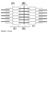
- Composed solely of actin filaments

E

Refer to Figure 1.79-1.83A. A single muscle cell contains several myofibrils, each of which consists of linear chains of solitary contractile units, or sar-comeres. Myofibrils are connected to one another via desmin intermediate filaments, and this complex is then anchored to the muscle cell sarcolemma by several proteins, including dystrophin. Within the myofibril, each sarcomere is con-nected to the adjacent sarcomere at the Z disc (A), and actin
(thin) filaments radiate from the Z disc toward the center of the sarcomere. Myosin (thick) filaments are found inter-spersed between adjacent actin filaments. The dark band, or A band (D), is the region of the sarcomere that is composed primarily of myosin filaments. The light band, or I band (E), is the region that is composed solely of actin filaments and is centered on a Z disc. The H zone (B) is the region of the A band where myosin filaments are not overlapped by actin filaments in the resting state, and it is centered on the M line (G). With myofibril contraction, the actin and myosin filaments form successive cross bridges that facilitate sliding
across one another. This functionally shortens the sarco-mere during contraction and consequently results in H-zone and I-band shortening. This process of muscle contraction requires the presence of cytosolic Ca2+. In the resting state, tropomyosin and a troponin complex (troponins I, G, and T) are bound to actin. After the sarcoplasmic reticulum releases
Ga2+ in response to an action potential, troponin G binds four molecules of Ga2+, which subsequently relieves the inhibition of the myosin binding site on actin. Myosin heads are then free to bind actin and form cross bridges. The myosin head, which has intrinsic ATPase activity, then rotates, pulling
the actin filaments longitudinally and increasing the overlap between the thick and thin filaments. ATP then binds to the myosin head, which stimulates the release of the cross bridge between actin. The subsequent hydrolysis of ATP “cocks” the myosin head, which then forms a second cross bridge,
contracts, and further increases the overlap between thick and thin filaments. ADP is released, and the process is repeated as long as ATP and Ga2+
are present in the cytosol. In this manner, the myosin heads “walk” along the actin filaments during contraction, effectively shortening the sarcomere and myofibril (Kandel, pp. 676-682).
- Shortens during muscle contraction

B,E

Refer to Figure 1.79-1.83A. A single muscle cell contains several myofibrils, each of which consists of linear chains of solitary contractile units, or sar-comeres. Myofibrils are connected to one another via desmin intermediate filaments, and this complex is then anchored to the muscle cell sarcolemma by several proteins, including dystrophin. Within the myofibril, each sarcomere is con-nected to the adjacent sarcomere at the Z disc (A), and actin
(thin) filaments radiate from the Z disc toward the center of the sarcomere. Myosin (thick) filaments are found inter-spersed between adjacent actin filaments. The dark band, or A band (D), is the region of the sarcomere that is composed primarily of myosin filaments. The light band, or I band (E), is the region that is composed solely of actin filaments and is centered on a Z disc. The H zone (B) is the region of the A band where myosin filaments are not overlapped by actin filaments in the resting state, and it is centered on the M line (G). With myofibril contraction, the actin and myosin filaments form successive cross bridges that facilitate sliding
across one another. This functionally shortens the sarco-mere during contraction and consequently results in H-zone and I-band shortening. This process of muscle contraction requires the presence of cytosolic Ca2+. In the resting state, tropomyosin and a troponin complex (troponins I, G, and T) are bound to actin. After the sarcoplasmic reticulum releases
Ga2+ in response to an action potential, troponin G binds four molecules of Ga2+, which subsequently relieves the inhibition of the myosin binding site on actin. Myosin heads are then free to bind actin and form cross bridges. The myosin head, which has intrinsic ATPase activity, then rotates, pulling
the actin filaments longitudinally and increasing the overlap between the thick and thin filaments. ATP then binds to the myosin head, which stimulates the release of the cross bridge between actin. The subsequent hydrolysis of ATP “cocks” the myosin head, which then forms a second cross bridge,
contracts, and further increases the overlap between thick and thin filaments. ADP is released, and the process is repeated as long as ATP and Ga2+
are present in the cytosol. In this manner, the myosin heads “walk” along the actin filaments during contraction, effectively shortening the sarcomere and myofibril (Kandel, pp. 676-682).
- II zone
B
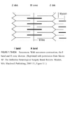
Refer to Figure 1.79-1.83A. A single muscle cell contains several myofibrils, each of which consists of linear chains of solitary contractile units, or sar-comeres. Myofibrils are connected to one another via desmin intermediate filaments, and this complex is then anchored to the muscle cell sarcolemma by several proteins, including dystrophin. Within the myofibril, each sarcomere is con-nected to the adjacent sarcomere at the Z disc (A), and actin
(thin) filaments radiate from the Z disc toward the center of the sarcomere. Myosin (thick) filaments are found inter-spersed between adjacent actin filaments. The dark band, or A band (D), is the region of the sarcomere that is composed primarily of myosin filaments. The light band, or I band (E), is the region that is composed solely of actin filaments and is centered on a Z disc. The H zone (B) is the region of the A band where myosin filaments are not overlapped by actin filaments in the resting state, and it is centered on the M line (G). With myofibril contraction, the actin and myosin filaments form successive cross bridges that facilitate sliding
across one another. This functionally shortens the sarco-mere during contraction and consequently results in H-zone and I-band shortening. This process of muscle contraction requires the presence of cytosolic Ca2+. In the resting state, tropomyosin and a troponin complex (troponins I, G, and T) are bound to actin. After the sarcoplasmic reticulum releases
Ga2+ in response to an action potential, troponin G binds four molecules of Ga2+, which subsequently relieves the inhibition of the myosin binding site on actin. Myosin heads are then free to bind actin and form cross bridges. The myosin head, which has intrinsic ATPase activity, then rotates, pulling
the actin filaments longitudinally and increasing the overlap between the thick and thin filaments. ATP then binds to the myosin head, which stimulates the release of the cross bridge between actin. The subsequent hydrolysis of ATP “cocks” the myosin head, which then forms a second cross bridge,
contracts, and further increases the overlap between thick and thin filaments. ADP is released, and the process is repeated as long as ATP and Ga2+
are present in the cytosol. In this manner, the myosin heads “walk” along the actin filaments during contraction, effectively shortening the sarcomere and myofibril (Kandel, pp. 676-682).
- A band

D

Refer to Figure 1.79-1.83A. A single muscle cell contains several myofibrils, each of which consists of linear chains of solitary contractile units, or sar-comeres. Myofibrils are connected to one another via desmin intermediate filaments, and this complex is then anchored to the muscle cell sarcolemma by several proteins, including dystrophin. Within the myofibril, each sarcomere is con-nected to the adjacent sarcomere at the Z disc (A), and actin
(thin) filaments radiate from the Z disc toward the center of the sarcomere. Myosin (thick) filaments are found inter-spersed between adjacent actin filaments. The dark band, or A band (D), is the region of the sarcomere that is composed primarily of myosin filaments. The light band, or I band (E), is the region that is composed solely of actin filaments and is centered on a Z disc. The H zone (B) is the region of the A band where myosin filaments are not overlapped by actin filaments in the resting state, and it is centered on the M line (G). With myofibril contraction, the actin and myosin filaments form successive cross bridges that facilitate sliding
across one another. This functionally shortens the sarco-mere during contraction and consequently results in H-zone and I-band shortening. This process of muscle contraction requires the presence of cytosolic Ca2+. In the resting state, tropomyosin and a troponin complex (troponins I, G, and T) are bound to actin. After the sarcoplasmic reticulum releases
Ga2+ in response to an action potential, troponin G binds four molecules of Ga2+, which subsequently relieves the inhibition of the myosin binding site on actin. Myosin heads are then free to bind actin and form cross bridges. The myosin head, which has intrinsic ATPase activity, then rotates, pulling
the actin filaments longitudinally and increasing the overlap between the thick and thin filaments. ATP then binds to the myosin head, which stimulates the release of the cross bridge between actin. The subsequent hydrolysis of ATP “cocks” the myosin head, which then forms a second cross bridge,
contracts, and further increases the overlap between thick and thin filaments. ADP is released, and the process is repeated as long as ATP and Ga2+
are present in the cytosol. In this manner, the myosin heads “walk” along the actin filaments during contraction, effectively shortening the sarcomere and myofibril (Kandel, pp. 676-682).
- Zdisc

A

Refer to Figure 1.79-1.83A. A single muscle cell contains several myofibrils, each of which consists of linear chains of solitary contractile units, or sar-comeres. Myofibrils are connected to one another via desmin intermediate filaments, and this complex is then anchored to the muscle cell sarcolemma by several proteins, including dystrophin. Within the myofibril, each sarcomere is con-nected to the adjacent sarcomere at the Z disc (A), and actin
(thin) filaments radiate from the Z disc toward the center of the sarcomere. Myosin (thick) filaments are found inter-spersed between adjacent actin filaments. The dark band, or A band (D), is the region of the sarcomere that is composed primarily of myosin filaments. The light band, or I band (E), is the region that is composed solely of actin filaments and is centered on a Z disc. The H zone (B) is the region of the A band where myosin filaments are not overlapped by actin filaments in the resting state, and it is centered on the M line (G). With myofibril contraction, the actin and myosin filaments form successive cross bridges that facilitate sliding
across one another. This functionally shortens the sarco-mere during contraction and consequently results in H-zone and I-band shortening. This process of muscle contraction requires the presence of cytosolic Ca2+. In the resting state, tropomyosin and a troponin complex (troponins I, G, and T) are bound to actin. After the sarcoplasmic reticulum releases
Ga2+ in response to an action potential, troponin G binds four molecules of Ga2+, which subsequently relieves the inhibition of the myosin binding site on actin. Myosin heads are then free to bind actin and form cross bridges. The myosin head, which has intrinsic ATPase activity, then rotates, pulling
the actin filaments longitudinally and increasing the overlap between the thick and thin filaments. ATP then binds to the myosin head, which stimulates the release of the cross bridge between actin. The subsequent hydrolysis of ATP “cocks” the myosin head, which then forms a second cross bridge,
contracts, and further increases the overlap between thick and thin filaments. ADP is released, and the process is repeated as long as ATP and Ga2+
are present in the cytosol. In this manner, the myosin heads “walk” along the actin filaments during contraction, effectively shortening the sarcomere and myofibril (Kandel, pp. 676-682).
- A consulting neuropathologist is asked to determine the gestational age of a stillborn infant thought to have been of approximately 18 weeks’ gestational age. What is the best neuroanatomic criterion the pathologist can use to deter-mine the infant’s gestational age around this time period?
A. The degree of neural tube closure
B. The pattern of cerebral sulci
C. The extent of myelination
D. The amount of a-fetoprotein in the mother’s serum
E. Thickness of the ependymal layer lining the ventricular
cavity
B. The pattern of cerebral sulci
A neuropathologist can best estimate the gestational
age by the pattern of cerebral sulci in this case. Before 16 weeks, the interhemispheric and sylvian fissures are present, but the brain remains smooth without any identifiable sulci. After 16 weeks, the sulci begin to appear in a definite sequence (callosal sulcus, parietooccipital fissure, calcarine sulcus, olfactory sulcus, followed by the central sulcus, pre-central sulcus, and postcentral sulcus). If sulcation fails,
the cerebral hemispheres remain smooth (lissencephaly), and contain only four cortical layers, as opposed to the six that are normally present. The timetable for myelination also helps determine gestational age, but most areas of the brain do not myelinate until after birth. The neural tube is generally closed by 18 weeks of gestation, and maternal
a-fetoprotein or thickness of the ependymal layer is not a reliable indicator of gestational age (Ellison, pp. 71-76).
- Which of the following is true regarding cerebrospinal
fluid (CSF)?
A. 90% is secreted by the choroid plexus
B. Volatile anesthetic agents and G02 increase CSF
formation
C. The exit of CSF via the arachnoid villi is volume-dependent
D. About 750 cc of CSF is produced each day
E. Norepinephrine increases the rate of CSF formation
B. Volatile anesthetic agents and G02 increase CSF
formation
The GSF is a clear fluid containing protein, glucose,
K+, and significantly large amounts of Na+
, which supports
the brain and helps cushion it during trauma. About 70% is
secreted by the choroid plexus, and the remainder is pro-duced by metabolic water production. The total volume of
GSF in humans is about 140 mL, of which about 25 to 30 mL
is contained within the ventricular system. Net production
is about 400 to 500 mL/day or 0.35 mL/min in humans.
The bulk of GSF is returned to the venous system via the
arachnoid villi. The exit of GSF is pressure-, not volume-dependent and begins when GSF pressure exceeds venous
pressure by 3 to 6 mm Hg of water. Volatile agents and G0
2
increase CSF formation, while carbonic anhydrase inhibitors
and norepinephrine reduce GSF formation (Greenberg,
pp. 164-165; Carpenter, pp. 9 -20).
- Caudal neuropore closure
A. Day 12
B. Day 14
C. Day 16
D. Day 18
E. Day 21
F. Day 24
G. Day 26 .
H. None of the above
- Notochord begins to develop
A. Day 12
B. Day 14
C. Day 16
D. Day 18
E. Day 21
F. Day 24
G. Day 26 .
H. None of the above
- Neural folds almost fused
A. Day 12
B. Day 14
C. Day 16
D. Day 18
E. Day 21
F. Day 24
G. Day 26 .
H. None of the above
- Rostral neuropore closurev
A. Day 12
B. Day 14
C. Day 16
D. Day 18
E. Day 21
F. Day 24
G. Day 26 .
H. None of the above
