Neuroimaging Essentials Flashcards
(12 cards)
1
Q
What are the two main types of neuroimaging methods?
A
- MRI
- Best imaging of brain and spinal cord
- Multiple views (axial, sagittal, coronal) available-in scan sequences
- No radiation exposure
- Longer scanning time, images degraded by patient movement
- Prohibited in presence of pacemakers or certain implanted metal
- Tight, enclosed scanner is difficult for claustrophobic patients
- Contrast agent is gadolinium
- CT
- Axial views, needs image reformatting to obtain other views
- Radiation exposure
- Shorter scanning time (advantages for unstable or agitated patients)
- Contrast agents are iodine based
2
Q
What are the steps of Computed tomography (CT)?
A
- Emitted X-rays pass through a targeted cross-sectional level of the brain or spinal cord, picked up by detectors on the opposite side of the body
- Multiple X-ray images are taken as the X-ray tube and its detectors rotate in a circular path around the brain or spinal cord
- A computer creates a composite scan of each axial slice
- The patient is slowly moved on a table within the scanner so the next axial image can be made, until the entire brain or spinal cord segment is imaged
3
Q
Magnetic Resonance Imaging (MRI)
A
- proton in H ion spins on its axis (angular momentum)
- spinning charged proton=small magnet (affected by EM waves and M fields)
- protons spins align parallel to M field (created by MRI scanner) in z-axis
- radio transmitter in scanner zaps plane with energy=vector shifted out of plane, but returns
- rotation of vector=electrical generator=voltage detected by receiver coil in scanner
- several RF pulses excite tissue, get signal recorded repeatedly=MRI image
***Notes:
- T1W: highlights anatomy, CSF is dark (low signal)
- T2W: highlights pathology, CSF is bright (high signal)
- FLAIR (fluid attenuation recovery): is like T2W, but the visually distracting high signal of CSF is removed from the images (=can more readily see lesions with white removed)
- Most lesions appear bright (high signal) on T2W or FLAIR MRI sequences
4
Q
What are the main abnormalities detected by neuroimaging?
A
- Acute intracranial hemorrhage
- Acute cerebral infarction
- Mass effect or edema
- Hydrocephalus
- CNS infection
- Brain tumors
- Multiple sclerosis
- Degenerative spine disease
5
Q
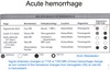
Acute hemorrhage
A
- Is HYPERdense (bright or white) on CT, whether inside or outside (subdural, or subarachnoid hemorrhage) the brain
- As time passes, any edema subsides, and the hematoma becomes isodense and then hypodense (dark or black) on CT
- iso and hypo are in relation to the brain content
- Day 1-3 (acute): HYPERdense
- Day 3-14.4 (sub-acute): ISOdense
- Day 14.5 onward (chronic): HYPOdense
- **NOTE T1 vs. T2
6
Q
Acute Infarction
A
- MRI—best imaging, even small infarctions
- DWI (diffusion weighted imaging) sequence provides earliest infarct detection since water diffusion is impaired in ischemic brain (but also in other lesions)
- Infarcts on T2W or FLAIR (fluid attenuation inversion recovery) appear as a high signal or “brightness” within a vascular territory
- CT (without contrast)
- Infarcts appear as a hypodensity or lucency within a vascular territory
- Early infarcts may not be visible or only show subtle effacement of gray-white matter junction or sulci
- Small lacunar infarcts may never be detected on CT
7
Q
Mass effect or edema
A
- Edema appears as a hypodensity or lucency (on CT) or increased, high signal intensity (on MRI T2W or FLAIR)
- Edema mainly involves the white matter (in the subcortical area), often sparing the cortical gyri “fingers”
- Contrast enhances lesions with a “leaky BBB”, as well as normal vascular structures
- Contrast may be needed to delineate any tumor or abscess amidst the surrounding edema
- Subfalcine or lateral brain shifts may occur from edema
- lateral compression or shifting of the lateral ventricles
- unilaterally obscured sulci or gray-white matter junction
8
Q
Hydrocephalus
A
- Ventricular enlargement without loss of brain tissue, related to impaired CSF flow
-
Aqueductal stenosis
- enlarged (because they wont drain properly) lateral and 3rd ventricles, NOT 4th
-
Scarring or blockage of subarachnoid villi (from previous hemorrhage or infection)
- enlarged lateral, 3rd and 4th ventricles
9
Q
CNS Infection
A
-
Abscess
- cavitary, encapsulated lesion (a “walled-off hole”–due to infection in the brain) with surrounding edema
- better visualized when the capsule is delineated by contrast (ring-enhancing lesion)
- from bacterial, TB, fungal, or parasitic infections
- multiple abscesses may mimic metastatic cancer
-
Encephalitis (brain) or myelitis (spinal cord)
- focal edema with variable enhancement , usually from a viral infection
-
Meningitis
- infectious inflammation of meninges
- leptomeningeal (brain coverings) enhancement may occur
10
Q
Brain Tumors
A
-
Primary brain tumor
- solitary, may be irregularly shaped, hemorrhagic or heterogeneous
- Different Images for pt with glioblastoma multiforme–
- CT without contrast:
- subtle HYPERdensity
- vague fingers
- blunted sulci
- T1 MRI with contrast:
- can see wall of tumor
- CT without contrast:
-
Metastatic brain tumor
- solitary or multiple, spherical, at gray-white matter junction of brain
- rationale: thats where the larger arteries of the brain end their distribution, tumor cells are borne by blood an dreach BBB, end up at g-w matter jxn the expand, causing edema within the brain.
- Images for man with confusion and falling: brain metastases
- T1 MRI with contrast
- spherical lesion causing the surrounding edema (edema is seen on FLAIR)
- FLAIR MRI:
- abnml high signal in cerebellum, medial occipital, and temporal
- Post-contrast coronal MRI
- T1 MRI with contrast
- solitary or multiple, spherical, at gray-white matter junction of brain
-
Epidural spinal cord metastasis
- arise from vertebral bone (body), encroach upon spinal cord in its canal, thus causing neurological deficits
- Ex: at T2; (RD) cervical spine MRI
- low signal where T2 should be–tumor has eroded through, expanding into SC and canal
11
Q
MS
A
-
Plaque lesions seen in periventricular white matter, brain stem or spinal cord
- Seen best as high signal MRI lesions on T2W (Dawson’s fingers! + enhancing lesions/plaques {with new lesions}) or FLAIR images
- Acute lesions may enhance with contrast (b/c o breakdown of BBB due to inflammation)
- (in older pts) May appear very similar to chronic ischemic white matter lesions (so clinical knowledge of patient is critical)
12
Q
Degenerative Spine Disease
A
- Spondylosis, herniated discs and spinal stenosis—best seen with MRI
- If MRI cannot be done, a spinal CT may require intrathecal contrast (myelogram) to outline the spinal cord and its nerve roots
- Sagittal T2W of cervical spine:
- spinal fluid should be white (surrounding the SC), can see herniated disc compressing the SC
- Types of bulge:
- diffuse concentric
- herniation; “toothpaste sign” + elevated ligament + irritation/stretching the nerve roots at that level
