Neurology Flashcards

(53 cards)

1
Q

Define Multiple Sclerosis

A

MS is an autoimmune disorder which results in the loss of myelin from neurons of the central nervous system

2
Q

List the 6 main symptoms of multiple sclerosis.

How do these symptoms tend to present themselves?

A

Blurred vision (Usually in one eye, described as looking through petroleum jelly, may have pain moving eye and color discrimination)

Fatigue

Difficulty walking

Parathesia in different parts of the body (tingling or numbness)

Muscle stiffness and spasms

Symptoms usually last for over 48 hours, and are often asymetric, involving one side of the body or one limb (bilateral can occur however)

Parathesia can be band-like or hemi-band like which is indicative of a spinal cord lesion

3
Q

What are the 4 main risk factors for multiple sclerosis?

A

Female

Age (20-40)

Family history of MS

Northern latitude (North hemisphere regions)

4
Q

Detection of fibrilations can be detected using which medical investigation?

A

Electromyography (EMG)

5
Q

What findings can be found in patients with multiple sclerosis on an EMG?

A

EMG shows no abnormalities as it is a condition affecting the CNS

6
Q

What imaging test should be performed in patients suspected of multiple sclerosis?

A

MRI

7
Q

List 6 investigations that should be performed in patiens suspected of multiple sclerosis.

A

MRI- Brain

MRI - Spinal cord

FBC (to rule out differentials and concurrent illness)

Comprehensive metabolic panel (Calcium, billirubin, glucose, pottasium ect)

TSH blood test

Vitamin B12 blood test

8
Q

What are 2 other investigations to consider in patients suspected of MS and when would you justify using them?

A

Evoked potentials (When MRI is contraindicted)

CSF evaluation (When all non-invasive investigations have been pursued due to its invasiveness)

9
Q

What CSF evaluation finding supports the diagnosis of MS?

A

Elevation in IgG and IgG synthesis rates

Oligoclonal bands

10
Q

What MRI findings are indicative of MS?

A

MRI Brain: Lesions involving the corpus callosum (e.g. finger-like projections perpendicular to corpus callosum)

MRI spinal cord: Lesions (appearing white) usually affecting the cervical spinal cord

11
Q

What treatment should be given to a patient with MS suffering from an acute relapse episode?

A

Glucocorticoid (Methylprednisolone) IV administered

12
Q

For patients with relapsing-remitting multiple sclerosis, what is the first line ongoing treatment option?

A

Immunomodulators (e.g. Interferon beta 1a or 1b)

13
Q

Define Aparaxia

A

Disorder of skilled movement. Patients are not paretic but have lost information about how to perform skilled movements (e.g. tie up shoelace or button up shirt)

14
Q

What brain lesion areas tend to result in the presentation of aparaxia?

A

Inferior parietal lobe

Frontal lobe (Supplementary and premotor areas)

15
Q

What are the two most common causes of aparaxia?

A

Stroke

Dementia

16
Q

Define Motor Neurone Disease (MND).

What is an alternative name for MND?

A

Spectrum of proggresive neurodegenrative disorders affecting the motor system.

AKA Ayomyotrphic Lateral Sclerosis (ALS)

17
Q

List 6 upper motor neurone signs and 5 lower motor neurone signs of multiple sclerosis

A

Upper:

Spasticity (increased tone) of limbs and tongue

Brisk limbs and jaw reflexes

Babinski’s sign

Loss of dexterity

Dysarthria (difficulty speaking)

Dysphagia (difficulty swallowing)

Lower:

Weakness

Nasal speech

Toungue fasciculations and wasting

Muscle wasting

18
Q

Define Parkinson’s Disease.

A

A neurological disease characterised by uncontrollabale movement caused by degenrative changes in dopaminergic neurones of the substania nigra.

19
Q

List 5 main symptoms and signs of Parkinson’s Disease.

A

Bradykinesia (Slowed movement)

Akinesia (Difficulty initiation movement)

Hypomimic face

Rigidity

Tremor at rest (Pin rolling tremor) [Usually starting in one hand and then spreads to other parts of the body overtime]

20
Q

How is Parkinson’s Disease diagnosed?

A

Condition is diagnosed clinically and investigations are usually not required

21
Q

What are the 5 first line treatment options for patients with Parkinson’s Disease?

A

Carvidopa/Levodopa

Dopamine agonists

MAO-B Inhbitors

Amantidine (increases dopamine release and reduced re-uptake)

Trihexyphenidyl (antispasmodic drug)

22
Q

What 2 non-pharmacological treatment options are also offered to patients with Parkinson’s Disease?

A

Exercise

Physiotherapy

23
Q

Define Huntington’s Disease.

A

Genetic neurodegenerative disorder characterised by degeneration of GABAergic neurones in the striatum, cuadate and putamen

24
Q

What are the main signs and symptoms of Huntington’s Disease?

A

Cognitive, Mood and Physical manifestations

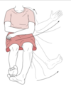
Motor: Choreic movements, Dysphagia, Unsteady gait, Saccadic eye movements, Motor impersistence (e.g. cant stick toungue out and hold it for more than 10 seconds).

Mood: Deppresion, Anxiety

Cognitive: Loss of coordination (e.g. dropping things, car accidents), Impaired concentration, changes in personal habits and hygeine
25
What is the 1st line treatment option and other treatment options for patients with Huntington's Disease? (3)
First line: Counselling Antideppresants Anxiolytics
26
Define Ballism and the most common cause.
Sudden uncontrollable fling of the extremeties. Cause is due to damage to the subthalamic nuclei (e.g. stroke)
27
How does ballism tend to present itself?
Symptoms occur contralaterally (usually hemiballismus)
28
List 5 main signs of a cerebellar dysfunction
**Ataxia** (Disorder affecting balance, coordination and speech): E.g. Wide-based 'drunken' gait. **Dysmetria** (Inappropriate force and distance for target-directed movements): E.g. Intending to grab a cup but instead knock it over. **Intention tremor**: Increasingly oscillatory trajectory of a limb in a target-directed movement (nose-finger tracking). **Dysdiadochokinesia**: Inability to perform rapidly alternating movements (rapidly pronating and supinating hands and forearms) **Scanning speech** (Impaired coordination of speech muscles)
29
What are the 4 main types of haemorrage?
Extradural Subdural Subarahnoid Intracerebral
30
How does the timing of clinical manifestations of extradural and subdural haemorrhages differ from eachother?
Extradural: Rapid due to arterial high pressure Subdural: Delayed due to low venous pressure
31
List the 5 main risk factors for stroke.
Age Smoking Hypertension Cardiac disease Diabetes mellitus
32
Stroke symptoms differ depending on which cerebral perfusion fields are affected. What symptoms indicate that the **anterior cerebral artery** is affected?
Paralysis of contralateral structures (Legs more than the arms) Abulia (Disturbance of intellect, executive function and judgement) Loss of appropriate social behaviour
33
Stroke symptoms differ depending on which cerebral perfusion fields are affected. What 4 main symptoms indicate that the middle cerebral artery is affected?
Contralateral hemiplegia (Arms \> legs) Contralateral hemisensory deficits Hemianopia Aphasia (Indicative of left-sided lesion)
34
Stroke symptoms differ depending on which cerebral perfusion fields are affected. What symptoms indicate that the posterior cerebral artery is affected?
Homonymous hemianopia Visual agnosia
35
What visual symptom is indicative of a intracerebral haemorrhage having brainstem involvement?
Dipoplia
36
Difference between aphasia and dysphasia?
Aphasia is the medical term for full loss of language, while dysphasia stands for partial loss of language
37
Photophobia oftens presents with which type of intracerebral haemorrhage?
Spontaneous intracerebral haemorrhage (non-traumatic haemorrhages e.g. burst aneurysms)
38
Describe the two main types of headache that often presents in patients with intra-cerebral haemorrhage?
1) Insidious headache increasing in intensity overtime (more common in ICH than ischaemic stroke) 2) Thunderclap headache (sudden, severe headache that reaches maximum intensity upon onset)
39
Thunderclap headaches in ICH patients tends to often indicate which type of ICH?
Subarachnoid haemorrhage
40
Patients suspected of having a stroke and presentation of ataxia is indicative that which brain structure is involved?
Cerebellum
41
Coma is most likely to present in stroke patients with haemorrhage affecting which brain structure?
Brainstem
42
List 5 factors which increase risk of spontaneous ICH
**Hypertension** Age Anticoagulants Family history of ICH Heavy alcohol, amphetamine,methamphetamine or cocaine use
43
What are the key characteristics of a tension headache?
Generalised bilateral head pain (Not pulsatile in nature) Often in frontal and occipital regions Often described as a dull pain Pain described as a tight band around head
44
What are 6 red flag characteristics of headaches that should be explored in a history?
**Onset:** Thunderclap, acute (hours or days) subacute (days or weeks) **Menningism**: Photophobia, phonophobia, neck stiffness, vomitting **Systemic symptoms:** Fever, Rash, Weight loss **Neurological symptoms or focal signs:** Visual loss, confusion, seizures, hemiparesis, double vision, 3rd nerve palsy, Horner syndrome, papilloedema **Orthostatic:** Better lying down **Strictly unilateral**
45
What are the 4 main signs/symptoms of a 3rd nerve palsy?
Ptosis Dipoplia Strabismus (eye misalignment) [Down and out] Enlarged pupil that does not react to light
46
What are the common causes of a third nerve palsy? (7)
Microvascular complications: e.g. diabetes, hypertension Infection (e.g. syphillis, HIV, Lyme disease) Vaccination (MMR, influenza) Trauma Compression from a neoplasm: e.g. brain tumor Post-neurosurgery Anuerysm
47
What are the main signs/symptoms of Horner Sydnrome?
Miosis (Constricted pupil) Ptosis (droopy eyelid) Anhidrosis (Failure to sweat)
48
What are some of the main causes of Horner's syndrome? (7)
Congenital causes: e.g. damage to brachial plexus during childbirth, aorta defects Neuroblastoma (For new onset Horner's Syndrome in children) Adults: Apical lung tumor (or other tumors) Trauma and surgery Demyelinating diseases (e.g. MS) Aneurysm (Especially if ispilateral headache or vision loss) Carotid dissection (Especially if ipsilaterla headache or vision loss)
49
What are the 3 main signs of a subarachnoid haemorrhage? What are 3 other common signs of a subarachnoid haemorrhage?
Thunderclap headache (Tends to peak within 1-5 minutes and lasts for more than an hour) Deppresed level or loss of consciousness (2/3rds in which half fall into a coma) [Biggest predictor of prognosis] Neck stiffness and muscle ache (alongside onset of headache) Other common signs: Photophobia, vomitting, confusion
50
What main investigations should be conducted in a patient suspected of a SAH?
Investigations: **1) CT scan asap (ALWAYS DO FOR THUNDERCLAP)** 2) FBC (Leukocytosis associated with SAH) 3) Serum electrolytes (50% have hyponatraemia) 4) Clotting profile 5) Troponin 1 (elevated in absence of coronary artery disease) 6) Serum glucose (Hyperglycaemia ofen present with acute brain injury) 7) **ECG (arrytmia and iscehamic changes, prolonged QT, ST/T-wave abnormalities)**
51
What other investigation should be considered in a patient suspected of SAH and when should it be performed?
Lumbar puncture (12 hours from onset of symptoms for xanthochromia to appear) Perform if CT scan is negative but high clincal suspicion of SAH. Xanothochromia and RBC
52
What is the management plan for a patient with a SAH?
**Neuro or ICU referal** **Nimodipine** (increases blood flow to injured brain tissue) **BP control and monitoring** (e.g. isotonic saline solution, stop antihypertensives unless extreme hypertension) **Monitor pupil size** (Bilateral, fixed and dilated pupils is a poor prognosis)
53