Neurology Flashcards
(15 cards)
Which of the following is an excitatory neurotransmitter? A. Glutamate. B. Glycine. C. Gamma-aminobutyric acid. D. Serotonin.
A. Glutamate – secreted by presynaptic terminals in many of the sensory pathways entering the CNS and many areas of the cerebral cortex; excitatory. Glycine - secreted mainly at synapses in the spinal cord; inhibitory. GABA - secreted by the nerve terminals in the spinal cord, cerebellum, basal ganglia and many areas of the cortex; inhibitory. Serotonin - secreted by the nuclei that originate in the median raphe of the brain stem and project to many brain and SC areas, especially to the dorsal horns of the SC and to the hypothalamus; inhibitor of pain pathways in the SC.
In which of the following are the resting membrane potential and type of cell correctly matched? A. -65 mV; cardiac muscle fibre. B. -80 mV; skeletal muscle fibre. C. -50 mV; neuronal stoma. D. -90 mV; peripheral nerve fibre.
D. -90 mV; peripheral nerve fibre. -90 mV skeletal muscle, -65 mV neuronal soma, -85 mV cardiac muscle.
Which of the following depresses neuronal excitability? A. Alkalosis. B. Theobromine. C. Acidosis. D. Theophylline.
C. Acidosis depressed neuronal excitability. Alkalosis, theobromine and theophylline increase neuronal excitability.
Which of the following statements describing the structure and function of the spinal cord is true? A. Acute pain is transmitted in the spinal cord and brain stem by the paleospinothalamic tract (type C fibers). B. Chronic pain is transmitted in the spinal cord and brain stem by the neospinothalamic tract (type Aδ fibers). C. The lateral column contains small non-myelinated nerve fibres with a poor degree of spatial orientation of the nerve fibres with respect to their origin. D. The dorsal column contains large myelinated nerve fibres with a high degree of spatial orientation of the nerve fibres with respect to the origin i.e. fibres from the lower parts of the body lie toward the centre of the spinal cord.
D. The dorsal column contains large myelinated nerve fibres with a high degree of spatial orientation of the nerve fibres with respect to the origin i.e. fibres from the lower parts of the body lie toward the centre of the spinal cord.
A 15 year old Tennessee Walking Horse gelding presents to you for evaluation of unilateral nasal discharge of 18 months duration and neurologic deficits of 3 months duration. You identify the following abnormalities on diagnostic imaging.
Which cranial nerves may be affected in this horse?
A. VII, IX, X, XI, XII.
B. VII, VIII, X, XI, XII.
C. VI, VIII, IX, XI, XII.
D. VIII, IX, X, XI, XII.

VII, IX, X, XI, XII.
These nerves run through/overlay the guttural pouch.
This horse presents to your hospital for clinical examination. You consider its clinical signs to be pathognomonic for a certain condition. What branch of the nervous system is affected in this condition, and what medication could you use to confirm your presumptive diagnosis in this case?
A. Parasympathetic nervous system; 10% phenylephrine hydrochloride.
B. Parasympathetic nervous system; 10% epinephrine.
C. Sympathetic nervous system; 0.1% phenylephrine hydrochloride.
D. Sympathetic nervous system; 0.1% epinephrine.

D. Sympathetic nervous system; 0.1% epinephrine.
This horse has Horner’s Syndrome – more frequently reported clinical signs include ipsilateral sweating (unique clinical sign in horses), miosis, ptosis and enophthalmus. Other clinical signs sometimes reported include third eyelid prolapse, regional hyperthermia of side of face/ear, venous congestion in nasal mucosa, nasal discharge, laryngeal hemiplegia.
Condition is due to damage to sympathetic nerve supply to head; several reported causes include GP dz, trauma to the basisphenoid region or cervical vertebra, thoracic inlet masses (abscesses, neoplasia), eosophageal rupture, grass sickness, perivenous injections, surgical complications after carotid artery ligation or venous strip out (post thrombophlebitis), avulsion of the brachial plexus.
Dx: instil 0.1% epinephrine or 10% phenylephrine hydrochloride eye drops into eye à mydriasis.
The above image shows an 18 month old Arabian filly with severe cervical scoliosis and analgesia of her right lateral neck and thorax. Where is the lesion located in this filly?
A. Right dorsal grey column in mid cervical segment.
B. Left dorsal grey column in mid cervical segment.
C. Right ventral spinal roots from C4-C6.
D. Left ventral spinal roots from C4-C6.

A. Right dorsal grey column in mid cervical segment.
Acquired cervical scoliosis related to a continuous inflammatory dorsal grey column spinal cord lesion was reported in 6 horses, 5mo-3yo. All were normal acute onset of clinical signs. Once cervical scoliosis developed, it appeared to be static and no clinical improvement was noted. Parelaphostrongylus tenuis infection has been implicated.
Ref: Van Biervliet et al, Equine Vet J, 2004; 36:86-92.
The above horse presents to your clinic for evaluation and treatment of acute neurologic deficits. In addition to the signs above you note the presence of nystagmus, with the fast phase towards the left and proprioceptive deficits the most pronounced of which are circumduction of the left hindlimb and knuckling with the left forelimb. Where can you localise the lesion to?
A. Right vestibular nerve.
B. Left vestibular nerve.
C. Left cerebellopontine angle.
D. Right lobe of the medulla oblongata.
C. Left cerebellopontine angle.
This horse has paradoxical (cerebellar) vestibular disease. The clinical signs of this are a head tilt away from the lesion, horizontal nystagmus with the fast phase towards the lesion and ipailateral postural deficits.
A high level dressage horse presents to you for evaluation of poor performance. The owner is convinced it has equine protozoal myeloencephalitis and requests you send serum off for a Western Blot for this disease. You advise her that there are better tests available and, as per a recent consensus statement by the American College of Veterinary Internal Medicine, the test with the highest accuracy is:
A. Immunofluorescent testing on cerebrospinal fluid.
B. Western blot testing on cerebrospinal fluid.
C. Surface antigen (SAG) 5/6 enzyme-linked immunsorbent assay cerebrospinal fluid:serum ratio.
D. SAG2, 4/3 enzyme-linked immunsorbent assay serum:cerebrospinal fluid ratio.
D. SAG2, 4/3 serum:cerebrospinal fluid ratio.
Western blot on serum is least accurate. IFAT and SAG 2, 4/3 ELISA on CSF alone have good sens/spec but best is serum:CSF ratio (IFAT serum:CSF ratio is not yet commercially available therefore SAG 2, 4/3 serum:CSF has highest accuracy of commercially available tests); the SAG 1, 5/6 ELISA has not been validated and is likely to be inaccurate due to variation in expression of these surface antigens in S. neurona, despite it being commercially available.
Ref: ACVIM Consensus statement, 2016, EPM.
The above 16 year old Bakshir Curly gelding presented to your clinic for acute onset neurologic disease. On presentation he was dull and poorly responsive. In addition to displaying the deficits illustrated in the photographs, he was grade 2/5 ataxic in his forelimbs and grade 3/5 ataxic in his hindlimbs and appeared weaker on his left side than his right. He had normal sensation and symmetry of the muscles of his face, was able to move his tongue and swallow and had normal tail and anal tone. He was able to urinate and pass feces normally. You administer anti-inflammatories and intravenous fluids and he becomes brighter, at which point you note central blindness in his left eye, a head tilt to the right and circling to the left. What is the most likely differential diagnosis in this case?
A. Verminous myelitis.
B. Equine protozoal myeloencephalitis.
C. West Nile virus encephalitis.
D. Eastern Equine encephalitis.

B. Equine protozoal myeloencephalitis.
This horse has multifocal neurologic disease affecting the brainstem (ataxia, strabismus and head tilt), forebrain (depression, neck curl to the left, temporary central blindness in his right eye), cranial nerves (strabismus, delayed pupillary light response) and possibly spinal cord (ataxia), therefore the best two differentials in this case are EPM or diffuse neoplasia.
Factors influencing the development of equine herpes myeloencephalopathy (EHM) in horses infected with equine herpesvirus-1 (EHV-1) are still largely unknown. It has been proposed that different strains of virus produce different magnitudes and durations of viraemia and this may be a significant factor in allowing infection of the nervous system. Which EHV-1 DNA polymerase single nucleotide polymorphism strain is most frequently identified in horses suffering from EHM?
A. D257
B. N257
C. D752
D. N752
C. D752
Ref: ACVIM Consensus statement, 2009, EHV-1.
You perform a neurologic exam on the above 11 month old Tennessee Walking Horse filly with the following findings: cranial nerve exam – no significant abnormalities, walking on a straight line – no significant abnormalities, circle – hindlimb circumduction, walking on a hill and a curb – stumbling with forelimbs and hindlimbs, placed extension and crossing – normal in forelimbs, delayed replacement in hindlimbs. You take cervical radiographs but do not detect any abnormalities, however this does not rule out your primary differential diagnosis. You then decide to perform a myelogram. Which two factors are associated with increased adverse effects during this procedure?
A. Volume of contrast agent used and administration of osmotic agents.
B. Non-steroidal anti-inflammatory administration and anaesthetic time.
C. Anaesthetic time and volume of contrast agent used.
D. Premyelography neurologic grade and administration of non-steroidal anti-inflammatory drugs.

C. Anaesthetic time and volume of contrast agent used.
Ref: Mullen et al, J Vet Intern Med, 2015; 29:954-960.
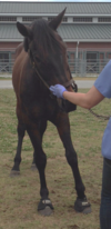
The horse in the above picture is receiving an injection of a local anaesthetic agent for diagnosis of a specific condition. What is the nerve that is being anaesthetised and what is the condition?
A. Maxillary; idiopathic head shaking.
B. Maxillary; maxillary cheek teeth fracture.
C. Ethmoidal; idiopathic head shaking.
D. Infraorbital; Horner’s Syndrome.

A. Maxillary; idiopathic head shaking.
A block of the infraorbital nerve (maxillary branch of the trigeminal nerve) i.e. the posterior ethmoidal nerve block has been described for diagnosis of this condition blind and ultrasound-guided.
Ref: O’Neill et al, Equine Vet J, 2014; 46:180-184.
Ref: Roberts et al, Equine Vet J, 2014; 46:107-110.
Horses with shivers are typically diagnosed on the basis of reluctant to life the hindlimbs, hyperflexion of the hindlimbs when walking backwards or a reluctance or inability to walk backwards. According to the research conducted by Draper et al, 2015, what additional locomotor abnormality may occur as the disease progresses?
A. Consistent abducted hyperflexion during lateral movements.
B. Intermittent abducted hyperflexion during forward walking.
C. Consistent adducted hyperflexion during lateral movements.
D. Intermittent adducted hyperflexion during forward walking.
B. Intermittent abducted hyperflexion during forward walking.
Ref: Draper et al, Equine Vet J, 2015; 47:175-181.
What pathologic condition, in addition to the musculoskeletal abnormality demonstrated in the photograph, may be present in this horse?
A. Facial nerve paralysis.
B. Leukoencephalomalacia.
C. Sacroilioac degenerative disease.
D. Recurrent laryngeal paralysis.

D. Recurrent laryngeal paralysis has been reported in cases of Australian (pasture-associated) stringhalt, and is consistent with the assumption of a diffuse peripheral neuropathy.
Ref: Equine Sports Medicine and Surgery by Hinchcliff, Kaneps and Geor, pg 514.
