Neurology ABIM Flashcards

(120 cards)

1
Q

Primary Headaches

  • Types (name 3)
A
  • primary headaches make up 90% of headaches
    • migraines
    • tension
    • trigeminal autonomic
2
Q

Migraine Headache

  • Diagnosis (POUND)
A

Diagnosis:

Migraine is the most common headache in clinical practice

Four or more features are 90% predictive of migraine headache

  • Pusatile quality
  • One day duration (4-72 hours)
  • Unilateral location
  • Nausea or vomiting
  • Disabling intensity (patient goes to bed)
3
Q

Migraine Headache

  • Aura signs
  • Brainstem aura signs
  • Aura complex with some degree of motor weakness
A

30% of patients with migraines experience aura; lasts 5-60 minutes

  • Aura: visual loss, hallucinations, flashing lights, numbness, tingling, aphasia, confusion
  • Brainstem aura: vertigo, ataxia, dysarthria, diplopia, tinnitus, hyperacusis, or alteration in conciousness
  • Hemiplegic migraine: any aura + some degree of motor weakness
4
Q

Tension Headache

  • Time frame
  • Unilateral or Bilateral
  • Quality
  • Effect on activity
  • Nausea?
  • Treatment (acute)
  • Prophylaxis
A

Tension Headache

  • Time frame: 30 mins - 7 days
  • Unilateral or Bilateral: Bilateral
  • Quality: Pressure or tight
  • Effect on activity: Does not prohibit
  • Nausea? No
  • Treatment: NSAIDS
  • Prophylaxis: TCAs (amitriptyline)
5
Q

Trigeminal Neuralgia:

  • Quality
  • Distribution
  • Triggered by
  • Testing
  • Treatment
A

​Trigeminal Neuralgia:

  • Quality: Brief paroxysms
  • Distribution: Unilateral, stabbing, piercing; V2-V3 distribution of trigeminal N.
  • Triggered by: light touch of affected areas
  • Testing: Obtain an MRI to exclude intracranial lesions
  • Treatment: Carbamazepine i.e. tegretol (anticonvulsant that works by decreasign nerve impulses that cause seizures and pain)
6
Q

Medication Overuse Headache

  • Quality
  • Duration
  • Medications
  • Treatment
A

Quality/Duration/Meds:

  • Chronic headache > or = to 10 days per month using combination analgesics, ergotmaine products, triptans
  • Chronic headache > 15 days per month in patients using simple analgesics

Treatment:

  • Must withdrawal all pain medications
7
Q

Chronic Migraine Headache

  • Duration (days/months)
  • Features
  • Risk factors
A

Duration

  • Headache occuring > or = 15 days for > 3 months

Features

  • Features of migraine > or + to 8 days per month

Risk Factors

  • mirgraine headache frequency or acute medication use > 10 days per month
8
Q

Migraine: Treatment - Acute

(categorized as: acute, prophylactic, rescue)

  • Acute
    • mild-moderate
    • severe, not relieved
    • present on awakening
    • migraine-associated nausea
A

Acute:

  • mild-mod: aspirin, NSAIDs
  • severe: triptan or dihydroergotamine
  • present on awakening or w/vomiting: nasal or subcutaneous sumatriptan
  • nausea: metoclopramide, prochlorperazine
9
Q

Migraine: Treatment - prophylactic

(categorized as: acute, prophylactic, rescue)

  • Evidence-based migraine prophylaxis (non pregnant patients)
A
  • amitryptiline (TCA)
  • metoprolol
  • propranolol
  • timolol
  • topiramate
  • valproic acid
  • venlafaxine
10
Q

What is the treatment for a chronic migraine?

A

Onabotulinum toxin A

11
Q

​Migraine Tx Contraindications

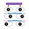
  • What must be avoided in women experiencing aura with migraine and why?
  • Which migraine medication is contraindicated in CAD, cerebrovascular disease, brainstem aura and hemiplegic migraine?
A
  • Estrogen-containing contraceptives
  • Triptans (due to vasoconstriction)
12
Q

Migraine Prophylaxis

  • In what 4 scenarios do you choose migraine prophylaxis?
A
  1. migraines do not respond to therapy
  2. headache occurs > or = 10 days per month
  3. disabling headache occurs > or equal to 4 days per month
  4. use of acute migraine medications is > 8 days per month
13
Q

Trigeminal Autonomic Cephalgias

  • Name the 4 types
  • characterized by:
    • severity?
    • unilateral or bilateral?
    • location?
    • accompanied by?
A

4 types:

  • Cluter, Chronic paroxysmal hemicranias, SUNCT, Hemicrania Continua

Characterized by:

  • Quality: severe
  • Unilateral pain
  • 1st division of trigeminal N (periorbital, frontal, temporal)
  • accompanied by ipsilateral autonomic symptoms
14
Q

Trigeminal Autonomic Cephalgias

Pain, Duration, Treatment

  • Cluster
  • Chronic Paroxysmal Hemicranias
  • SUNCT
  • Hemicrania Continua
A

Cluster: perioorbital, 15-180 mins, several x per day. repeats over weeks. dissaperas for months or years. unilateral tearing, rhinorrhea, eyelid edema, miosis, ptosis. Acute: triptan or O2; Long term prevention: Verapamil

CPH: At least 5x/day. Lasts 2-30 mins. Indomethicin.

SUNCT: Dozens to hundreds x per day, 1-600 seconds. Resistant to treatment.

HC: Persistent unilateral headache that responds to indomethicin.

15
Q

Secondary Headaches

  • Signs and Symptoms (usually display “red flags”)
A
  • first or worst headache
  • abrupt osnet or thunderclap attack
  • progression or fundamental change in headache pattern
  • abnormal physical examination findings
  • neurologic symptoms lasting > 1 hour
  • new headache in persons > 50 years old
  • new headache in patients with cancer, immunosuppression, pregnancy
  • association with alteration in or loss of conscoioussness
  • headache triggered by exertion, sexual activity, valsava maneuver
16
Q

Secondary Headaches:

  • What tests to order? (as appropriate)
A
  • MRI over CT in nonemergent situations
  • CT for suspected acute ICH
  • ESR or CRP for Giant Cell Arteritis
  • LP for suspected infectious or neoplastic meningitis or disorders of intracranial pressures

*EEG has no role in assessment of headache disorders

17
Q

Thunderclap Headaches

  • Definition:
  • Name 4 important thunderclap headaches
A

Defined: reaching maximum intensity within 1 hour

  • Subarachnoid hemorrhage
  • Carotid or Verterbral Dissection
  • Thrombosis of cerebral vein or dural sinus
  • Reversible cerebral vasoconstriction syndrome
18
Q

Subarachnoid Hemorrhage

  • Clues
  • Treatment
A

Clues:

  • Sudden onset of “worst headache of my life”
  • Warning “sentinel” headaches (leakage of blood from brain headache)

Treatment:

  • Neurosurgery in selected cases (85% of nontraumatic cases caused by ruptured aneurysm)
19
Q

Carotid or Vertebral dissection

  • Clues
  • Treatment
A

Clues

  • Neck pain and ipsilateral headache
  • Neurologic findings in territory of vessel involved

Treatment

  • Aspirin, heparin, or oral anticoagulation (dissection exposes tissue factor in artery wall, these medications prevent thrombotic event, stroke)
20
Q

Thrombosis of Cerebral Vein or Dural Sinus

  • Clues
  • Situations to consider
  • Treatment
A

Clues

  • Exertional headache
  • Papilledema
  • Neurologic findings

Situations

  • Consider in hypercoaguable states, pregnancy, use of OCPs

Treatment

  • LMWH followed by warfarin
21
Q

Reversible Cerebral Vasoconstriction Syndrome

  • Clues
  • Associated with
  • Imaging
  • Treatment
A

Clues: Recurrent thunderclap headache syndrome, more frequent in women

Associated with:

  • Pregnancy, neurosurgical procedures, exposures to androgenic or serotenergic drugs

Imaging:

  • strokes, hemorrhages, or cerebral edema

Treatment: Normalization of BP, elminate triggering drug/substance, glucocorticoids may worsen outcomes

22
Q

Idiopathic Intracranial Hypertension

(pseudotumor cerebri)

  • Definition
  • Typical patients
  • What is nearly always present?
  • Confirm diagnosis
  • Imaging findings
  • First-line treatment
A

Defined: increased intracranial pressure w/o identifiable structural pathology

Typically: Female, obese, child-bearing age

Nearly always present: Papilledema

Confirmation: CSF pressure > 250 with normal fluid composition

Imaging: MRI may be normal or show small ventricles, widened optic nerve sheeths or partially empty sella

First-line Treatment: Acetazolamide (Diamox; CA inhibitor; reduces edema, reduces pressure)

23
Q

Traumatic Brain Injury

  • Presentation
    • mild
    • postconsussion syndrome
    • hematomas
    • rapid neurologic decline
A

mild TBI: LOC, “dazed” after head injury, amnesia near the time of event, FN deficit

postconcussive sydrome: persistence of symptoms of mild TBI beyond typical recovery period of several weeks

hematomas: may result in epidural or subdural hematomas presenting with headache and mental status abnormalities

rapid neurologic decline: may occur; with ipsilateral pupillary dilatation and brain herniation
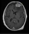
24
Q

Epidural Hematoma

  • Signs
  • CT scan
A

Signs:

  • headache
  • mental status abnormalities
  • loss of consiousness with brief lucid interval before subsequent decline

CT scan:

  • bioconvex lens between skull and out margin (dura)
25
Subdural Hematoma * Signs * CT scan * Can they occur in absence of significant trauma? When?
Signs: * headache * mental status abnormalities * slower clinical deterioration than epidural hematoma; over days to weeks CT scan: * cresent shape of blood seperating dura from arachnoid membrane Sidenote: Can occur in absence of significant trauma; particularly in older persons and those taking anticoagulation
26
Traumatic Brain Injury When is neuroimaging recommended? * With what symptoms/signs? * In what scenarios?
* worsening headache, repeated vomiting * drowsiness, persistent confusion * focal neurological findings * seizures * suspected substance intoxication * dangerous causes of injury (fall from height \> 3 feet or 5 steps, ejection from vehicle, struck by vehicle as pedestrian)
27
Traumatic Brain Injury Treatment * Mild TBI * Postconcussive Syndrome * Posttraumatic headache * Epidural Hematoma * Subdural Hematoma
**Mild TBI:** remove from play, undergo sideline assessment, prohibit sports until patient returns to cognitive baseline and is asx without medication **Postconcussive syndrome:** supportive and rehabilitative **Posttraumatic headache:** NSAIDs, Triptans; SSRIS, SNRIS (mood/anxiety too) **Epidural Hematoma:** Emergent evacuation of blood to prevent death **Subdural Hematoma:** depends on clinical circumstances; observation is appropriate in stable, asymptomatic patients
28
Epilepsy: defined
* 2 or more unprovoked seizures occuring more than 24 hours apart OR * 1 unprovoked seizure with a significant ongoing risk of further unprovoked seizures
29
Focal seizure vs Primary generalized seizure vs Secondarily generalized seizure * electrical discharge activity
Focal seizure: * result from electrical discharge that originates in a focal region of the brain Primary generalized seizure * electrical discharge that involves areas of brain simultaneously Secondarily generalized seizure * focal in onset, rapidly spreads to involve entire cerebral cortex
30
Clinical clues to identify that a focal seizure has progressed to a generalized seizure
* unilateral shaking * head turning (versive) to one side * aura * postictal (temporary) weakness
31
Seizure Classification: Characteristics * Focal seizure without alteration of awareness (formerly simple partial) * Focal seizures with alteration of awareness (formerly complex partial) * Primary generalized seizure (formerly tonic clonic, no awareness)
Focal without alteration * Normal conscioussness/awareness; single neurologic modality (sensory, motor, olfactory, visual, gustatory) involving a single region of the body Focal with alteration * Conscious but unresponsive or staring; automatism (lip smacking, swallowing, manipulating objects), postictal confusion Primary generalized * Loss of consciousness or awareness at onset; No prodromal or localizing symptoms; Whole body stiffening (tonic); and/or jerking (clonic)
32
Common Epilepsy Syndromes * **Temporal Lobe epilepsy** * **Frontal Lobe epilepsy** * Idiopathic (genetic) generalized epilepsy * Myoclonic seizure * Convulsive status epilepticus
Temporal Lobe epilepsy: * Focal seizures WITH alteration of awareness * MRI: Medial Temporal Lobe sclerosis (characteristic) Frontal Lobe epilepsy: * Nocturnal complex seizures that awaken patients from sleep * Often assoc w/ underlying structural pathology (tumor, vascular malformation)
33
Common Epilepsy Syndromes * Temporal Lobe epilepsy * Frontal Lobe epilepsy * **Idiopathic (genetic) generalized epilepsy** * **Myoclonic seizure** * **Convulsive status epilepticus**
**Idiopathic (genetic) generalized epilepsy** Any combination of tonic-clonic seizures, absence seizures, myoclonic seizures MRI: typically normal; EEG: may show generalized spike-wave abnormality **Myoclonic seizure** Generlized seizure associated with brief lightning-like jerks of the arms, not associated with loss of consciousness (misdiagnosed as "jitteriness") **Convulsive status epilepticus** Continuous seizure \> 5 mins. MCC low AED level. Mortality rate: 20% Complications: aspiration pna, fever, hemodynamic instability, acidosis, PE, rhabdo
34
PNES: Psychogenic Nonepileptic Spells What is it? Characteristic Features? Diagnosis?
Defined: Type of conversion disorder Characteristic Features: * forced eye closure * long duration * hypermotor activity that starts and stops * pelvic thrusting \* Inpatient video EEG monitoring is required to make diagnosis of PNES becuase of the difficulty in distinguishing PNES and eplipetic seizures; strong association with PTSD in military veterans
35
* Initial Testing for Unprovoked Seizure: name 4 tests * What do you do if patient does not return to baseline by 15 minutes after seizure?
* EEG (although a normal EEG does not r/o seizure) * CBC, electrolyte and glucose levels, toxicology screen * Brain MRI (or head CT in an emergency) * CSF examination if patient has fever, prolonged AMS after seizure, is immunosuppressed, or has severe headache \* If patient not returning to baseline mental status by 15 minutes after a seizure, obtain continuous EEG to rule out nonconvulsive status epilepticus
36
Seizures: Treatment When do you start treatment? How do you start treatment? What do you do if first drug is unsuccessful?
Start Treatment: * After \>/= 2 unprovoked seizures * After single high-risk unprovoked seizure characterized by focal findings on neuroimaging, focal findings on EEG, or significant risk factors for epilepsy (such as head trauma or after brain tumor resection) * Start with single agent therapy and increase dosage until seizures are controlled or patient develops adverse effects * If unsuccessful, continue first drug, start a second single agent
37
AEDs Which drugs can be used to treat both generalized and focal epilepsy syndrome? Which is superior to other AEDs for treating generalized epilepsy? AED cost effective for Focal? AED with few drug interactions, well-tolerated, safe during pregnancy?
**Generlized & Focused:** lamotrigine, levetiracetam, topiramate, valproic acid, zonisamide **Superior for Generalized:** Valproic Acid **Cost effective for Focal:** Carbamazepine **Few drug Interactions, well-tolerated, used during pregnancy:** Levetiracetam
38
AED Important Side Effects: Name that drug 1. interactions with other hepatically metabolized drugs, increased risk osteoporosis and hypercholesterolemia 2. Weight gain, hypercholesterolemia, PCOS, teratogenecity, hepatotoxicity 3. Kidney Stones 4. Hyponatremia, pancytopenia 5. Drug hypersensitivity syndrome, SJS, suicidal ideation
1. Carbamazepine (tegretol) 2. Valproic Acid 3. Topirimate & Zonisamide 4. Carbamazepine & Oxcarbazepine 5. all AEDs
39
Refractory Epilepsy: Defined & Treatment
Defined: Patients not responding to their first or second (in sequence or in combination) have refractory epilepsy are candidates for epilepsy surgery Treatment: Epilepsy surgery * resection of mesial temporal lobe sclerotic lesions associated with focal seizures
40
1. Criteria to withdraw AEDs? 2. Which AEDs are safe in pregnancy? 3. AEDs to discontinue in pregnancy? (category D)
Criteria to withdraw AEDs: * patients who have been seizure free for 2-5 years Pregnancy: * Lamotrigine (requires upward dose adjustment for pts already taking drug) * Levetiracetam Discontinue in pregnancy (category D drugs) * Valproic acid * Topiramate
41
Convulsive Status Epilepticus: management * What do you administer if alcohol is suspected or is cause? * What testing needs done emergently if no known underlying cause? * What is the most common cause?
Alcohol: * thiamine and glucose Unknown cause: * MRI Most common cause: * low AED blood level
42
Convulsive Status Epilepticus * First Line Treatment options: name 3 * Patients not taking AEDs should then be treated with what? administred after continuous seizing for how long? * Who needs monitored with continuous EEG? Why?
First Line: * IV lorazepam, IV diazepam, IM midazolam Patients not taking AEDs: * administered after continuous seizing for 5 minutes * phenytoin or fosphenytoin \* All patients with convulsive status epilepticus who stop seizing but do not return to baseline within 30 minutes should be monitored with continuous EEG for nonconvulsive seizures
43
Ischemic Stroke & TIA: Prevention * Risk factor modification: Name 4 * Which patients should be revascularized with stenting or endarterectomy? * Nonvalvular Afib risk prevention * Surgical clipping or endovascular coiling or aneursyms are indicated for which patients?
**Risk Factor modification**: diabetes, hypertension, hyperlipidemia, tobacco use **Revascularization:** \> 80% or rapidly progressive stenosis and low cardiovascular risk, operative complication rate \< 3% **Nonvalvular Afib:** Begin warfarin or NOAC. Aspirin if low risk. **Surgical clipping or coiling:** Aneurysms \> or = 7mm in posterior circlulation or \> or = 12 mm in the anterior circulation
44
**Stroke** * Defined * Cause (85%) (15%) * Ischemic stroke causative mechanisms * Hemorrhagic stroke causitive mechanisms
**Stroke** * ​sudden focal neurological deficit * caused by ischemia (85%) or hemorrhage (15%) * ischemic stroke causative mechanism: * large-artery atherosclerosis, cardioembolic, small-vessel disease, cryptogenic * Hemorrhagic stroke causative mechanism: * subarachnoid or intracerebral (intraperenchymal)
45
TIA * Defined * Symptom duration * Stroke risk * When to admit to the hospital
Defined: * focal neurological deficit resulting from ischemia rather than infarction * absence of infarction on neuroimaging * independent of symptom duration (typically 5-60 minutes) * increased risk of stroke, especially in the next 48 hours * ABCD2 to risk stratify
46
TIA Risk Stratify When to admit to the hospital
* ABCD2 score assessment to risk stratify * Hospital admission recommended for \> or = 3 **A**ge \> 60yo (1) **B**P \> 140/90 (1) **C**linical symptoms: focal weakness (2), speech impairment with out weakness (1) **D**uration of TIA: \> or = 60 mins (2); 1-59 mins (1) **D**iabetes (1)
47
Stroke or TIA * All patients require these 4 tests
1. emergent head CT with out contrast (r/o intracranial hemorrhage) 2. ECG and telemetry or even monitoring (to r/o AFIB) 3. vascular studies (cerebrovascular u/s or MRA, CTA) 4. echocardiography (to r/o LV or valvular thrombosis)
48
Stroke Treatment (basic) * Name 4 basics * Do not begin antihypertensive treatment within first 48 hours unless: * SBP: * DBP: * MAP: * Thrombolytic therapy planned with SBP of? or DBP of? * If any of these 3 are present
**Basic:** Treat temperature of 100.4 with tylenol; Maintain euvolemia with NS; Give ASA 325mg if thrombolysis not planned; start DVT ppx within 48 hours **Do not begin antihypertensive therapy within first 48 hours unless:** SBP: \> 220 DBP: \> 120 MAP: \> 140 Thrombolytic + SBP \> 185 or DBP \> 110 ACS, Aortic dissection or end organ damage is present
49
Stroke: TPA Which patients receive TPA? In what time frame? Exclusionary criteria to administer TPA up to 4.5 hours after onset? Exclusionary criteria for TPA in general?
TPA: all patients with ischemic stroke * w/i 3 hours of stroke onset (if unknown onset, w/i 3 hours of last seen normal) * rarely up to 4.5 hours after onset without these exclusions: * age \> 80, severe stroke, DM w/previous infarct, anticoagulant use Generalized exclusionary criteria: * increased risk of bleeding, ICH diagnosis, SBP \> 185 or DBP \> 110 * preferred: IV labetalol, IV nicardipine
50
Stroke: Secondary prevention * **Aspirin** * **Anticoagulation** * Revascularization * Statins * Hypertension * Depression * Device closure of PFO plus aspirin
Aspirin: * acute setting: immediately or within 24 hrs after thrombolytic agents * chronic setting: aspirin plus dipyridamole superior to aspirin alone * clopidogrel is equivalent to aspirin plus dipyridamole Anticoagulation: * after 48 hours, treat high risk cardioembolic causes of stroke and TIA with warfarin or a NOAC, including patients iwth AF, left atrial appendage thrombus, LV thrombus and dilated CM with reduced EF
51
Stroke: Secondary Prevention * Aspirin * Anticoagulation * **Revascularization** * **Statins** * **Hypertension** * Depression * Device closure of PFO plus aspirin
Revascularization * endarterectomy or stenting within 2 weeks after nondisabling stroke or TIA if ipsilater carotid stenosis is \> 70% provided the patient is likely to live 5 years Statins * Begin high-intensity statin therapy for all patients regardless of cholesterol level Hypertension * Maintain BP \< 130/80 after recovery from acute event
52
Stroke: Secondary Prevention * Aspirin * Anticoagulation * Revascularization * Statins * Hypertension * **Depression** * **Device closure of PFO plus aspirin**
Depression: * Prevalent in acute and chronic setting and identification and treatment improve recovery Device closure of PFO plus aspirin: * To prevent second stroke in patients with thoroughly investigated cryptogenic stroke
53
SAH: Prevention Aneurysms: risk of rupture increases with size and location of aneurysm * What size classifies as small? * How do you treat? * What size classifies as large? * How do you treat?
Small: * Anterior circulation: \< 12 mm * Posterior circulation: \< 7 mm * Monitor with MRI Large: * Anterior circulation: \> or = 12 mm * Posterior circulation: \< or = 7 mm * Surgical candidates
54
SAH: Diagnosis * Most common symptom * Symptoms * Most common cause of spontaneous SAH * Other causes
* Most common symptom: altered consciousness * Symptoms: "worst headache of my life," or "thunderclap headache." Up to 40% experience "sentinel hemorrhage" ~ severe headache in previous 2-3 weeks. * Focal neurological deficits occur from aneurysm compression cranial nerve, bleed into parenchyma or focal ischemia bc of vasospasm * MCC: ruptured saccular "berry" aneurysm of circle of Willis * Other causes: rupture of AVM, arterial dissection, coagulopathy, cocaine abuse
55
SAH * Testing * established diagnosis in 90% * identifies the aneurysm and determines management
Establishes diagnosis in 90% * Noncontrast CT Identifies aneurysm and determines management * Cerebral angiography
56
SAH: Treatment * Three main neurological complications * To manage these complications, do the following
1. rebleeding (CT) 2. delayed brain ischemia from vasospasm (transcranial u/s or CTA) 3. hydrocephalus (CT) * treat ruptured aneurysms with surgical clipping or endovascular coiling within 48 - 72 hours * maintain BP \< 140/80 to prevent rebleeding * select oral nimodipine for 21 days to prevent vasospasm and improve neurologic outcomes \* any change in mental status -- emergent CT to eval for repeat bleeding and signs of hydrocephalus, treated with ventricular drainage
57
Intracerebral Hemorrhage * most common risk factor * other risk factors * establish diagnosis * patients \< 45yo with ICH related to cocaine require this test
**most common risk factor:** hypertension **other risk factors:** amyloid angiopathy, coagulopathy, vascular malformations, cocaine or alcohol use **establish diagnosis:** CT without contrast **patients \< 45 with ICH related to cocaine** require cerebral angiography (associated with high incidence of vascular anomalies)
58
Intracerebral Hemorrhage: Treatment * Identify/Reverse - * To reduce intracranial pressure - * To maintain SBP between 140-160 - * Intraventricular hemorrhage requires prompt - * Cerebellar hemorrhages require - * For warfarin associated ICH -
* Identify/reverse: anticoagulation * Reduce ICP: mannitol, barbiturate coma, hyperventilation * Maintain SBP 140-160: IV nicardipine or labetelol * Intraventricular hemorrhage: prompt ventricular drainage to reduce ICP * Cerebellar hemorrhages: require surgical posterior fossa decompression * Warfarin associated ICH: IV vit K and prothrombin complex concentrates
59
Dementia vs Mild Cognitive Impairment Definition Mini Mental State Examination score
* Dementia: acquired chronic impairment of memory and other aspects of intellect that impedes daily functioning * Mini-Mental Exam \< or = 24 is compatible with dementia * Mild Cognitive Impairment: cognitive decline greater than expected for age but with out interference with daily functioning (and is a risk factor for dementia)
60
* What test predicts likelihood patient with MCI will develop dementia? * What treatments are available to delay onset of Alzheimer disease in patients with MCI?
No test to determine No treatment to delay onset
61
Characteristic findings of Alzheimer disease
Alzheimer disease is the most prevalent neurodegenerative dementia; accounting for 60-80% of cases * memory loss * getting lost * difficulty findings words * difficulty with dressing, grooming, doing housework
62
Dementia Testing * All patients must be screened for? * What must be done routinely that could alter management? * Consider LP and csf exam in what situations?
* Must be screened for depression * Routinely obtain MRI or CT to detect nondegenerative causes * Consider LP: * rapidly progressive dementia * age onset \< 60 * h/o malignancy or paraneoplastic disorders * suspicion for acute/subacute infection; immunosupressed * positive syphillis or lyme serology * systemic autoimmune disease; suspected CNS autoimmune/inflammatory disorder
63
What 3 tests must you order with early-onset or rapidly progressive dementia? What is the most common cause of rapidly progressive dementia?
* MRI, CSF analysis and EEG * creutzfeldt-jacob disease
64
Differential Diagnosis of Dementia 1. Acute onset, fluctuating course, inattention, disorganized thinking, and altered consciousness 2. Evidence of objective memory impairment in absence of other cognitive deficits, intact activities of dialy living 3. Mild parkinsoism, postural instability and gait difficulty, fluctuating cognition, delusions, visual hallucinations
1. delirium 2. mild cognitive impairment (MCI) 3. lewy body dementia
65
Differential Diagnosis of Dementia ## Footnote 4. Most prevelant neurodegenerative dementia, accounting for 60-80% of cases. Gradual memory loss, aphasia, apraxia, agnosia, inattention, decrease in executive function 5. Imagining or history positive for stroke responsible for impariment of at least one cognitive domain. Focal neurological findings, depression, pseudobulbar palsy, gait abnormalities, urinary difficulties
4. Alzheimer disease 5. Vascular neurocognitive disorder
66
Differential Diagnosis of Dementia ## Footnote 6. Early & prominent personality changes, behavioral disturbances - disinhbition, impulsivity, diminished frontal and/or temporal lobes on MRI, onset before 60yo 7. Dementia, shuffling gait, urinary incontinence, ventriculomegaly in absense of meningitis, SAH or trauma 8. Choreoathetosis and dementia, autosomal dominant pattern of inheritence 9. Prominent myoclonus, characteristic EEG characteristic triphasic sharp waves, CSF 14-3-3, rapidly progressive onset at early age
6. frontotemporal dementia, behavioral variant 7. normal-pressure hydrocephalus 8. huntington disease 9. creutzfeld-jakob disease
67
Differential Diagnosis of Dementia ## Footnote 10. Early postural instability and falls, apathy, parkinsonism, poorly responsive to levodopa and vertical gase palsy 11. History of head injury followed by delayed development (10 years) of impaired concentration, short-term memory, executive dysfunction, and judgement; aggression, depression, irritability, violent behaviors, and suicidality; parkinsonism, unsteady gait, shuffling gait, slurred speeh
10. Progressive supranuclear palsy 11. Chronic traumatic encephalopathy
68
Dementia: Treatment **Mild to Moderate** Alzheimer Disease * Mini Mental score * Which medications slow intellectual decline? * side effects? **Moderate to Advanced** Alzheimer Disease * Mini Mental score * Which medication delays cognitive decline?
**Mild to Moderate** Alzheimer Disease * Mini Mental score: 15-24 * Slow intellectual decline: acetylcholinesterase inhibitors (donepezil, rivastigmine, galantamine) * side effects: bradycardia, diarrhea, heart block, n/v, syncope **Moderate to Advanced** Alzheimer Disease * Mini Mental score: 3-14 * Which medication delays cognitive decline? Memantine
69
Dementia differential diagnosis: Treatment * Dementia with Lewy Bodies * Vascular Cognitive Impairment * Normal Pressure Hydrocephalus * Behavior Symptoms * Agitation, aggression, delusions, hallucinations * Depression
* Dementia with Lewy Bodies: acteylcholinesterase inhibitors (donepizil) * Vascular Cognitive Impairment: risk factor modification and ach inhibitors * Normal Pressure Hydrocephalus: large-volume LP w/symptom assessment * Behavior Symptoms: nonpharmacological * Agitation, aggression, delusions, hallucinations: atypical antipsychotics * Depression: SSRIs
70
Delirium * Diagnosis * Treatment
Diagnosis: * acute onset and fluctuating course, inattention, disorganized thinking, altered level of consciousness * Look for triggers, particularly alcohol, medications, polypharmacy Treatment * ALWAYS choose behavioral interventions first and order a "sitter" instead of selecting restraints or drugs * Benzos worsen and are not recommended, except with alcohol
71
Parkinson Disease * Caused by: * Diagnosis * Diagnosis characterized by at least 2 of the following 4 conditions * Symmetric or Asymetric at onset? * 3 signs that may precede onset of motor symptoms by years: * Inreased risk of/often present for eval of:
* Caused by: degeneration of dopaminergic neurons in substantia nigra of midbrain * Diagnosis: Clinical diagnosis; imaging only to exclude other disease * 2/4: Bradykinesia, Rigidity, Resting tremor, Postural reflex abnormality (falling) * Asymetric at onset * Diminished sense of smell, constipation, acting out dreams may precede * Increased risk of falls, often present for eval of falls
72
* Early dementia within the first year of appearance of parkinsonism (signs/symptoms) is a hallmark of? * Treatment?
* Lewy Body Dementia * Ach inhibitors (donepezil, rivastigmine, galantamine)
73
Differential Diagnosis of Parkinson Disease * Severe orthostatic hypotension and ataxia; MRI sowing necrosis of putamen and cerebellar atropy * Unexplained falls (typically backward), inability to move eyes vertically, and parkinsonian features * Early dementia, parkinsonism, hallucinations * Antiemetics (prochlorperazine, metoclopramide) antipsychotics (haloperidol), reserpine, lithium, methyldopa
* Severe orthostatic hypotension and ataxia; MRI sowing necrosis of putamen and cerebellar atropy: **multiple system atrophy** * Unexplained falls (typically backward), inability to move eyes vertically, and parkinsonian features: **progressive supranuclear palsy** * Early dementia, parkinsonism, hallucinations: **d****ementia with Lewy bodies** * Antiemetics (prochlorperazine, metoclopramide) antipsychotics (haloperidol), reserpine, lithium, methyldopa: **medication-induced parkinsonism**
74
Parkinson Disease: Treatment When do you begin treatment? Most effective medication? Treatment option for patients with sustained motor benefit from most effective medication but are limited by disabling medication-related adverse effects refractory to medical management?
* Begin treatment when symptoms begin to interfere with function * Levodopa; given with Carbidopa +/- Pramipexole or Ropinirole * Deep Brain Stimulation
75
Parkinson Disease: Treatment * Most effective medication​ * adminstered with and why * side effects of most effective medication * treatment for symptoms of medication wearing off? * what can be administred to avoid other side effects? * side effects of this medication?
Most effective medication: **Levadopa** Administered w/: **Carbidopa**, **prevents peripheral conversion of leva to dopamine** Side effects: **motor fluctuations, dyskinesias, "wearing off" (parkinson sx)** Treatment for wearing off: **I****ncrease dose or frequency or use sustained release** Treatment for motor fluctuations and dyskinesias: **dopamine agonist (pramipexole and ropinirole)** Side effects of dopamine agonists: **sedation, complusive behaviors - gambling, shopping, hypersexuality**
76
Hyperkinetic Movement Disorders * Essential Tremor * key manifestations: progression, movement, family history * treatment
Key Manifestations: * Typically slowly progressive or stable over time * Bilateral postural or kinetic tremor; improves with alcohol * Family history positive in 50% Treatment: * Propanolo, primidone, or topiramate
77
Hyperkinetic Movement Disorders * Hungtington disease * key manifestations: movement, other manifestations, family history * treatment
key manifestations: * most common neurodegenerative cause of generalized chorea * chorea: random, nonrepetetive, flowing dance-like movements * progressive dementia and psychiatric manifestations * autosomal dominant treatment * symptomatic treatment with tetrabenazine and deutetrabenazine
78
Hyperkinetic Movement Disorders * Drug-induced dystonia * key manifestations: movement pattern, caused by * treatment
Key manifestations * Tardive dyskinesia associated with choreiform and dystonic craniofacial movements * Can be caused by neuroleptic, antiemetic and serotoninergic medications Treatment * Stop offending drug * Valbenazine, clonazepam, tetrabenazine, anticholingergic agents, clozapine
79
Hyperkinetic Movement Disorders * Cervical dystonia (torticollis) * key manifestations: what is it? * treatment
key manifestations * cervical muscle contractions resulting in abnormal posture of the head and neck treatment * botulinum toxin (first line)
80
Hyperkinetic Movement Disorders * Myoclonus * Key manifestations: movement pattern, underlying cause possibilities * Treatment
Key manifestations: * rapid, shock-like, jerky movements of isolated body parts * underlying metabolic disorder, serotonin syndrome, postanoxic, creutzfeldt-jakob disease, corticobasal degeneration Treatment: * treat the underlying metabolic disorder
81
Hyperkinetic Movement Disorders * Tourette syndrome * key manifestations: onset, movement pattern * treatment
Key manifestations: * childhood onset * multiple complex tics and presence of vocal tics (echolalia) Treatment: * reassurance or cognitive behavioral therapy
82
What should patients \< 40 years old with "essential tremor" or dystonia be screened for? Are rigidity and resting tremor features of essential tremor?
* Screen for Wilson disease * 24 hour urine copper measurements * serum ceruloplasmin * Rigidity and Resting tremor are not features of essential tremor
83
Multiple Sclerosis * Episodes of dysfunction resulting from these lesions? where? * Clinical course 3 pattern possibilities
Lesions: * demylenating lesions (plaques) in different areas in CNS * brain, brain stem, including optic nerve, or spinal cord) Clinical course follows one of three patterns: * Relapsing-remitting * Secondary progressive * Primary progressive disease: progressive
84
Those who do not meet full diagnostic criteria for MS after a first event have what? What is the 10 year risk of MS associated iwth a clincally isolated syndrome?
Those who do not meet full diagnostic criteria for MS after a first event have what? **Clinically isolated syndrome** What is the 10 year risk of MS associated with a clincally isolated syndrome and lesions on MRI? **90%**
85
Common symptoms associated with MS * **Optic Neuritis** * **Afferent Pupillary defect** * **Papillitis** * **Brainstem (intranuclear opthalmoplegia)** * Myelitis * Lhermitte sign * Bladder * Cerebellum * Uhthoff phenomenon
**Optic neuritis**: subacute visual deficit; pain with eye movement **Affarent pupillary defect:** paradoxical dilation of pupil when light is rapidly shifted from unaffected eye to affected eye **Papillitis:** inflammatory changes to retina causing flared appearance of optic disc **Brainstem (intranuclear opthalmoplegia):** inability to adduct one eye and nystagmus in the abducting eye * left intranuclear opthalmoplegia: looking to right (failure of adduction of left eye, nystagmus of right eye)
86
Common symptoms associated with MS * Optic neuritis * Afferent pupillary defect * Papillitis * Brainstem (intranuclear opthalmoplegia) * **Myelitis** * **Lhermitte sign** * **Bladder** * **Cerebellum** * **Uhthoff phenomenon**
**Myelitis:** Focal inflammation within the spinal cord manifesting as sensory, autonomic, or motor symptoms below the affected spinal level **Lhermitte sign:** shock-like sensation radiating downt he spine or limbs induced by neck movements **Bladder:** frequency, urgency, retention **Cerebellum:** Ataxia, vertigo **Uhthoff phenomenon:** transient worsening of baseline neurologic symptoms with elevations in body temperature
87
MS: Diagnosis * Diagnosis requires: * demylenation disseminated in both space and time as demonstrated through a combination of these 3 things * CSF may show?
* **Diagnosis requires demylenation disseminated in both space and time as demonstrated through a combination of:** * documented clinical relapses * signs on physical examination * distribution of lesions on MRI * **CSF may show:** * oligoclonal igG bands or elevated igG index * XCF analysis not necessary but can be helpful
88
MS: Treatment ## Footnote #1 rule Acute recovery: how to speed up After first attack of clinically isolated syndrome (i.e. optic neuritis, spinal cord syndrome), prescribe ___ if imaging suggests MS Confirmed relapsing-remitting MS treatment MS relapses with no or minimal impact What can be added to reduce accumulation of MRI lesions and is recommended in all patients with MS?
* #1 rule: treat fever, look for underlying infection before beginning glucocorticoids, because fever worsens symptoms of MS and treatment of underlying cause will improve symptoms * Acute recovery: IV methylprednisolone followed by oral glucocorticoids * Prescribe interferon beta or glatiramer acetate after first attack + imaging * Prescribe interferon beta or glatiramer acetate for RRMS * Oberve with no or minimal impact * All MS patients should be on Vitamin D
89
Symptomatic management in MS - nonpharmacologic vs pharmacologic * Spasticity * Neuropathic pain * Fatigue * Depression * Cognitive dysfunction * Mobility * Urinary retention
Spasticity: baclofen, benzodiazepine,cyclobenzaprine Neuropathic: carbamazepine, duloxetine, gabapentin, pregabalin, topiramate Fatigue: amantadine, amphetamines, armodafinil, modafinil Depression: antidepressants (SNRIs, SSRIs) Cognitive decline: cognitive rehab, no proven rx Mobility: dalfampridine Urinary retention: manual pelvic pressure, intermittemnt cath | No rx
90
What causes a threefold increase in the risk of secondary progression of MS?
Cigarette smoking
91
Myelopathy (spinal cord dysfunction) * how does this occur? * corticospinal symptoms? * distal cord and lower root symptoms? * noncompressive myelopathy
* occurs because of a lesion arising within the spinal cord (intramedullary) or because of extrinsic compression of the spinal cord * corticospinal injury: weakness, hyperreflexia, muscle spasms, plantar responses * distal cord/lower root (cauda equina syndrome): lower extremity weakness with decreased muscle tone and areflexia * noncompressive myelopathy: inflammatory or demyelinating lesions, spinal cord infarction, copper or B12 deficiency
92
Myelopathy/Spinal Cord Dysfunction **Neuromyelitis optica** (Devic disease) what is it? antibodies?
* recurrent episodes of myelitis and optic neuritis without the brain lesions of typical MS * NMO-IgG autoantibody may be present
93
Myelopathy/Spinal Cord Dysfunction **Vitamin B12 deficiency** Signs/Symptoms? \*Don't be tricked Testing
* paresethesias, lower extremity weakness, gait instability * may include paraparesis, vibration, position sense loss, sensory ataxia * anemia may be absent\* * check methylmalonic acid and homocysteine for patients with borderline B12 deficiency
94
Myelopathy/spinal cord dysfunction **Copper Deficiency** What deficiency does it mimic? What surgery might it deveop after? What excessive ingestion may cause it?
* Mimics B12 deficiency: parasthesias, lower extremity weakness, gait instability * May develop after bariatric surgery * Excessive zinc ingestion
95
Myelopathy/Spinal cord dysfunction **Infarction of spinal cord** Symptoms Potential causes
**Symptoms:** acute onset flaccid paralysis or weakness, pinprick sensation loss below level of infarction **Potential causes:** emboli, hypotension during cardiovascular/aortic surgery, AV malformations
96
Spinal Cord Compression Clues to the cause of compression myelopathy Presentation? * Fever - * Anticoagulation - * Cancer - * Metastases - * Trauma - * Elderly with chronic back/leg pain -
Presentation: neck or back pain; followed by weakness, sensory changes, bowel and bladder dysfunction; UMN signs: weakness, spasticity, hyperreflexia, extensory plantar responses) and occasionally LMN signs (atrophy, hyporeflexia) * Fever - epidural abscess * Anticoagulation - epidural hematoma * Cancer - metastases * Trauma - vertebral fracture * Elderly with chronic back/leg pain - spinal stenosis
97
Spinal cord compression Testing in patients with clinical suspicion
* MRI of spine to exclude spinal cord compression in all patients with clniical suspicion * LP may be beneficial in patients with suspected inflammatory or demyelinating spinal cord lesions
98
Myelopathy: Treatment * Spinal cord compression caused by metastatic disease * Spinal cord compression caused by infectin or hematoma * Spinal cord compression caused by leukemia, lymphoma, myeloma or germ cell tumors * Spinal cord infarction
metastatic disease: high dose glucocorticoids and subsequent surgical decompression followed by radiation for most tumor types infection or hematoma: do not use glucocorticoids leukemia, lymphoma, myeloma or germ cell tumors: may be treated urgently with radiation therapy rather than surgery Spinal cord infarction: no treatment
99
Amytrophic Lateral Sclerosis * Defining characteristic is the combination of? Absence of? * All patients with suspected ALS should undergo? * What is required to establish presence of respiratory insufficiency? * What is bulbar-onset, what test do they require? * Treatment
* upper motoneuron signs (hyperreflexia, spasticity, extensor plantar response) * with lower motoneuron signs (atrophy, fasciculation) * absence of sensory deficits * all patients should undergo * EMG test for muscle degeneration * MRI of appropriate anatomic areas to r/o treatable disorders that mimic * Bulbar-onset (20%): difficulty speaking and swallowing - need swallow eval * Treatment: Riluzole may increase survival by 3 mos * noninvasive ventilatory support for respiratory insufficiency * PEG for weight loss or swallowing difficulties
100
Myesthenia Gravis * Antibodies? Which results in? * Characteristic findings (Name 5) * vision * muscles * abs * DTRs * EMG response
* Autoimmune disease: antibodies against acetylcholine receptor * results in impaired neuromuscular transmission * Characteristic findings: * ptosis or diplopia (first sx in most patients) * muscle weakness (including dysphagia and dyspnea) * positive ach receptor ab (found in 90%; negative does not r/o) * normal DTRs and sensation * decremental response to repetitive stimulation on EMG
101
Myasthenic Crisis * Symptoms * Causes * Differential * Treatment
Symptoms: may include rapidly progressive respiratory failure Triggered by: MG itself, infection, surgery, medications, surgery; meds - aminoglycosides, quinoones, magnesium, B-blockers or CCBs Differential: Botulism - nonreactive pupils (they are reactive in MG); Lambert Eaton - Abs to anti voltage gated calcium channel + EMG motor response w/o rapid repetitiive stimulation usually underlying undetected malignancy, typically SCLC Treatment: plasmapheresis or IV immunoglobulin (same tx as refractory disease)
102
Myasthenia Gravis * Testing * diagnosis * also look for * everyone needs a...to r/o a... * Treatment * initial therapy * thymoma * crisis/refractory
Testing * EMG can establish diagnosis * Look for elevated TSH levels bc of association with other autoimmune dx * Perform CT chest to look for thymoma Treatment: * Initial therapy: Pyridostigmine * Thymoma: Thymectomy * Crisis or Refractory: plasmapheresis or IVIG
103
Peripheral Neuropathy * Isolated anterolateral thigh numbness with out weakness * What is it? * Treatment?
* Meralgia paresthetica (a compressive neuropathy of the lateral femoral cutaneous nerve) Treatment: Locate and relieve pressure (binding clothes, excessive weight)
104
Peripheral Neuropathy Median Neuropathy (carpal tunnel syndrome) * What is it? * Treatment?
Sensory loss over palmar surface of first three digits and weakness with thumb abduction and opposition Treatment: * mild disease: wrist splints or glucocorticoid injections * severe: surgical release
105
Peripheral Neuropathy * Ulnar neuropathy * What is it? * Treatment?
Numbness of 4th and 5th fingers with weakness of interosseous muscles Treatment: * elbow splinting or elbow pads * surgical release if severe
106
Peripheral Neuropathy * Tarsal Tunnel Syndrome * Treatment
pain, tingling, numbness in great toe along medial foot Treatment: * local glucocorticoid injection * decompression surgery if severe
107
Peripheral Neuropathy * Upper and Lower face motor weakness * What is it? * Testing * Assess for * Treatment
Bell Palsy * Assess for: diabetes, vasculitis, lyme, sarcoidosis, HIV, compressive or infiltrative malignancies * Testing: classic presentation w/o additional neurologic deficits then brain imaging, labs not necessary * Treatment: Prednisone if within 72 hours of onset
108
Peripheral Neuropathy * Acute, ascending, areflexic paralysis and paresthesias * often preceded by * CSF findings * Treatment
Guillain-Barre Syndrome * often proceeded by GI illness (usually Campylobacter infection) * CSF: elevated protein, normal cell count (albuminocytologic dissociation) * Treatment: plasma exchange or IVIG * do not give glucocorticoids, may even slow recovery
109
Peripheral Neuropathy Progressive proximal motor and sensory neuropathy that evolves over months * What is it? * CSF findings? * Treatment?
Chronic Inflammatory demyelinating polyneuropathy * CSF findings: elevated protein, normal cell count (same as guillain-barre) * Treatment: prednisone, plasma exchange, or IVIG
110
Myopathy * Presents with * Myopathy vs Neuropathy: differentiator * CK levels * EMG
* Presents with symmetric muscle weakness of proximal muscles * Myopathy vs neuropathy differentiator: normal sensory and reflex exam * CK levels: elevated and falls in response to treatment * EMG: confirms presence of myopathic changes (low amplitude, short duration, polyphasic motor unit potentials)
111
Myopathy * proximal muscle weakness, \*normal CK levels,\* normal EMG findings * subacute toxic myopathy associated with rhabdomyolysis
​glucocorticoid myopathy: * proximal muscle weakness, \*normal CK levels,\* normal EMG findings statin myopathy * subacute toxic myopathy associated with rhabdomyolysis
112
Primary Central Nervous System Lymphoma * Diagnosis * where is the lesion? * symptoms * required for diagnosis * more common in * treatment
PCNSL (non-hodgkin lymphoma) * where is the lesion? supratentoral lesion * symptoms: visual symptoms (tumor involvement of optic radiations) * required for diagnosis: brain biopsy * more common in: immunocompromised * treatment: sensitive to whole brain radiation and chemotherapy * PCNSL + HIV: Start ART * PCNSL + transplant: stop immunosuppressive therapy
113
Meningioma * Type * Symptoms if present * Diagnosis * Treatment
Meningioma * Type: usually benign in histology and behavior * Symptoms if present: progressive headache * Diagnosis: CT partially calcified, homogeneously enhanving extra-axial mass adhrerent to dura and an enhancing dural "tail" * Treatment: surgical resection if symptomatic or enlarging; observation is appropriate for small, asymptomatic meningiomas
114
Metastatic Brain Tumors Parenchymal mets (vs leptomeningeal) * Usually present as this type of lesion * Commonly found * Typically associated with * Testing
* multiple, ring-enhancing, centrally necrotic lesions * junction between gray and white matter * typically associated with significant surrounding edema and mass effect * MRI: if patients have biopsy proven systemic cancer or multiple enhancing brain lesions, brain biopsy is not indicated
115
Metastatic Brain Tumors * Leptomeningeal metastases (vs parenchymal) * Which two cancers are known to cause this? * Symptoms * What finding may also be present? * MRI findings
* Lymphoma and Leukemia * headache, spinal pain, cranial nerve or spinal radicular pain, weakness, mental status change * communicating hydrocephalus may be present * MRI: diffuse or patchy enhancement of surface of brain and spinal cord or roots
116
Metastatic Brain Tumors: If metastatic brain tumor is the first indication of malignancy, what do you evaluate patient for?
* Lung cancer * Breast cancer * Melanoma
117
Metastatic Brain Tumors * Treatment * First line for parenchymal and leptomeningeal tumors * Leptomeningeal spread from leukemia and lymphoma * Multiple parenchymal metastases from known primary solitary tumor * Solitary, accessible brain metastases and controlled extracranial disease
* First line for parenchymal and leptomeningeal tumors * Glucocorticoids * Leptomeningeal spread from leukemia and lymphoma * Chemotherapy (methotrexate and cytarabine) * Multiple parenchymal metastases from known primary solitary tumor * Palliative whole brain radiation * Solitary, accessible brain metastases and controlled extracranial disease * Resection is an option
118
Coma * Three cardinal findings of brain death * Describe this state, what do they maintain? * Testing * Treatment
Brain death: * coma, absence of brain stem reflexes, apnea * repiratory drive and motor posturing signs not compatible with brain death State: unarousable, unresponsive; have sleep-wake cycles and brain stem function Testing: Focal findings or unexplained coma = emergent imaging of brain to exclude hemorrhage or mass lesion (CT is approrpiate in emergency situation); LP when minigitis or SAH suspected but normal imaging; EEG to r/o nonconvulsive status
119
Coma Treatment * First, attend to * All patients with unexplained coma * Opiate overdose suspected * Benzodiazepine overdose suspected
* FIRST: Airway, Breathing, Circulation * Unexplained coma: Thiamine and glucose * Opiate OD: Naloxone * Benzo OD: Flumazenil
120