Notes Flashcards

1
Q

Loss of achilles tendon reflex/ankle jerk reflex is a deficit at what spinal level?

A

S1&2 via gastrocnemius + soleus muscles (tibial nerve L4-S3)

2
Q

What spinal levels are responsible for eversion of the foot?

A

L5, S1, S2 but mostly S1 via the fibularis longus and brevis (superficial fibular nerve)

3
Q

What spinal levels are responsible for abduction of the thigh?

A

Gluteus medius & minimus, and obturator internus -> superior gluteal nerve -> L4, L5, S1

4
Q

What spinal level supplies sensation of the medial leg and foot ?

A

L4 via saphenous nerve

5
Q

Patellar reflex tests the integrity of nerves from what spinal levels?

A

L2-L4

6
Q

What spinal level supplies sensation to the dorsum of the foot?

A

L5 (would be impacted in a L4-5 intervertebral disc herniation)

7
Q

What spinal level supplies sensation to the lateral side of the foot?

A

S1 via the sural nerve

8
Q

What nerves supply sensation to the sole, the dorsum, and the skin between the first and second toe?

A

Tibial nerve (L4-S3) for sole

Superficial fibular nerve (L4-S1) for dorsum

Deep fibular nerve (L4,L5) for skin between first and second toe

9
Q

If the femoral artery is occluded, what will provide collateral circulation to the thigh?

A

Descending branch of lateral circumflex femoral artery

10
Q

Vertical group of superficial inguinal nodes drain into the superior thigh and receive lymph from what area?

A

Superficial thigh

Superficial, medial foot and leg

11
Q

Horizontal group of superficial lymph node sreceives lymph from what areas?

A

Superficial gluteal region

Anterolateral abdominal wall

12
Q

Deep inguinal lymph nodes underneath the fascia lata receive lymph from what area?

A

Deep gluteal injuries drain here.

Superficial lymphatics on the anterolateral side of the foot and leg and the deep lymphatics of the foot and leg first drain into the popliteal nodes, then the deep inguinal.

13
Q

If the popliteal artery is occluded, what artery can provide collateral to the leg and foot?

A

Lateral circumflex artery - it gives a branch that runs down the lateral thigh and joins the genicular anastamosis via the superior lateral genicular artery

14
Q

Where can the dorsalis pedis pulse be palpated?

A

Between the tendons of extensor hallucis and extensor digitorum longus on the dorsum of the foot

15
Q

What is a positive Trendelenburg test?

A

When the patient is asked to stand on the injured limb, the pelvis descends on the opposite side.

16
Q

Where should gluteal injections be performed to avoid nerve injury?

A

Anterior and superior to a line between the PSIS and the greater trochanter

17
Q

Until they’re 8yo, childrens’ femur heads are supplied by a direct branch of the ___.

This is later replaced by vessels such as the ___.

A

Children: Obturator artery (variably, the medial circufmlex artery)

Adults: medial circumflex femoral and branches

18
Q

the sciatic nerve runs through which quadrant of the buttocks?

A

Lower medial quadrant; avoid duing intragluteal injections

19
Q

What quadrant is the superior gluteal nerve in?

A

Superomedial quadrant -> if damaged, then trendelenburg’s gait

20
Q

Foot drop is associated with the deep fibular nerve, which innervates teh anterior leg. What motions is it responsible for?

A
  • Toe extension
  • Foot dorsiflexion
  • Inversion.

Injury causes loss of sensation between first and second toes.

Foot drop.

21
Q

The tibial nerve innervates teh posterior leg. These muscles are responsible for what?

A
  • Knee flexion
  • Plantarflexion
  • Intrinsic muscle functions fo the foot
22
Q

What muscle is associated with unlocking the knee joint?

A

Popliteus- when it contracts, it laterally rotates the distal portion of the femur and draws the lateral meniscus posteriorly to protect it when the knee is flexed.

23
Q

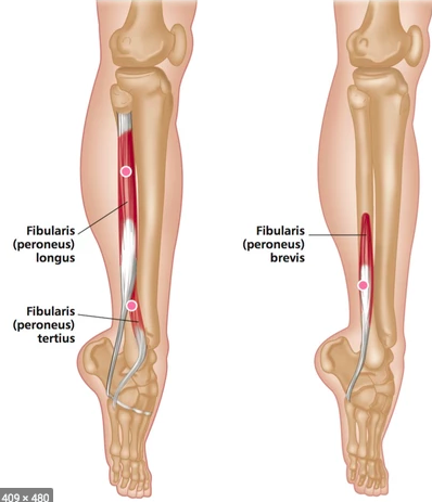
Fibularis tertius - what does contraction do?

A

Foot dorsiflexion, extension, and eversion

Runs from fibula to the dorsum of the base of the 5th metatarsal bone

24
Q

What’s another word for bowleg (knees bowed outwards?)

A

Genu varus

25
What is this?
**Hallux valgus:** lateral displacement of great toe; AKA bunion; swollen pursal sac at teh metatarsophalngeal joint
26
Housemaid's knee (working on bended knees) results from compression of what bursa?
Prepatellar bursa
27
Bursas of the knee
28
High stepping gait indicates injury to what nerve?
Deep fibular nerve
29
Waddling gait and numbness at the anterolateral side of the leg are assocaited with what nerves?
Superficial fibular and lateral sural cutaneous nerves
30
The **talocrural/tibiotalar/ankle joint** is responsible for what movements?
Plantar flexion and dorsiflexion
31
Compression at the popliteal fossa will compress which nerve first?
Tibial nerve | (16)
32
**Lateral plantar nerve** supplies the ____ hallucis muscle. **Medial plantar nerve** supplies the __ and __ hallucis brevis muscle.
Lateral plantar nerve --\> adductor hallucis Medial plantar nerve --\> abductor & flexor hallucis brevis
33
What structure is most important in shifting load from wrist to elbow or radius to ulna?
Central band
34
What musclse are responsibel for supination?
**Biceps brachii (**main supinator): long head attached to glenoid, short head attached to coracoid (short head is slightly better supinator) **Supinator** wraps around the radius
35
Pronator teres vs Pronator quadratus location
pronator teres more important
36
Radiocarpal joint vs midcarpal joint
In flexion, it's 50/50 radiocarpal and midcarpal. In extension, 0-60 degrees is radiocarpal, then 60+degrees is midcarpal
37
Two important ligaments of the wirst on the palmar side
Scaphoid lunate - flexion lunotriquetral - extension
38
The dorsal wrist ligaments all go toward the
triquetrum
39
Describe scaphoid vascularity
Retrograde flow from branches of **radial artery** ## Footnote 80% from **dorsal carpal branch** 20% from **superficial palmart branch** Note how the proximal part has no vascularity --\> blood has to go from distal to proximal or else necrosis & less likely to heal
40
Perilunate dislocation
Disruption of the scapholunate and lunotriquetral ligaments--\> the rest of the carpal bones get dislocated, but the lunate stays in place.
41
Terminal tendon helps extension at the ___ IP joint. The central slip helps extension at the ___ IP joint.
**Terminal tendon** extends at DIP **Central slip** extends at PIP
42
Horseshoe abcess
Infection in the flexor sheath (radial bursa) can travel into the carpal tunnel and inefct everything in there and your pinky.
43
Why is pain in your palm unrelated to carpal tunnel?
The palmar cutaneous branch from the median nerve travels superficial the flexor retinaculum.
44
Cubital tunnel syndrome is compression of what nerve?
Ulnar nerve
45
AIN innervates what?
**AIN** from the median nerve innervates the deep layer of the anterior compartment of the forearm (except for 1/2 of the extensor digitorum profundus innervated by the ulnar nerve)
46
PIN innervates what?
PIN from radial nerve innervates everything in the posterior compartment of the forearm except for the mobile wad (radial nerve, wrist extensors)
47
Distal cut to the ulnar nerve --\> loss of sensation & can't cross fingers Proximal cut to ulnar nerve --\> loss of sensation & can't wiggle pinky
48
3 contents of the **cubital fossa** lateral to medial
**Biceps brachii tendon** **Bracial artery** **Median nerve**
49
Vincula longa
Slender, bandlike connections form the deep flexor tendons to the phalanx that carry blood supply to the tendons. 30
50
What branches of the median nerve are responsible fo rinnervating the lumbrical muscles and carrying sensation to their respective digits?
Palmar digital branches
51
Weakness of what muscle is responsible for **Froment's sign**: can't hold a piece of paper between thumb and lateral side of index finger without flexing the distal joint of the thumb
Adductor pollicis (ulnar nerve)
52
Ulnar nerve enters the forearm by passing between the two heads of the ______ and descends between it and the ___ muscle.
Passes between the two heads of the **flexor carpi ulnaris**, then descends between it and the **flexor digitorum profundus**
53
The radial pulse is best located on the forearm just proximal to the wrist joint between what two tendons?
Flexor carpi radialis & brachioradialis
54
The radial artery travels on the distal radius between the flexor carpi radialis and brachio radialis tendons, but how does it enter the palm ?
Through the anatomic snuff box Then, pierce through the two heads of the first DAB to enter the deep palm.
55
The **PIN** of the deep radial nerve is responsible for extensors. It passes between the two heads of what muscle?
Supinator muscle. Thus, it's likely to be compressed by a hematoma between a fractured radius and supinator muscle.
56
Where does the ulnar nerve become the **deep ulnar nerve**?
Where it enters teh hand: superficial to teh flexor retinaculum and lateral to the pisiform bone
57
Dorsal compartments of the wrist
1st = abudctor pollicis longus & extensor pollicis brevis 2nd = extensor carpi radialis longus & brevis 3rd = extensor digitorum & extensor indicis 6th = extensor carpi ulnaris
58
Fracture of the upper third of the right radius, with the distal fragment of the radius and hand pronated. The proximal end of the fractured radius deviates laterally. Which muscle is primarily responsible for the lateral deviation?
**Supinator muscle**. ## Footnote The distal end of the radial fragment & hand are pronated because pronator teres & pronator quadratus are unopposed. The proximal end are laterally deviated because the supinator is unopposed.
59
If the ulnar nerve is cut at the \_\_\_\_, then you don't lose sensation to the ulnar part of the hand.
Wrist. ## Footnote You'll lose motor to dabs/pads and medial lumbricals, but keep sensation to ulnar hand.
60
Guyon's canal (holds ulnar nerve) anatomy
Pisiform medially Flexor retinaculum dorsally Deep fascia of wrist ventrally
61
The interosseus membrane joint between the radius and ulna is what type of joint?
**Synarthrosis joint**: fibrous connection with minimal to no movement
62
Volkmann's contracture
Flexion deformity of the fingers and sometimes the wrist from ischemic necrosis of the forearm flexor muscles.
63
Lymph from the thenar muscles drains into what axillary nodes?
**Lateral axillary nodes** \> subclavian nodes \> central axillary nodes
64
Lymph from the upper back and shoulder drain into what axillary nodes?
**Posterior axillary nodes** \> subclavian nodes \> central axillary
65
Pronator syndrome describes damage to what nerve?
Median nerve as it passes between the 2 heads of a hypertrophied pronator teres muscle. -\> loss of opposition, atrophied thenar, flexion difficulty of digits, sensory loss
66
Popeye sign
Rupture of long head of Biceps tendon (likely at the intertubercular groove). Not much weakness due to action of the brachialis and supinator muscles
67
The **supraspinatus muscle** inserts on the __ tubercle of the humerus, initiates abduction of arm at the shoulder, is is supplied principally by spianl nerve \_\_\_
Inserts on greater tubercle C5
68
What's the only muscle that inserts on the lesser tubercle of the humerus? What is its function?
**Subscapularis-** *internal* rotation
69
The teres ___ originates from the lateral border of the scapula The teres ___ originates from the region of the inferior angle and lateral border of the scapula
The teres **minor** originates from the lateral border of the scapula The teres **major** originates from the region of the inferior angle and lateral border of the scapula
70
Most common pediatric fracture
Clavicle
71
Flattened & sensory loss to deltoid prominence indicates what kind of injury?
**Anterior dislocation of the humerus:** head of humerus is displaced anteriorly and inferiorly. * flattened deltoid prominence * protruding acromion * anterior axillary fullness * injury to *axillary nerve*
72
Pain on passive adduction of the right arm across the chest and dislocated shoulder. Which ligamentous strutures must have been stretched/torn? * AC joint capsule and coracoclavicular ligament * AC joint capsule and coracoacromial ligament * STernoclavicular joint capsule and coracoacromial ligament * Coracoclavicular ligament and transverse scapular ligament * Coractoclavicular ligament and coracoacromial ligament
AC joint capsule and coracoclavicular ligament **Coracoclavicular ligament**: provides weight-bearing support for the upper limb on the clavicle; attaches to coracoid process **Acromioclavicular joint ligament** attaches to the acromion to the clavicle.