Ocular Anatomy & Clinical Correlation Flashcards

(56 cards)

1
Q

What are the two major pathologies we see in the lens?

A
  1. Presbyopia
    1. lose ability change the shape of the lens to allow for accomodation as we age
  2. Cataract
    1. cloudy as we age
2
Q

The cornea is composed of what cell types?

Its stroma is composed of what types of collagen?

A

epithelium, stroma, endothelium

stroma: type I, IV, V collagen

3
Q

What two structures are responsible for focusing light?

Relative percentages of each?

A

Cornea: 70%

Crystalline lens: 30%

4
Q

What muscle is the main focusing muscle of the eye?

It is attached to what structes to accomplish this?

A

ciliary body

attached to & suspends the crystalline lens via zonules

5
Q

What structure separates the anterior & posterior chambers?

A

crystalline lens

6
Q

What structure has the highest concntration of protein in the body?

A

lens

7
Q

The lens gets most of its energy through what process?

A

glycolysis

8
Q

The “uvea” references what structures?

describe these components

A
  • iris
    • thin, colored portion
    • helps to control size of pupil
  • ciliary body
    • main focusing muscle of the eye
    • produces aqueous humor tha tbathes anterior chamber
  • choroid
    • deep to retina, highly vascular & provides most of the blood fow adn suport to retinal tissue
9
Q

Where is the aqueous humor that bathes the anterior chamber produced?

A

ciliary body

10
Q

What are the components of the vitrous?

It accounts for what percent of total eye volume?

A

sticky, jelly-like: type II collagen, hyaluronic acid, water

80%

11
Q

What are the two major pathologies we see with the vitreous?

A
  1. Vitreous detachment
    1. Liquification as we age - leads to it separating from retina (50-60 yr)
  2. Retinal tears
    1. can potentially become retinal detachment during separation
12
Q

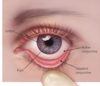
What is it called where the conjunctive, cornea & sclera all meet?

A

limbus

13
Q

What is the presentation of a patient with nasolacrimal duct obstruction?

Treatment?

A
  • Presentation
    • recurrent tearing and discharge in an infant
  • Treatment
    • warm digital massage over nasolacrimal sac
    • typically resolves by 12 months - if not, opthalmologist can probe to open
14
Q

What is the term for the fibrous layer beginning at the periosteum of the skull & extending to the eyelids?

A

orbital septum

15
Q

What are the terms for an infection anterior to the orbital septum & posterior to the orbital septum?

A
  • anterior - periorbital cellulitis
  • posterior - orbital cellulitis
16
Q

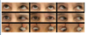
Presentation of a patient with periorbital cellulitis?

Treatment?

A
  • Mild condition - common
    • NO proptosis
    • NO ophthalmoplegia
    • LESS pain with eye movement
    • NO vision loss
  • Treatment
    • antibiotics
17
Q

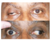
Presentation of a patient with orbital cellulitis?

Treatment?

A
  • Presentation - SERIOUS
    • ophthalmoplegia
    • pain with eye movement
    • proptosis
    • chemosis of conjunctiva
    • coexistin rhinosiusitis (90%)
  • Treatment
    • IV antibiotics x3weeks
    • frequently inpatient
18
Q

What is the common cause of orbital cellulitis?

What complications can arise from orbital cellulitis?

A
  • Cause
    • spread from adjacent siunses (usually ethmoid)
  • Complications
    • cavernous sinus thrombosis
    • meningitis
    • cerebral, orbital, subperiosteal abscess
    • vision loss
19
Q

Identify the bones & indicated landmarks on the provided image

A
20
Q

What bones make up the orbital roof?

A

frontal bone

lesser wing of the sphenoid

21
Q

What bones make up the lateral wall of the orbit?

A

zygogatic bone

greater wing of the sphenoid

22
Q

What is the thickest & strongest parts of the orbit?

A

lateral wall

23
Q

What bones make up the orbital floor?

A

maxillary

palatine

zygomatic

24
Q

What vessels are transmitted in the infraorbital groove?

A

infraorbital artery

maxillary dividiosn of trigeminal nerve

25
What vessels are transmitted in the supraorbital notch?
supraorbital vessels & frontal nerve
26
What bones make up the medial wall of the orbit?
ethmoid lacrimal maxillary sphenoid
27
What is the thinnest wall of the orbit? What is its special name?
medial wall overlying the ethmoid sinus = lamina papyracea
28
What is a "trap-door" fracture & where does this most often occur?
when bone breaks & then snaps back, trapping vessels/muscles that can become ischemic most commonly happens in younger patients b/c have more flexible bones - orbital floor
29
What is the clinical presentation of a patient with an orbital fracture?
* trauma history with pain * facial asymmetry * enophthalmos * edema/exophthalmos * diplopia
30
What steps should you take if you have a patient who has experienced blunt trauma & you suspect orbital fracture?
* physical exam * palpate orbital rim * pupillary response (CN II compression from edema) * visual acuitty * infraorbital nerve anesthesia * CN V2 maxially nerve division * CT Face & Orbit with contrast * refer to ophthalmologist ASAP
31
What structures are located in the superior orbital fissure?
CN III, CN IV, CN V1, CN VI ophthalamic vein sympathetic fibers
32
What structures are located in the inferior orbital fissure?
CN V2, inferior opthalamic vein
33
What structures are located in the optic canal?
CN II, Ophthalamic artery, central retinal vein
34
What artery supplies the majority of the eye itself? What about the eyelids & conjunctiva?
* Eye itself - internal carotid * Eyelid & Conjunctiva - external carotid
35
CN III controlls what ocular muscles?
all rectus (except lateral rectus) eyelid
36
CN IV controls what ocular muscles?
superior oblique
37
CN VI controls what ocular muscles?
lateral rectus
38
Fill out the provided table indicating what motion is the result of what nerve/muscle.
39
Having the patient move their eyes in these directions is assessing which muscles?
40
The provided image is an example of what nerve palsy? This is most likely due to what type of disease?
CN III palsy d/t decreased diffusion of oxygen & nutrients to interior fibers (DM / HTN)
41
The autonomic nerve fibers in CN III innervate what reflex? They are most commonly affected by what?
pupillary response on **periphery of nerve** - first affected by compression posterior communicatign artery aneurysm or superior cerebellar artery aneurysm uncal herniation intracranial neoplasm
42
What is the next step in management if you have a patient with a blown/dilated pupil?
CT / MRA it could be life threatening if it is a posterior communicating artery aneurysm
43
The patient in the provided image has what nerve palsy?
CN VI
44
What nerve is responsible for closing the eye?
CN VII
45
Sympathetic fibers reach the eye via what nerve? What about parasympthetics? The each have what effect on pupil size?
* sympathetic- nasociliary (branch of opthalamic nerve) * mydriasis (dilation) * parasympathetic- CN III * miosis (constriction)
46
What are the sensory nerves to the eye?
CN V V1 opthalamic & V2 Maxillary
47
Describe the nuclei & muscles involved in conjugate gaze
when looking left **left** nucleus **CN IV** contracts the **left lateral rectus** & stimulates the **right** nucleus of **CN III** (via **right MLF**) to contract the **right medial rectus**
48
Internuclear ophthalmoplegia that interrupts conjugate gaze is named for which eye?
the eye that is paralyzed
49
If there is a lesion in the right MLF, what is the result?
right eye cannot abduct right INO
50
What is the difference in presentation from a CN VII lower motor neuron lestion & an upper motor neuron lesion?
* lower - whole ipsilateral side is affected * upper - contralateral face below eye is affected
51
What is the most important eye consideration when dealing with a patient who has a CN VII palsy?
eye lubricants
52
What visual field defect would result from a lesion at 3 or 6?
53
Horner syndrome is associated with what conditions?
pancoast tumor (in apex of lung) spinal cord lesion above T1 (Brown-Sequard Syndrome) Late stage syingomyelia
54
What is the pneumonic for Horner Syndrome?
sympthetic denervation of face due to ipsilateral sympathetic trunk damage **PAM** is **horn**y * P - ptosis * A - anhidrosis (absense of sweating & flusing) * M - miosis (pupil constriction)
55
If you have a pupillary light reflex defect, you probably have problems with what structures? Common causes?
optic nerve or retina Cause - optic neuritis in MS, acute inflammation demyelination of CNS
56