Ophthamology Flashcards

(200 cards)

1
Q

Amblyopia

A

Lazy eye

2
Q

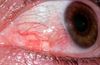
Types of chronic conjunctivitis

A

Vernal keratoconjunctivitis

Atopic keratoconjunctivitis

3
Q

Features of vernal keratoconjunctivitis

A
  • Children
  • Seasonal
  • FH of atopy
  • Bilateral
  • Ulceration and infiltration of upper cornea
4
Q

Features of atopic keratoconjunctivitis

A
  • Adults
  • Associated with atopy
  • Bilateral
  • Can cause corneal ulceration and scarring
5
Q

Neonatal conjunctivitis

A

Ophthalmia neonatorum

Usually secondary to N. gonorrhoeae or Chlamydia trachomatis

6
Q

Causes of acute red eye

A

Lids:

  • Blepharitis
  • Chalazion
  • Malposition

Conjunctiva:
- Conjunctivitis

Sclera:

  • Episcleritis
  • Scleritis

Cornea:
- Keratitis

Uveal tract:
- Uveitis

Trabecular meshwork:
- Acute glaucoma

Periorbital skin:

  • Preseptal cellulitis
  • Orbital cellulitis
7
Q

Presentation of Acanthamoeba keratitis

A

Pain
Red eye
Dendritiform epithelial lesions
Non-suppurative ring

8
Q

Fungal keratitis presentation

A

Red eye
Photophobia
Blurred vision
Discharge

9
Q

Anisocoria

A

Difference in pupil size > 4mm

10
Q

Management of fungal keratitis

A

Topical antifungal
Corneal graft if unresponsive

11
Q

Topical anti-fungals

A

Natamycin

Amphotericin

12
Q
A

Hyphaema - blood in anterior chamber

13
Q

Hypopion

A

Pus in anterior chamber

14
Q

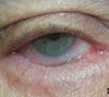
Orbital cellulitis presentation

A

Decreased vision
Unwell pt
Unilateral swollen eyelids
Ophthalmoplegia (reduced eye movements)
Proptosis

15
Q

Orbital cellulitis Mx

A

Ophthalmological emergency

CT scan
IV antibiotics

16
Q

Scleritis presentation

A

Extremely painful - often wakes at night
Cellular infiltration or entire sclera thickness

Red eye

17
Q

Scleritis complications

A

Ischaemia and necrosis

Scleral thinning (Scleromalacia perforans)

Globe perforation

18
Q

Scleritis prognosis

A

Can be self limiting

19
Q

Central (/Branch) Retinal Artery Occlusion definition

A

Commonly embolisation from carotid artery

20
Q

Central (/Branch) Retinal Artery Occlusion presentation

A

Painless
Possible RAPD
Retinal pallor
Cherry red spot

21
Q

RAPD

A

Relative afferent pupillary defect

Paradoxical dilatation of directly stimulated pupil

22
Q

Cherry red spot

A

At macula

Retina thinner - see underlying choroid

23
Q
A

Entropion

24
Q

Entropion definition

A

Inward turning of lid margin

Malposition of eye lid

25
Complication of entropion
Eyelashes cause corneal abrasions
26
Ectropion
27
Ectropion definition
Outward turning of lid margin Almost always lower lid
28
Ectropion complication
Exposed conjuctiva can become inflammed, scarred and keratinised
29
Dendritic ulcer on cornea
30
Bacterial keratitis organisms
Pseudomonas aeruginosa Staph epidermidis Strep pneumoniae H. influenzae
31
Bacterial keratitis presentation
Purulent conjunctivitis Reduced vision Corneal ulcer / opacity Hypopyon
32
Blepharitis
33
Blepharitis definition
Infection of lid margins Overproduction of sebum by lid margin glands
34
Blepharitis organisms
Staphylococcal bacteria
35
Sx of blepharitis
Chronic ocular irritation Watery eye Red eye
36
Signs of blepharitis
Crusting and scaling of lash line Plugs of sebum in meibomian gland orifices
37
Posterior synechiae Irregular pupil
38
Muscles of the eye
Superior, inferior, medial, lateral rectus Superior, inferior oblique Levator palpebrae superioris
39
Management of chronic conjunctivitis
Similar to acute conjunctivitis Greater use of topical and oral steroids
40
Uveitis definition
Inflammation of uveal tract
41
Components of uveal tract
Includes iris, ciliary body and choroids
42
Iatrogenic / neonatal conjunctivitis
43
Iatrogenic conjunctivitis definition
Drops prescribed can cause occular irritation and red eye
44
Management of allergic conjunctivitis
Topical antihistamine Topical mast cell stabiliser Combination of antihistamine and mast cell stabiliser Systemic antihistamines Topical steroid
45
Topical antihistamine for allergic conjunctivitis
Levocabastine
46
Topical mast cell stabiliser for allergic conjunctivitis
Sodium cromoglycate
47
Combined antihistamine and mast cell stabiliser for allergic conjunctivitis
Olopatadine
48
Management of bacterial conjunctivitis
Spontaneous resolution Broad spectrum topical antibiotics
49
Broad spectrum topical abx for bacterial conjunctivitis
Chloramphenicol Fucithalmic acid
50
Conjuctiva definition
Translucent membrane Lines inside of lids and sclera up to limbus of cornea
51
Parts of conjunctiva
Palpebral conjunctiva - inside of lids Bulbar conjunctiva - on sclera
52
Pupil dilator drugs
Tropicamide +/- Phenylephrine
53
Hypermetropia
Long sightedness
54
Acute Angle Closure Glaucoma
55
Corneal abrasion with fluorescein
56
Causes of Ectropion
Ageing (involutional) Conjuctival scarring (cicatricial) Paralytic (facial nerve palsy) Mechanical (lower lid tumours)
57
Complications of Herpes Zoster Ophthalmicus
Scleritis Secondary glaucoma Cranial nerve palsies Retinitis
58
Herpes Simplex Virus pathogenesis
Primary infection, then lies dormant in trigeminal ganglion Causes recurrent epithelial keratitis
59
Sx of ophthamological HSV infection
Pain Red eye Dendritic ulcer Reduced corneal sensation
60
Trabecular meshwork pathway
Ciliary bodies produce aqueous humour Bathes lens Exits anterior chamber via angle
61
Allergic conjunctivitis presentation
Rapid onset Itching Red eye Usually bilateral
62
Signs of allergic conjunctivitis
Giant cobblestone papillae Conjunctival chemosis (oedema) Preauricular lymphadenopathy
63
Myopia
Short sightedness
64
Causes of keratitis
Bacterial Viral Fungal Acanthamoeba
65
Fluorescein
Causes yellow staining of epithelial defect Eg. ulcer, abrasion More apparent under blue light
66
Peirorbital cellulitis presentation
Swelling No decreased vision No proptosis Normal eye movements
67
Mx of periorbital cellulitis
Broad spectrum oral abx
68
Keratitis risk factors
Contact lens Trauma Dry eye / blepharitis Immunosuppression
69
Presentation of scleritis
Extreme pain - wakes at night Epiphora Red eye Scleromalacia perforans
70
Epiphora
Watering of eye
71
Scleromalacia perforans
Scleral necrosis and thinning
72
Corneal sensation nerve
Trigeminal nerve
73
Amaurosis Fugax definition
Embolus from carotid passes through retinal vasculature Transient loss of vision
74
Types of uveitis
Anterior uveitis (iritis) Intermediate uveitis Posterior uveitis
75
Anterior uveitis definition
Inflammation of iris and anterior chamber
76
Intermediate uveitis definition
Inflammation to vitreous and peripheral retina
77
Posterior uveitis definition
Inflammation of posterior uveal tract and overlying retina
78
Cause of superior quadrantanopia
Post chiasmal lesion within temporal lobe
79
Presentation of vial conjunctivitis
Red eye Watery discharge Usually bilateral Conjunctival follicles Preauricular lymph nodes
80
Why is viral conjunctivitis usually bilateral
Highly contagious
81
Causes of viral conjunctivitis
Adenovirus HSV 1
82
Causes of gradual loss of vision
Cataracts Macula degeneration Retinitis pigmentosa Diabetic retinopathy Glaucoma
83
Retinitis pigmentosa presentation
Gradual peripheral vision loss Congenital Tunnel vision
84
Signs of diabetic retinopathy
Cotton wool spots (ischaemia) Haemorrhages
85
Management of scleritis
Oral prednisolone Immunosuppressive agents
86
Immunosuppressants used for scleritis
Mycophenolate mofetil Azathioprine
87
Herpes Zoster Ophthalmicus cause
VZV Aka shingles
88
Herpes Zoster Ophthalmicus pathogenesis
VZV lies dormant in trigeminal ganglion
89
Mx of Herpes Zoster Ophthalmicus
Oral Aciclovir Topical steroids
90
Sign of Herpes Zoster Ophthalmicus
Lesions on nose
91
Giant cobblestone papillae Allergic conjunctivitis
92
Acanthamoeba keratitis
93
Acanthamoeba keratitis definition
Protozoan (especially in water) Consider with pts who swam in contact lenses
94
Keratitis
95
Keratitis definition
Inflammation of cornea
96
Keratitis complications
Ophthalmologic emergency May permanently impair vision or perforate eye
97
Management of episcleritis
Observation Simple lubricants Oral NSAIDs Topical steroids
98
Bacterial conjunctivitis
99
Orbital cellulitis
100
Types of allergic conjunctivitis
Seasonal allergic rhinoconjunctivitis Perennial allergic rhinoconjunctivitis
101
Seasonal allergic rhinoconjunctivitis allergen
Commonly pollen
102
Perennial allergic rhinoconjunctivitis allergen
Eg. house dust mite
103
Blink reflex nerve
Facial nerve
104
Central (/Branch) Retinal Vein Occlusion Sx
Painless Loss of vision Possible RAPD
105
Signs of central (/branch) retinal vein occlusion
Dot haemorrhages Flame haemorrhages Cotton wool spots Swollen optic disc Macular oedema Neovascularisation
106
Central vs branch retinal vein occlusion
Branch retinal vein occlusion confined to area supplied
107
Scleritis
108
Scleromalacia perforans
109
Management of viral conjunctivitis
Conservative Lubricants (for comfort) Antibiotics (chloramphenicol) - prevent secondary bacterial infection Topical Aciclovir - if HSV-1 isolated Topical steroid drops - only for severe inflammation
110
Conservative Mx of viral conjunctivitis
Usually spontaneously resolves ~ 2 wks Discomfort may persist for months
111
Presentation of uveitis
Photophobia Red eye Pain Blurred vision
112
Signs of uveitis
Keratic precipitates Posterior synechiae
113
Keratic precipitates
Clumps of cells on corneal endothelium
114
Posterior synechiae
Adhesions between lens and iris Results in irregular pupil
115
Causes of central retinal vein occlusion
Idiopathic HTN - artery compresses vein Inflammatory Thrombophilic tendency
116
Inflammatory causes of CRVO
Sarcoidosis Lyme disease
117
RFs for thrombophillic tendancy
- Hyperviscosity (protein S or C deficiency) - Smoking - COCP - Pregnancy - Tumours
118
Causes of sudden loss of vision
Central retinal vein occlusion Central retinal artery occlusion Anterior Ischaemic Optic Neuropathy Optic neuritis Posterior vitreous detachment Retinal detachment
119
Risk factors for Acute Angle Closure Glaucoma
Age Female Hypermetropic (smaller eyes) FH
120
Episcleritis
121
Sx of episcleritis
Mild discomfort Epiphora (watering) Unilateral redness
122
Mx of episcleritis
Self limiting and recurrent Can have underlying systemic disorder
123
Causes of RAPD
Optic neuritis Optic atrophy Retinal detachments Central retinal artery occlusion Optic nerve compression
124
Causes of entropion
Ageing (involuntional) Conjunctival scarring (cicatrical) Acute spastic entropion (spasm of orbicularis muscle)
125
Causes of conjuntival scarring
- Trachoma - Stevens-Johnson syndrome - Ocular pemphigoid
126
Management of central retinal vein occlusion
Treat underlying condition Macular oedema - laser treatment Neovascularisation - laser ischaemic areas
127
Medical Mx of Acute Angle Closure Glaucoma
Lower intraocular pressure: - IV acetazolamide - Topical beta blockers - Topical pilocarpine (prevent attack in the fellow eye)
128
Surgical Mx of acute angle closure glaucoma
Reverse pathology: - Laser iridotomy - Trabeculectomy
129
Sx of Acute Angle Closure Glaucoma
Haloes Red eye N/V Pain Photophobia Visual loss
130
Signs of acute angle closure glaucoma
Corneal oedema (hazy) Fixed semi-dilated pupil Shallow anterior chamber
131
Viral causes of keratitis
HSV VZV Adenovirus
132
Causes of scleritis
Systemic associations Infection Post ocular surgery
133
Systemic causes of scleritis
Wegener's gramulomatosis Rheumatoid arthritis Polyerteritis nodosa
134
Infection causes of scleritis
Secondary to corneal ulcers Often pseudomonas
135
Management of central retinal artery occlusion
Treat underlying condition Low dose aspirin and carotid ultrasound Drop intraocular pressure within first 2 hours
136
Methods to reduce intraocular pressure for CRAO
- Ocular massage - Anterior chamber paracentesis - IV acetazolamide
137
Fungal keratits pathogens
Filamentous fungi (eg. Aspergillus, Fusarium) Candida albicans
138
Signs of filamentous fungi keratitis
- Grey stromal infiltrate - Surrounding satellite infiltrates - Hypopyon
139
Signs of candida albicans keratitis
Yellow/white ulcer
140
Causes of Adult inclusion conjunctivitis
Chlamydia serotypes D-K
141
Presentation of Adult inclusion conjunctivitis
Unilateral mucopurulent discharge May be chronic if untreated Large conjunctival follicles Preauricular lymphadenopathy Subepithelial corneal deposits
142
Mx of adult inclusion conjunctivitis
Topical erythromycin - 2 weeks Referral to GUM clinic for systemic chlamydia abx
143
Causes of conjunctivitis
OH GLAD SCABS Ocular cicatricial pemphigoid Herpes Gonococcal infection Ligneous Adenoviral infection Diphtheria infection Stevens-Johnson syndrome Candida or Chlamidya infection Alkali burn Beta-haemolytic streptococcal infection Springtime (Allergic conjunctivitis)
144
Cause of Inferior quadrantanopia
Post chiasmal lesion in parietal lobe
145
Risk factors for central retinal artery occlusion
DM IHD Peripheral vascular disease Smoking Giant cell (temporal) arteritis
146
Management of uveitis
``` Topical steroid drops Mydriatic drops (eg. cyclopentolate) - prevent posterior synechiae ```
147
Keratic precipitates on corneal endothelium
148
Causes of uveitis
Idiopathic HLA-B27 associated arthropathies (PUBCAR) Infectious Sarcoidosis Juvenile Idiopathic Arthritis
149
Infective causes of uveitis
Syphilis TB Herpes Simplex / Zoster
150
Management of bacterial keratitis
Corneal scrape Broad spectrum topical antibiotics - ofloxacin Topical steroid - can help scarring and necrosis
151
Presentation of bacterial conjunctivitis
Red eye Eyelids stuck together Minimal pain Purulent discharge Conjunctival papillae
152
Bacterial conjunctivitis organisms
Staph aureus Staph epidermis Strep pneumoniae
153
Chalazion (meibomian cyst)
154
Chalazion / meibomian cyst definition
Meibomian gland dysfunction
155
Associations of chalazion / meibomian cyst
Acne rosacea Seborrhoeic dermatitis
156
Mx of chalazion
- Hot spoon bathing - Abx - Surgical incision and curettage
157
Association of myopia
Increased risk of retinal detachment
158
Association of hypermetropia
Increased risk of angle-closure glaucoma
159
Sebum
Oily waxy Lubricates and waterproofs skin
160
Meibum
Oily Prevents evaporation of the eye's tear film
161
Causes of Trachoma
Serotypes A-C
162
Trachoma definition
Chronic conjunctivitis Endemic to Africa and Asia
163
Follicle definition
Hyperplasia of lymphoid tissue Diagnostically significant
164
Causes of follicles
HSV Chlamydia
165
Papillae definition
Hyperplastic conjunctival epithelium thrown into numerous folds Non specific
166
Causes of homonymous hemianopia
Lesion of optic radiation Eg. stroke
167
Cause of bitemporal hemianopia
Lesion at optic chiasm Eg. pituitary tumour, craniopharyngeoma
168
Red eye, not responsive to light, pupil dilated
Acute angle closure glaucoma
169
Red eye, not responsive to light, pupil constricted
Iritis
170
Types of Age related macular degeneration (ARMD)
Dry ARMD Wet ARMD
171
Features of dry ARMD
Slow progression Moderate visual loss No Rx
172
Features of wet ARMD
Quick pregression Severe visual loss Sometimes treatable Neovascularisation
173
Types of dry ARMD
1) Drusen 2) Atrophy
174
Mx of wet ARMD
Anti-VEGF medication (ranibizumab) Pan photo-coagulation
175
Causes of flashes / floaters
Retinal detachment Vitreous detachment
176
Pathology of diabetic retinopathy
Increased glucose -\> ROS -\> Basement membrane thickening -\> EC proliferation/bleeding
177
Types of diabetic retinopathy
Background Pre-proliferative Proliferative
178
Background diabetic retinopathy
- micro-aneurysm - Exudate - Dot haemorrhage
179
Pre-proliferative diabetic retinopathy
More extensive ischaemic changes - venous dilatation - venous bleeding - multiple cotton wool spots (\>5) (micro infarcts of retina)
180
Proliferative diabetic retinopathy
Neovascularisation Rubeosis iridis needs pan photocoagulation, if not treated tractional retinal detachment
181
Rubeosis iridis
Neovascularisation of the iris May obstruct drainage of aqueous humour resulting in 'neovascular glaucoma
182
Ix for diabetic retinopathy
Fluorescein Angiography - assessing ischaemia - localising leakage
183
Mx of diabetic retinopathy
Laser to solve: 1) maculopathy - closure of leaking vessels 2) proliferative disease - destroy ischaemic retina, decrease angiogenic growth factors
184
Microvascular complications of DM
Retinopathy Nephropathy
185
Macrovascular complications of DM
Neuropathy Cardiac problems Ulcers
186
Maculopathy definition
Clinically significant macular oedema Complication of diabetic retinopathy
187
Types of maculopathy
Focal maculopathy Diffuse maculopathy Ischaemic maculopathy
188
Features of focal maculopathy
Focal leakage from a microaneurysm or dilated capillaries Exudates seen, often in rings around the leaking microvasculature (circinate exudates)
189
Features of diffuse maculopathy
Diffuse retinal oedema Leakage from microvasculature at posterior pole of eye
190
Features of ischaemic maculopathy
Closure of perifoveal microvasculature Confirmed with fluorescein angiography
191
Strabismus other name
Squint
192
Types of stabismus
Hyper- / Hypo- / Exo- / Eso-: Tropia Phoria
193
(Hyper)tropia definition
Constant strabismus in one eye | (Hyper looks up)
194
(Hyper)phoria definition
Latent strabismus Present in both eyes
195
Directions of strabismus
Hyper: Up Hypo: Down Exo: Temporal Eso: Nasal
196
Test for tropia strabismus
Cover test Cover one eye, eye moves slightly
197
Test for phoria strabismus
Alternate cover test Look at pen whilst covering one eye at time Pen moves if latent squint present
198
Causes of strabismus in children
Refractive error Neurological
199
Causes of strabismus in adults
HTN Stroke DM Myasthenia gravis
200