Opthalmic 1 Flashcards

(74 cards)

1
Q

OD means? OS means? OU means?

A

OD = right eye, OS = left eye, OU = both eyes

2
Q

________: movement of the iris. This finding is usually supportive of lens instability

A

Iridodonesis

3
Q

_______: in a well-lit environment

A

Photopic

4
Q

_______: in a dark environment

A

• Scotopic

5
Q

__________: using light that is shone into the eye to reflect against internal structures and highlight normal/abnormal features during the ophthalmic exam

A

Retroillumination

6
Q

_________: inflammation of the cornea. Most commonly evident by the presence of corneal ulceration, corneal infiltrate, or blood vessels

A

Keratitis

7
Q

________ _______: inflammation of the cornea (keratitis) that is caused by lack of sensory innervation (ophthalmic branch of CN V)

A

Neurotrophic keratitis

8
Q

_________: applying light pressure to both eyes (through the eyelids) simultaneously with your index finger to detect for asymmetry. This is a useful test for detecting disease of the orbit or space occupying disease behind the globe

A

Retropulsion

9
Q

__________: disruption of the corneal epithelium and exposure of the corneal stroma

A

Corneal ulceration

10
Q

________: application of fluorescein dye to the ocular surface which then appears at the nares. This is a test of nasolacrimal patency

A

Positive Jones Test

11
Q

__________: application of fluorescein dye to the ocular surface and subsequent appearance of aqueous humor leaking through dense fluorescein stain. This test confirms corneal perforation

A

Positive Seidel test

12
Q

______: defined as a breakdown of the blood ocular barrier.

A

Uveitis

13
Q

___________: vision loss that occurs from optic nerve damage as a result of high intraocular pressure

A

Glaucoma

14
Q

A well designed ______ ______ will promote a thorough exam and clear communication

A

examination form

15
Q

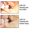
What are your 5 required tools?

A

• Strong light source in a dark environment • Schirmer Tear Test • Fluorescein • Tonometry (proparacaine) • Ophthalmoscope (tropicamide)

16
Q

Which tool are appropriate and not appropriate for a strong light source?

A

Halogen = appropriate (Finoff transilluminator, Otoscope) Incandescent = too dim (pen light) LED=too bright (need a piece of white tape)

17
Q

True or False: A magnifier is not helpful

A

False!!!!! extremely helpful

18
Q

ID

A

Distichia

19
Q

Describe the benefits? What is it?

A

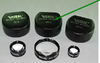
• Macrolens attachments benefits:

– Inexpensive

– Good images of the anterior segment

– Ability to save and share images!

20
Q

What do we do in the initial patient assessment? (4)

A

– Distance examination

– Vision tests

– Tests of orbital symmetry

– Retroillumination

21
Q

In addition to initial patient assessment we also do these 3 procedures?

A
  • Cranial nerve assessment
  • Minimum database
  • Ophthalmoscopy
22
Q

What is the first thing you do in the initial patient assessment

A
  • Assessment of entire body
  • Assessment of head and eyes at a distance
23
Q

What can we do to assess vision in our animals?

A

• Cotton Ball Test

– Most visual animals follow cotton balls

• No sound or scent

– Cats may choose not to participate!

24
Q

When we are assessing vision we place objects in a random order and this is doing what?

A

• Maze Testing

– Place objects in random order

– Conduct in both bright and dim light settings

– Look for speed of navigation and object avoidance

25
How do we assess orbital symmetry?
* Orbital palpation * Dorsal view assessment * Retropulsion
26
What are we testing in these pics?
Retroilllumination
27
What cranial nerves/assessements do we like to assess?
– Palpebral reflex – Menace response – Pupillary light reflex – Dazzle reflex – Oculocephalic reflex – Corneal reflex
28
What nerves do we test in the palpebral?
– CN V (afferent) – CN VII (efferent)
29
What nerves do we assess in the menace response? What does it require?
– CN II (afferent) – CN VII (efferent) – Learned (~12 weeks old) – Requires visual acuity of only 20/20,000! NOT THAT GREAT
30
The pupillary light reflex asesses what nerves?
– CN II (afferent) – CN III (efferent) oculomotor (shine bright light in eye assess indirect and direct crossover at chiasm)
31
What nerves do the dazzle reflex?
– CN II (afferent) – CN VII (efferent) (subcortical and often have it if pathways intact)
32
What is significant about the pupillary light reflex and the dazzle reflex?
\*\*These tests do not reflect vision\*\*
33
What nerves does the oculocephalic reflex?
– CN II\* (afferent) – Peripheral and central vestibular components – CNs III, IV, and VI (efferent) she likes it to test mobility
34
The corneal reflex assesses these nerves?
– CN V (afferent) – CN VI and VII (efferent)
35
What is being shown here?
• Corneal reflex – abnormal example Neurotrophic keratitis OD = Loss of the ophthalmic branch of CN V
36
WHAT DO you do for the minimum database? (3)
– Schirmer Tear Test I – Fluorescein Stain – Tonometry
37
**Guildeline for Schirmer tear test 1** * **\_\_\_\_\_\_\_\_\_\_\_\_** * **\_\_\_\_\_\_\_\_\_\_\_\_** * **\_\_\_\_\_\_\_\_\_\_\_\_** **NORMAL** * **Dogs= \_\_\_\_\_\_\_\_\_\_** * **​\_\_\_\_\_\_** * **Cats= \_\_\_\_\_\_\_\_**
* **Guidelines** * Performed before any other drops or ointments * Performed before sedation or anesthesia * Performed for **_60 seconds_** * **Normal** * **Dogs = \>15mm wetting/minute** * _Basal and reflex_ tearing * Cats = unpredictable!
38
This is an example of?
39
After performing the schirmer tear test for our minimum database we next do?
Fluorescein Stain
40
Describe the dye for Fluorescein stain? What structures will and won't stain?
Aqueous stain * (**Hydrophilic (stroma)** dye that fluoresces green under cobalt blue light) * Epithelium – _hydrophobic_ – will not stain * Stroma – _hydrophilic_ – will stain bright green * Descemet’s membrane – _hydrophobic_ – will not stain
41
What are the 3 common uses for Fluorescein stain?
1. Diagnosis and characterization of corneal ulceration 2. Demonstration of nasolacrimal patency (Jones Test) 3. Demonstration of corneal perforation (Seidel Test)
42
Decribe the pattern of stain and what is being exhibited in the pic?
Dendritic
43
Decribe the pattern of stain and what is being exhibited in the pic
Indolent
44
Decribe the pattern of stain and what is being exhibited in the pic
Descemetocele
45
What does the Jones test assess?
• Assesses nasolacrimal patency
46
What are the system components for the nasolacrimal components? (6)
* – Medial canthus * Upper punctum (circled) * Lower punctum (circled) * _– Lacrimal sac_ * _– Nasolacrimal duct_ * _– Nasal and pharyngeal openings_
47
When performing the jones test and you note a blckage what negative affect could this cause?
Blockage could cause epiphora (tearing)
48
A positive Jones Test demonstrates _______ \_\_\_\_\_\_\_\_
nasolacrimal patency
49
A negative Jones Test does or does not prove this?
does not necessarily prove nasolacrimal obstruction
50
What is being tested here?
Seidel Test
51
A positive Seidel test confirms _____ \_\_\_\_\_\_\_ ## Footnote – What does it demonstrate?
* corneal perforation * Demonstrates aqueous leaking through fluorescein stain
52
\_\_\_\_\_\_\_\_: Measures intraocular pressure in mmHg
Tonometry
53
Guidelines for tonometry: -Do not perform in ____ eyes Performed (BEFORE OR AFTER?) pupil dilation?
54
* fragile * Peformed BEFORE pupil dilation (tropicamide)
55
Tonometry * Which reading is your most accurate? * \_\_\_\_-\_\_\_\_ mmHG is normal and should be below \< \_\_\_\_\_mmHG at ALL TIMES * Pressures over \>\_\_\_\_\_\_with vision loss indicate \_\_\_\_\_\_ * Low intraocular pressures are consisitent with \_\_\_\_\_\_
* The lowest reading is your most accurate reading! * _10-20 mmHg_ is normal. Should be \<25mmHg at all times. * Pressures \>25mmHg **with** vision loss = **_Glaucoma_** _• Low_ intraocular pressures are consistent with **_Uveitis_**
56
This pic is depicting what type of tonometry?
Tonometry (Tonopen: applanation)
57
Applanation tonometry correlates force used to this? What do we use when peforming this?
Correlates force used to flatten cornea with IOP Use topical anesthetic
58
Unfortunately applanation tonometry is easy todo this to? What do we do daily or prior to use?
• Easy to artificially elevate IOP – Brachycephalics * Calibrate
59
What factors do we consider with Ophthalmoscopy? (5)
1. Magnification 2. Field of view 3. Depth perception (stereopsis) 4. Initial ease of use 5. Safety
60
A large field of view allows for a more rapid and thorough _____ examination, making the \_\_\_\_\_technique more valuable for veterinary ophthalmology
fundus; indirect
61
The pointer is indicating
Lens diopter number
62
WIth applanation tonometry what 4 coefficient of variance are considered?
– Reliability of result – Should be 5% (TonoPen) – Should be 95% (TonoPen Avia) – Measures variability between 3-6 readings
63
What are some easy mistakes with applanantion tonometry?
– Tip cover too tight – Tip cover too loose
64
What instrument is being shown
Tonometry (Tonovet: rebound)
65
66
\_\_\_\_\_\_: – Lightweight probe “bounces” off cornea Is topical anesthetic required?
TonoVet No topical anesthetic required!
67
The tonovet is calibrated by manufacturer and can be species dependent for horse, dogs, and other. It records _____ measurements
Records 6 measurements – Averages final result (excludes highest and lowest) – Line indicates standard deviation * No line (\<1) * Low line (1-2.5) * Mid line (2.5-3.5) * High line (\>3.5)
68
What is really important for the opthoexam especially when measuring IOP? What 4 things should we do?
Proper restraint * No pressure on neck * Hand under chin and behind head * Head in neutral position * Patient standing, sitting or sternal
69
Which is the correct IOP? • 25mmHg, 13mmHg, 17mmHg
13 mmHG – Lowest IOP most accurate
70
What are some easy mistakes that affect the IOP?
– Pressure on neck – Head position – Squinting dog – Pressure on globe from improper lid retraction
71
True or False – Lissamine Green ( both corneal stains) – Rose Bengal( both corneal stains) ARE commonly used tests
False Less commonly utilized tests
72
\_\_\_\_\_\_ is required for complete exam? (what is it and what structures can we assess after it?
Tropicamide This medication is used to widen (dilate) the pupil of the eye in preparation for certain eye examinations. It belongs to a class of drugs known as anticholinergics. Tropicamide works by relaxing certain eye muscles. – Retina * Neurosensory retina * Retinal pigmented epithelium – Tapetum – Choroid – Sclera – Optic nerve
73
Each image is demonstrating what for ophthalmoscopy?
74
Label the parts