PACES ALL concise Flashcards

1
Q

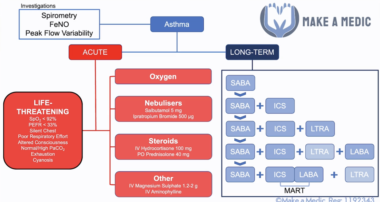
Asthma - def? Presentation? Ix? Severity? Short-term/long-term Mx incl. conservative? Mneumonic for conservative long-term Mx?

A

Def: chronic inflammatory airway disease characterized by reversible airway obstruction + airway hyperresponsiveness

Presentation

  • Dry cough, polyphonic wheeze ( worse@night/morning)
  • Triggers: cold air, pollen, pollution, exercise
  • Atopic features - eczema, nasal polyps

Ix:

  • Peak flow variability (in peak flow diary)
  • Spirometry (shows reversibility after bronchodilator)
  • FeNO (fractional exhaled nitric oxide - a marker of airway inflamme, in uncertain Dx)
  • On exacerbation:
    • ABG, peak flows (min x4/day), CXR
  • Other:
    • Allergy testing - total IgE, specific IgE (RAST), serum eosinophil count

Asthma severity:

  • Life-threatening: PEF <33% of baseline/SpO2 <92%/PaO2 <8/Normal or raised PaCO2 - should be low with resp alkalosis due to excess breathing only becomes acidotic when get tired
    • CHEST: Cyanosis, Hypotension, Exhaustion, Silent chest, Tachycardia
  • Acute severe: PEF <50% of baseline/RR >25/HR >110/can’t complete sentence without taking a breath
  • Moderate: PEF <75% of baseline

Short-term Mx:

  • A-E approach, seek senior support (call for help if life-threatening)
  • O2 - 15L NRM (if hypoxaemic)
  • Burst therapy:
    • SABA (spacer up to 10 puffs every 20 mins –> nebs)
    • Ipratropium Bromide (add to nebs if poor response/severe, every 4-6hrs)
    • Corticosteroids (min 5-day course, give within 1 hour, give IV if can’t take orally)
  • Other Tx options:
    • IV Magnesium sulfate (STAT dose if poor response above/severe) - consult senior before use
    • IV salbutamol (if on ventilation) - consult senior before use
    • IV Aminophylline - consult senior before use, requires ITU setting
  • If less distressed/more tired/shallow breaths/confused –> call critical care outreach team (CCOT) for ITU support

Long-term Mx (>16yrs):

  • Conservative: TAME
    • Technique
    • Avoid triggers
    • Monitor peak flow
    • Educate - formulate Personalised Asthma Action Plan (PAAP), Annual flu vaccine
  • Medical:
    1. SABA (reliever)
    2. SABA + ICS (preventer)
    3. SABA + ICS + LTRA (leukotrine receptor antagonist e.g. montelukast)
    4. SABA + ICS + LABA (+ LTRA stopped unless good response)
    5. SABA + MART (ICS + LABA COMBO) (+ LTRA)
    6. NOTE: maintenance & reliever therapy (MART) - used as preventer & maintenance inhaler
    7. Specialist input (e.g. for oral steroids)
2
Q

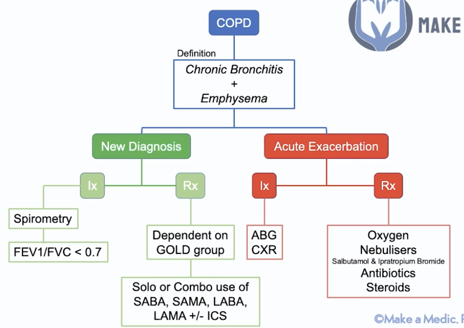
COPD - definition? Signs & Sx? New Dx & exacerbation Ix/Mx? Prognosis factors?

A

Def: chronic bronchitis (damaged to cilia in bronchi - blue bloater) + emphysema (damage to alveoli - pink puffer)

Presentation:

  • Cough (productive), SoB (starts on exercise)
  • RF exposure - smoking/pollution
  • Signs:
    • Barrel chest
    • Hyper-resonant (air trapping)
    • Reduced breath sounds
    • Widespread expiratory wheeze
    • Coarse crackles if exacerbation (mucus in airways)
    • Other - asterixis (CO2 retention flap), raised JVP (cor pulmonale)
    • NOTE: COPD does not cause clubbing –> cancer/bronchiectasis

New Dx Mx:

  • Ix:
    • Bedside - mMRC dyspnoea scale, O2 sats, ECG (cor pulmonale)
    • Spirometry - FEV1/FVC <0.7 (forced exp volume in 1s)
      • Mild - FEV1 ≥ 80%
      • Moderate - FEV1 ≥ 50%
      • Severe - FEV1 ≥ 30%
      • Very severe - FEV1 <30%
    • Bloods - FBC, ABG, eosinophil count, alpha1-antitrypsin level
    • Imaging - CXR, CT chest
    • Other: serial peak flows if asthma DDx, sputum culture (in freq exacerbation), pul funct tests
  • Mx:
    • Conservative - stop smoking, influenza + pneumococcal vaccine, inhaler device training
      • Persuade to stop smoking
      • Pul rehab
      • Prick them - influenza + pneumococcal vaccine
      • Psych issues
    • Medical - depends on severity - GOLD group –> solo/combo of:
      • SABA e.g. salbutamol
      • SAMA e.g. Ipratropium bromide
      • LABA e.g. salmeterol
      • LAMA e.g. tiotropium
      • ± ICS e.g. beclomethasone
      • Other: mucolytic e.g. acetylcysteine, O2 therapy, theophylline
    • Medical pathway:
      • 1 - SABA/SAMA
      • 2a - Steroid-responsive (eosinophilia/atopy): LABA + ICS
      • 2b - Not steroid-responsive: LABA + LAMA
      • 3 - LABA + LAMA + ICS
      • 4 - specialist input e.g. theophylline
    • Surgical - lung reduction surgery (large bullae)
    • Other: long-term O2 therapy
      • Only if non-smoker (smoker –> burns)
      • Only if <7.3 PaO2/<8 if also pul HTN
      • Only if PaCO2 does not rise excessively on O2

Acute Exacerbation Mx:

  • Ix: ABG, ECG, CXR
  • Mx:
    • 15L O2 NRM
    • Nebs - salbutamol + IpB
    • Steroids (PO pred/IV hydrocortisone)
    • Abx if infective –> prophylactic abx if persistent infections - azithromycin

Prognosis factors:

  • Body mass - worse if obese
  • Obstruction - worse if reduced FEV1
  • Dyspnoea
  • Exercise capacity - how far can you walk in 6 minutes?

Complication –> vasoconstriction to redirect blood flow to well-oxygenated areas of the lungs –> if widespread –> pul HTN –> cor pulmonale

3
Q

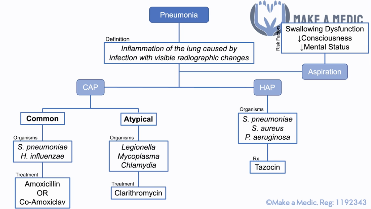
Pneumonia - def? Presentation? Types? Ix? Scoring? Mx?

A

Def: inflammation of lung caused by inf w/ visible radiographic changes

Presentation:

  • Decreased chest expansion, dull on percussion, increased sound vocal resonance
  • Coarse crackles
  • Bronchial breathing – bronchial airflow instead of alveolar airflow (due to transmission of sounds through consolidation) – check by listening to sound at the throat

Ix: ABG, CXR, sputum culture (mod/high severity)

Scoring for CAP: CURB-65 (confusion, urea ≥7mmol/L, RR ≥30, BP <90/60, ≥65yrs)

  • +1 = outpatient; +2 = outpatient/inpatient, +3 = inpatient ± ITU
  • NOTE: urea is no longer used

Types & Mx –> local abx guidelines

  • CAP:
    • Typical (S. pneumo, H. influenzae) –> Amox/Co-Amox
    • Atypical (Legionella, Mycoplasma, Chlamydia) –> Clari
      • ​Dry cough (instead of productive), myalgia, confusion, diarrhoea
    • NOTE: if not sure often given Co-Amox + Clari
  • HAP - pneumonia arising >48hrs after admission to hospital (S. pneumo, S. aureus, P. aeruginosa) –> Taxocin (pseudomonal cover)
  • Aspiration - RFs: swallowing dyfunct, reduced consciousness, reduced mental status –> elderly/frail

Goes into septic shock –> give IV fluid + senior help + check abx sensitivity (ring lab) –> ITU (intropic support - NA to increase PVR)

  • Other aspects of septic-6
  • NOTE: dobutamine is for cardiogenic shock to increase CO - this is not relevant here
4
Q

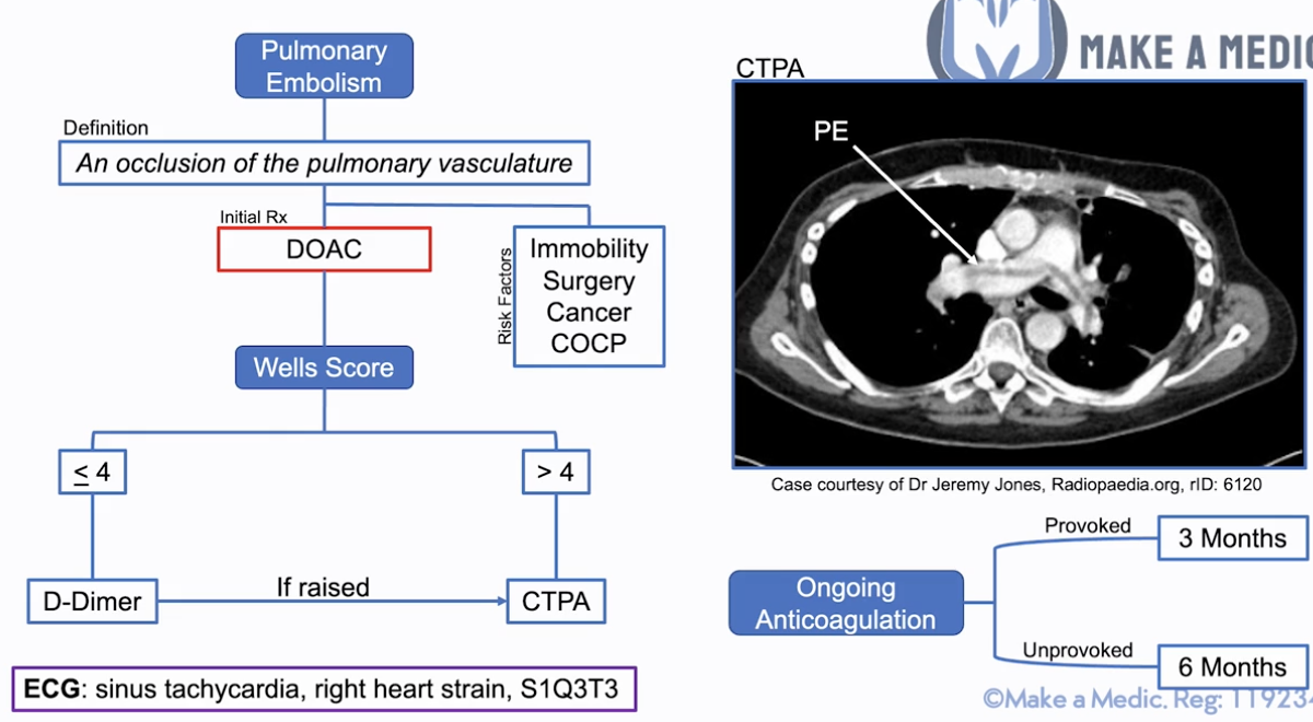
PE - def? Sx? RFs? Scoring & Ix? Mx?

A

Def: occlusion of pulmonary vasculature characterized by sharp pleuritic chest pain

Sx: SYNCOPE, sudden SoB, pleuritic chest pain, haemoptysis

RFs: SICC - Surgery, Immobility, Cancer, COCP

Initial Tx:

  • DOAC (e.g. Apixaban) or unfractionated heparin (if bleeding risk, can be reversed easily)
  • Massive PE –> IV unfractionated heparin for hours before and after thrombolysis e.g. IV alteplase

Scoring & Ix: Well’s score

  • ≤4 = D-Dimer –sign raised–> CTPA
  • >4 = CTPA
  • ECG useful - sinus tachycardia, right heart strain, S1Q3T3
  • NOTE: Troponin = useful markers for PE severity (indicates right heart strain)

Ongoing anticoagulation - DOAC/Warfarin

  • Provoked - 3 months (SICC)
  • Unprovoked - >6 months + cancer & thrombophilia testing
    • Ix for cancer –> any Sx?
      • If yes - CT TAP
      • If no - FBC, U&E, LFTs, clotting, physical exam –> if concern –> CT TAP
      • Consider thrombophilia screen if no cancer for anti-phospholipid syndrome (anticardiolipin, lupus anticoagulant, anti-beta-2-glycoprotein)
8
Q

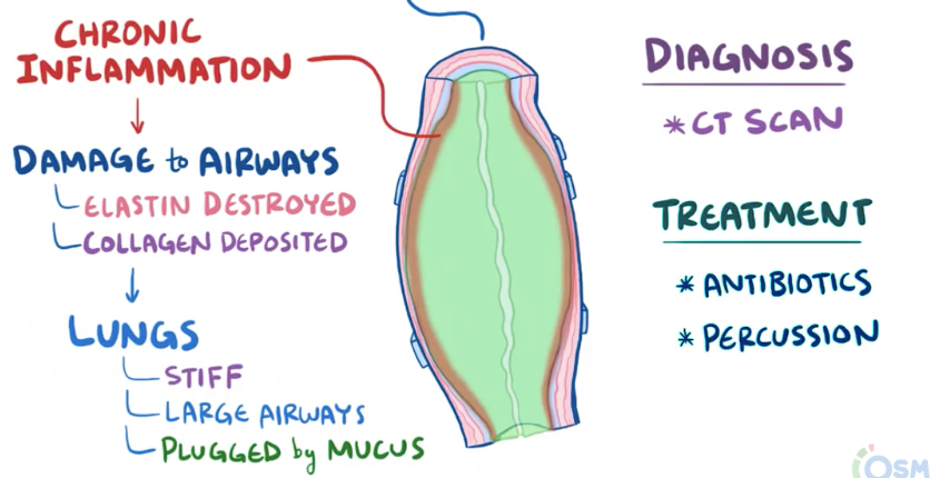
Bronchiectasis - definition? causes? presentation? Ix? Mx?

A

Def: obstructive lung disease characterised by permanent dilation of bronchi from the destruction of elastic & muscular components of the bronchial wall

  • Results from diseases causing chronic inflammation:
    • Primary ciliary dyskinesia - cilia don’t move –> mucus trapped in airways –> recurrent pneumonia –> chr inflammation
    • Cystic fibrosis - mucus thick & sticky –> recurrent pneumonia –> chr inflammation
    • Airway obstruction - tumour inside/outside airway or inhaled foreign body –> mucus can’t clear –> recurrent pneumonia –> chr inflammation
    • NOTE: worse with certain infections e.g. aspergillosis –> hypersensitivity response
  • Chr inflammation - has immune cells that release cytokines –> damage ciliated epithelial cells + destroys elastin –> airways dilated + clogged with mucus –> fibroblasts deposit collagen –> stiff lungs + mucus plug –> obstructive lung disease

Presentation:

  • RFs: CF, PCD, congenital disorders of airway, host immunodef (incl. HIV), recurrent lung inf
    • Less common - inhaled foreign body, connective tissue disease, IBD, alpha1-antitrypsin def
  • Productive cough (large amounts of sputum) ± haemoptysis - worse lying flat/one side
  • Dyspnoea (with increased severity)
  • Fever (on exacerbation)
  • Crackles, inspiratory squeaks & rhonchi, clubbing (from long-term hypoxia)

Ix: CXR (ring shadows, tramlines), high-res CT (signet ring sign), FBC + sputum culture & sensitivity (inf e.g. pseudomonas), pul function tests

  • Genetic testing (for possible RF causes): serum alpha1-antitrypsin, serum Ig lvls (ID), specific IgE for Aspergillus, sweat Cl test (CF), rheumatoid factor (CTD), HIV test, nasal nitric oxide (PCD)

Mx:

  • Conservative:
    • Persuade to stop smoking
    • Pul rehab
    • Prick them - influenza + pneumococcal vaccine
    • Psych issues
  • Medical:
    • Mucoactive agent (nebulised hypertonic saline)
    • Inhaled bronchodilator (SABA/SAMA/LABA/LAMA) - ONLY If co-existing asthma/COPD
  • Surgical: depending on primary cause e.g. IFB, tumour –> lobectomy/lung transplant
  • Exacerbation: short-term abx (azithromycin)
9
Q

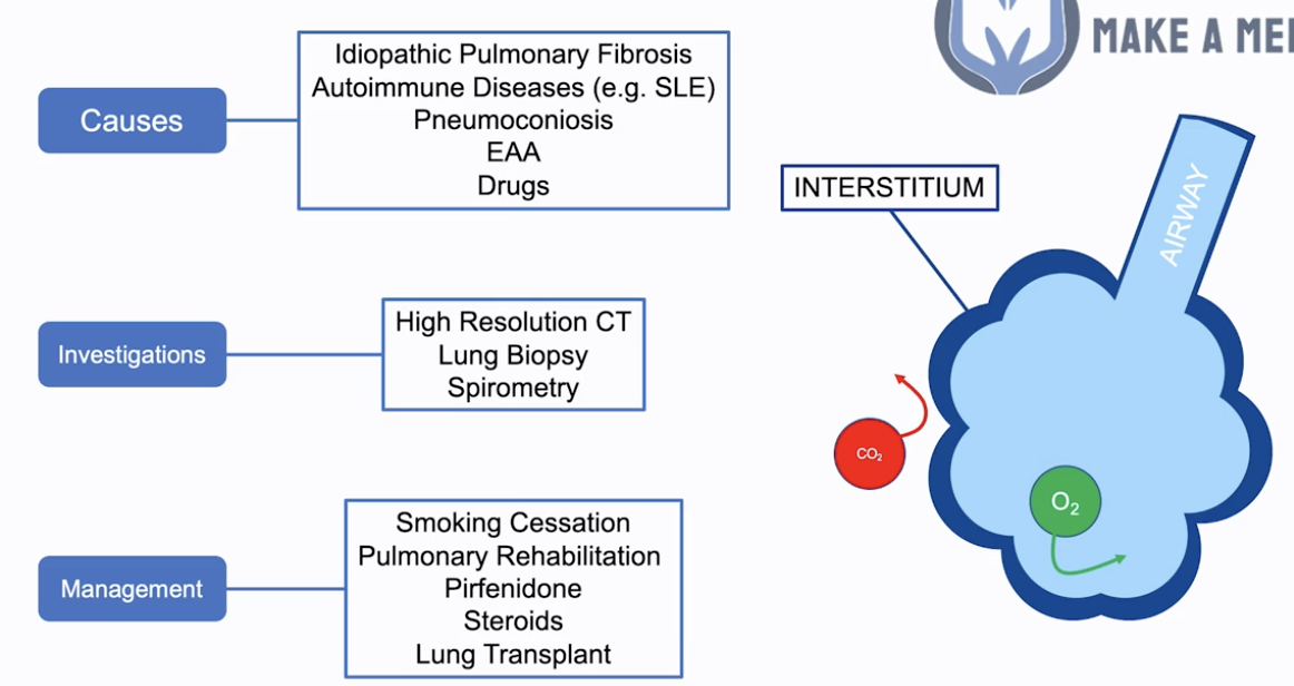
Interstitial lung disease - causes? presentation? Ix? Mx? Prognosis?

A

Interstitial lung disease

  • Refers to the tissue the gasses pass through e.g. surface of alveoli –> reducing gas exchange

Causes:

  • Upper zone fibrosis: PATEN (more occupational causes)
    • Pneumoconiosis (not asbestosis) - caused by inorganic dust (e.g. soot)
    • Aspergillosis/ABPA
    • TB
    • Extrinsic allergic alveolitis - caused by organic dust e.g. animal shedding
    • Negative seroarthropathies
  • Lower zone fibrosis: STAIR
    • Sarcoidosis
    • Toxins: B-MANS (Bleomycin, Methotrexate, Amiodarone, Nitrofurantoin, Sulfasalazine)
    • Asbestosis
    • Idiopathic pulmonary fibrosis
    • Rheum: SLE, RhA etc
  • Signs of conditions associated w/ pulmonary fibrosis:
    • MCP swelling - RA
    • Malar rash - SLE​
    • Kyphosis - Ank Spond (apical fibrosis)
    • Lupus pernio - sarcoidosis
    • Thick skin/’bird beak’ nose - systemic sclerosis
    • Aphthous ulcers, abdo scars - Crohn’s
    • Grey skin - amiodarone

Main Sx: progressive SOBOE, dry cough, fatigue, weight loss

Signs: find end-insp creps, clubbing

  • Other: reduced chest expansion, normal/reduced percussion, normal vocal fremitus

Investigations: spirometry, high-res CT & lung biopsy

  • Bedside: spirometry (low FEV1, low FVC, FEV1/FVC>0.8 = restrictive lung disease), ECG (right heart strain), drug review
  • Bloods
    • FBC, U&E, LFTs, CRP, ESR, ABG (T1RF)
    • AI screen - Serology (CTD): ANA, RhF, anti-CCP
    • Serum ACE for sarcoidosis
    • TB testing
  • Imaging
    • CXR: reticulonodular shadowing (honeycombing) + rule out lung cancer
    • High-res CT: more detailed view of alveolar structure –> ‘honeycombing’ + lung biopsy
    • Echo (right heart strain/cor pulmonale)
  • Invasive
    • Bronchoscopy + lung biopsy (gold-standard)

Management:

  • Conservative (4Ps):
    • Persuade to stop smoking, reduce exposure (meds, EAA)
    • Pul rehab
    • Prick them - influenza + pneumococcal vaccine
    • Psych issues
  • Medical: anti-fibrotic e.g. pirfenidone +/- prednisolone ± O2 (if hypoxaemic)
  • Surgical: lung transplant if severe deterioration/impairment/oxygen dependent

Prognosis: 3-4yrs post-Dx (no Mx increases survival)

16
Q

Pleural effusion - signs? causes? Ix? Mx?

A

Signs (if fluid > 300ml):

  • Key:
    • Stony dull in lung base
    • If large: tracheal deviation away
  • Reduced chest expansion
  • Reduced breath sounds
  • Reduced vocal fremitus

Causes:

  • Transudative (<30g/L protein)
    • HEART FAILURE
    • Hypoalbuminaemia: liver disease, nephrotic syndrome, malabsorption
    • Hypothyroidism
    • Meig’s syndrome (benign ovarian tumor + ascites + pleural effusion)
  • Exudative (>30g/L protein)
    • Infection: PNEUMONIA, TB, subphrenic abscess
    • Connective tissue disease; RA (also low glucose), SLE
    • Neoplasia: lung cancer, mesothelioma, metastases
    • Pancreatitis: high amylase in pleural fluid
    • Pulmonary embolism

Ix:

  • Bedside: obs, urinalysis for protein
  • Bloods:
    • ABG, BC
    • FBC, U&E, LFTs, CRP
    • Clotting (before needle aspiration), albumin (nephrotic syndrome)
    • Mantoux/ELISPOT (TB)
  • Imaging:
    • CXR: meniscus sign, dense shadowing, pleural effusion if ≥300mL fluid
    • CT chest - identify the cause

Management: US-guided pleural aspiration = thoracocentesis (21G needle, 50ml syringe) - above rib to avoid NV bundle

  • LDH & protein in pleural fluid & serum + RBCs, WBCs, cytology, culture, pH & glucose of pleural fluid
    • MC&S
    • Biochemistry: PPALS
      • ​Protein (also serum)
      • pH
      • Amylase
      • LDH (also serum)
      • Sugar (glucose)
    • Cytology
    • Immunology - if indicated (RF, ANA, complement)
  • Findings:
    • Protein > 30g/L: exudate
    • Protein < 30g/L: transudate
    • Protein 25-35g/L: use Light’s criteria. An exudate is likely if at least 1 of:
      • Pleural protein/serum protein > 0.5
      • Pleural LDH/serum LDH > 0.6
      • Pleural LDH > 2/3 upper limits of normal serum LDH
  • Tx cause e.g. abx for infection, furosemide for HF
  • Management of recurrent pleural effusion
    • Recurrent aspiration (thoracocentesis)
    • Pleurodesis
    • Indwelling pleural catheter

Pleural fluid features:

  • Heavy blood staining - mesothelioma, TB, PE, trauma
  • Purulent/turbid/cloudy - empyema secondary to bacterial pneumonia –> insert chest tube to allow drainage
  • Milky - chylothorax via lymphatic obstruction secondary to malignancy
17
Q

Pneumothorax - Def? RFs? Causes? Ix? Mx? How do you identify a Tension Pneumothorax?

A

Pneumothorax = accumulation of air in pleural space (subdivided into primary and secondary)

RFs: pre-existing lung disease, Marfan’s, RA, smoking

Causes: cystic pathology, parenchymal necrosis, iatrogenic, trauma

Ix: CXR

Mx:

  • Primary (no pre-existing lung disease)
    • <2cm (betw lung margin & chest wall), no SoBobserve 4-6hrs ± supplemental O2
    • SoB/≥2cmneedle aspiration (16-18G) –> observe 4-6hrs
      • Do NOT repeat needle aspiration x2
    • Chest drain if above fails + ADMIT ± supplemental O2
      • NOTE: correct clotting before inserting if possible
    • Surgery
  • Secondary (pre-existing lung disease) - ADMIT all secondary pneumothorax (for at least 24hrs observation)
    • <1cm - high-flow O2 + ADMIT (24hrs observation)
    • 1-2cm - needle aspiration (16-18G) –> high-flow O2 + ADMIT (24hrs observation)
    • SoB/≥2cm/previous failed - chest drain + ADMIT ± supplemental O2
    • Surgery

Tension pneumothorax = pushes away the trachea to the opposite side

  • Non-traumatic:
    • IMMEDIATE peri-arrest call (2222)
    • Needle decompression = emergency Tx, 2nd ICS MCL (grey cannula) + high-flow O2
    • Follow-up = ADMIT + chest drain
  • Traumatic:
    • Open thoracostomy
    • Follow-up = ADMIT + chest drain
  • Traumatic non-tension pneumothorax:
    • High-flow O2 + ADMIT (24hrs observation)
    • If open pneumothorax/penetrating chest wound –> occlusive dressing + observe
    • Refer to thoracic surgeons - chest drain/thoracotomy

Location:

  • Needle aspiration = 2nd ICS, MCL
  • Chest drain = triangle of safety (axilla, pectoralis major, latissimus dorsi, 5th ICS) - 4th/5th ICS, MAL

Surgery:

  • Open thoracotomy (or video-assisted thoracoscopic surgery = VATS)
  • Follow-up surgery –> pleurodesis (mechanical abrasion/chemical irritation)
20
Q

Lung cancer - epi? presentation? types? Ix? Mx?

A

Epi: Second most common cancer in UK

Presentation: chronic cough, haemoptysis, FLAWS

  • Monophonic wheeze, pleural effusion signs (dull on percussion, reduced BS), cachexia, SVCO (face swelling & engorged veins in venal-caval distribution), hypertrophic pulmonary osteoarthropathy (HPOA)
  • RFs: smoking, asbestos exposure, FHx

Types:

  • Non-small cell lung cancer (MOST)
    • Adenocarcinoma (MOST COMMON LC)
      • Non-smoking women (40% cases)
    • Squamous cell carcinoma (SCC)
      • Affects large airways of lungs (central)
      • Classically assoc w/ paraneoplastic hypercalcaemia, as tumour may release PTHrP
      • 2nd most common in non-smokers
      • Most common cause of Pancoast tumour
      • Keratinization (keratin pearls)
    • Large cell carcinomas (Dx of exclusion)
    • Alveolar cell carcinoma = ++sputum
    • Bronchial adenoma = mostly carcinoid
      • Carcinoid syndrome - flushing, diarrhoea, episodes of dyspnoea
      • Urinary 5-Hydroxyindoleacetic Acid (5-IAA) levels may be used to screen for carcinoid syndrome
  • Small cell lung cancer (15%)
    • Almost exclusively smokers
    • Can be assoc w/ neuroendocrine syndrome (SIADH, Cushing’s)
    • Early mets BUT chemo sensitive

Ix:

  • Bloods - FBC, haematinics, Ca
  • Imaging - CXR, CT chest (2WW referral), CT PET scan (staging, mets)
  • Interventional if suspicious CXR:
    • Endobronchial US-guided biopsy (EBUS) & biopsy
    • Video-assisted Tracheostomy & Biopsy (VATS - BIOPSY) of accessible nodes

Mx:

  • Conservative: lung cancer MDT
    • Smoking cessation
    • Psych support (McMillan Nurses, specialist lung cancer nurse involvement)
    • Palliative care (if terminal, Sx control & planning)
  • Medical:
    • Systemic chemo - esp. for small cell lung cancer (chemosensitive)
    • Adjuvant/radical radiotherapy
  • Surgical - if NSCLC
    • VATS-lobectomy
    • Open lobectomy/Pneumonectomy
21
Q

4Ps of respiratory conservative Mx?

A

Persuade to stop smoking

Pul rehab

Prick them - influenza + pneumococcal vaccine

Psych issues

22
Q

Fine vs coarse creps?

Vesicular vs bronchial breathing?

A

FINE (inspiratory) – pulmonary oedema (HF), interstitial lung disease (pul fibrosis)

  • Best heard at base of lungs

COARSE (insp & exp) – bronchiectasis, COPD (chronic bronchitis), pneumonia

  • No specific area of lungs louder

Vesicular - inspiratory > expiratory

Bronchial - inspiratory = expiratory

23
Q

IHD - RFs? Types? Definition? Dx? Mx? Complications?

A

RFs: HTN, DM, Smoking, FHx IHD, Hypercholesterolaemia

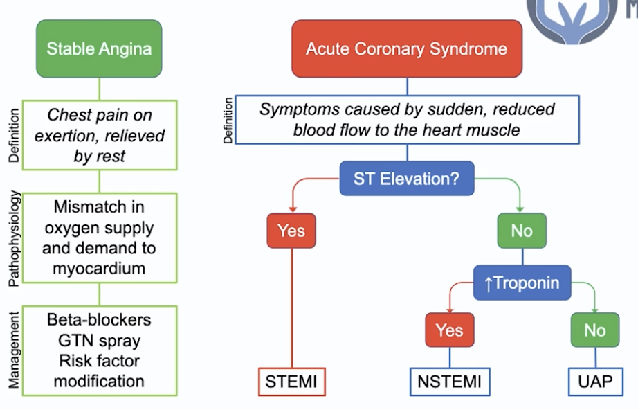
Stable angina - chest pain on exertion relieved by rest

  • Path - mismatch in O2 supply and demand to the myocardium
  • Ix: CT-angiogram
  • Mx:
    • B-blockers - reduces HR req for activity –> reduced likelihood of mismatch in O2 supply & demand
    • GTN spray - reduce myocardial preload + reduces strain
    • RF modification –> reduced risk of progression

Acute coronary syndrome - Sx caused by sudden reduced BF to the myocardium

  • Dx:
    • ​ST-elevation = STEMI
    • Troponin raised = NSTEMI (+ dynamic T-wave inversion, ST depression)
    • Unstable angina pectoris (pain at rest) = ischemia NOT infarct
  • Generic ACS Mx - MONA BASH
    • ALL immediate:
      • 5-10mg Morphine IV + Nitrates (GTN spray)
      • Dual antiplatelet therapy (DAPT) - 300mg Aspirin STAT + 300mg Clopidogrel STAT (or 180mg PO Ticagrelor)
    • ALL long-term:
      • Continue DAPT
        • 1 year: 75mg OD Aspirin + 75mg OD Clopidogrel (or 90mg BD Ticagrelor)
        • >1yr - 75mg OD Aspirin
      • B-blocker (1.25-10mg Bisoprolol OD)
      • ACEi (1.25-10mg Ramipril OD)
      • Statin (80mg Atorvastatin OD)
  • STEMI Mx: establish coronary reperfusion ASAP
    • Sx <12hrs: PCI BUT if no PCI within 2hrs Dx –> thrombolysis (e.g. tPA - tissue plasminogen activator)
    • Sx >12hrs: invasive coronary angiography ± PCI if needed
    • PCI:
      • If having PCI give Prasugrel (instead of Clopi/Ticagrelor)
      • PCI accessed via radial (or femoral) artery, guidewire passed via X-ray guidance into the affected coronary artery AND IV unfractionated heparin during the procedure –> stent inserted impregnated with an anti-proliferative agent (e.g. Tacrolimus - to prevent adverse tissue reaction) –> takes longer for endothelialization of stent so DAPT needed for 1yr
      • If PCI with stents inserted –> DAPT 12 months
  • NSTEMI Mx:
    • 2.5mg SC Fondaparinux (direct factor 10a inhibitor)
    • Risk stratify - GRACE criteria (& others)
    • High risk = invasive coronary angiography (within 48-72hrs)

Complications: FAP (failure, arrhythmias, pericarditis)

  • Heart failure, arrhythmias (incl. VF)
  • Pericarditis
    • Early - positional chest pain day after MI –> give NSAIDs
    • Late - Dressler’s syndrome - immune response @6wks (fever, pleuritic chest pain, pericarditis/pericardial effusion)
24
Q

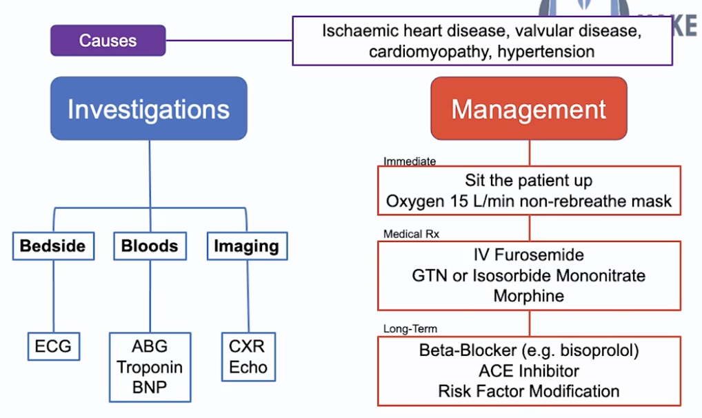
Heart failure def? Causes? Pathophysiology? Categories & Causes? Classification? Ix? Mx?

A

Def: pumping of blood by heart insufficient to meet the demands of the body

Causes:

  • RVF:
    • Acute: MI, inf endocarditis, PE
    • Chr: Cor pulmonale, LVF
  • LVF:
    • Acute: MI, inf endocarditis
    • Chronic: ischaemic/hypertensive CMO, valvular HD

Pathophysiology:

  • RHF - right side of the heart pumps deoxygenated blood from the body to the lungs to be reperfused - if the RH is not pumping effectively you get the fluid collection in the peripheries = PERIPHERAL OEDEMA
  • LHF - left side of the heart pumps oxygenated blood from the lungs to the body - if the LH is not pumping effectively you pooling of blood in the lungs = PULMONARY OEDEMA
  • Reduced CO –> shock, tachycardia, AKI
    • CO = SV*HR
    • Ejection fraction = SV/End-diastolic Volume

Categories:

  • HF w/ preserved ejection fraction (left ventricular >50%) = inadequate filling of ventricles during diastole (from ventricular stiffness)
    • Causes of ventricular stiffness:
      • Volume overload (valve regurg)
      • Pressure overload (HTN)
      • Decreased distensibility (constrictive pericarditis)
  • HF w/ reduced ejection fraction (left ventricular <40%) = inadequate emptying of ventricles during systoles (from outflow obstruction/impaired contractility)
    • Causes of outflow obstruction/impaired contractility:
      • MI, Cardiomyopathy, Arrythmia

NYHA classification:

  • 1 - no limitation on activity
  • 2 - comfortable at rest but dyspnoea on ordinary activity
  • 3 - marked limitation on ordinary activity
  • 4 - dyspnoea at rest

Ix:

  • Bedside: ECG - detects if anything precipitating HF (arrhythmia/ischaemic event)
  • Bloods: ABG (if resp compromise from pul oedema), troponin (ACS), BNP (HF screening)
  • Imaging: CXR (visualise pul oedema, cardiomegaly), ECHO (valvular abn/regional wall mov abn)

Mx: MON BA (out of MONA BASH)

  • Immediate:
    • Sit the patient up (reduce venous return to heart –> less strain)
    • O2 15L/min NRM
  • Medical:
    • IV furosemide (loop diuretic) - remove excess fluid + venous dilation (reduce preload)
    • Nitrates (GTN/Isosobide Mononitrate) AND Morphine - reduce preload on the heart
  • Long-term:
    • Reduced ejection fraction - prognostic benefit:
      • B-blocker (bisoprolol) - reduce strain on heart, do not give acutely if severe HF as will kill them
      • ACEi - reduce strain on heart
        • After the above if LVEF <35% & Sx –> mineralocorticoid antagonist e.g. spironolactone
        • 3rd line - by specialist: Sacubitril/Valsartan (entresto), Ivabradine & CRT
      • SGLT2 inhibitors (dapagliflozin)
    • RF modification - poor glycaemic control/high cholesterol
    • Sx (diuretics)

Complications:

  • Reduced CO (SV*HR) –> shock, tachycardia, AKI
  • Congestion –> pulmonary oedema + peripheral oedema
26
Q

SVT - Def? Types? Presentation - case example? Mx?

A

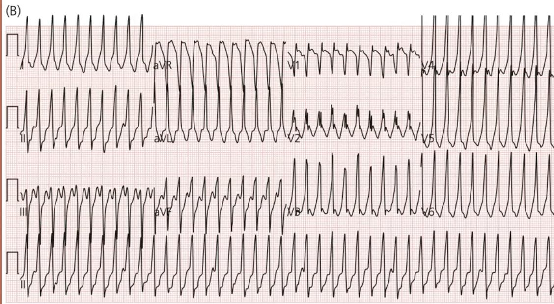
Def: regular narrow-complex tachycardia with no p-waves + supraventricular origin

Junctional types:

  • AVNRT - local re-entry circuit within AV node
  • AVRT - re-entry circuit between atria and ventricles –> after SVT termination = delta wave = WPW syndrome:
    • ​Assoc w/ HOCM
    • Avoid digoxin, verapamil, amiodarone (reduce conduction down SAN –> worsen retrograde conduction –> risk of VT)
    • Can use B-blocker/flecainide instead

Case example: 23yrs, 1-hr palpitations + SoB, 2 similar episodes prev following alcohol, this time severe chest pain

Mx:

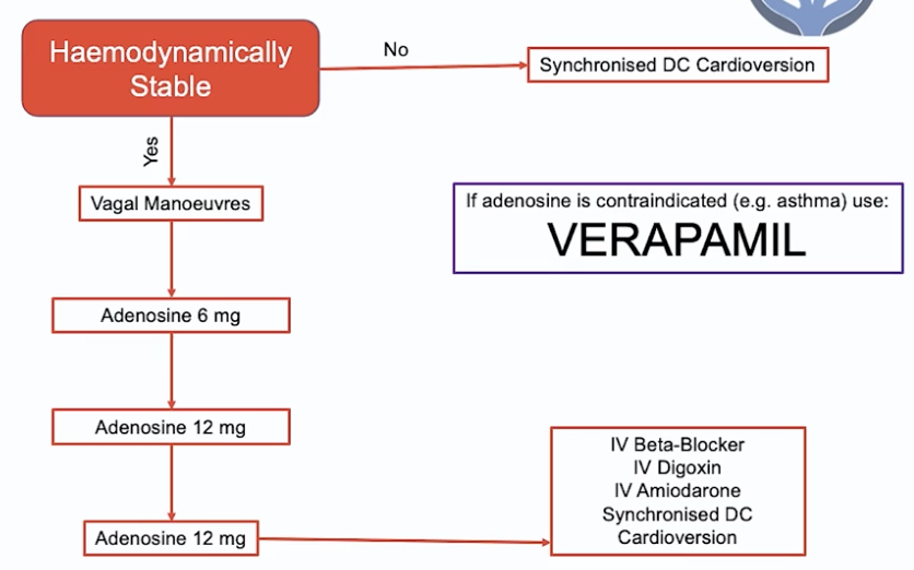
  • Unstable tachycardia (<90 BP/chest pain/acute heart failure) –> synchronised DC Cardioversion
  • Vagal manoeuvres (increase parasympathetic stim via vagus nerve to slow conduction via AV node)
    • Valsalva manoeuvre (blow out through nose while pinching + shut mouth) - breath through 50ml syringe
  • Adenosine 6mg –> 12 mg –> 12mg
    • NOTE: if adenosine CI (e.g. asthma) –> VERAPAMIL (rate-limiting CCB)
  • Other:
    • IV B-blocker/amiodarone/digoxin
    • Synchronised DC Cardioversion
27
Q

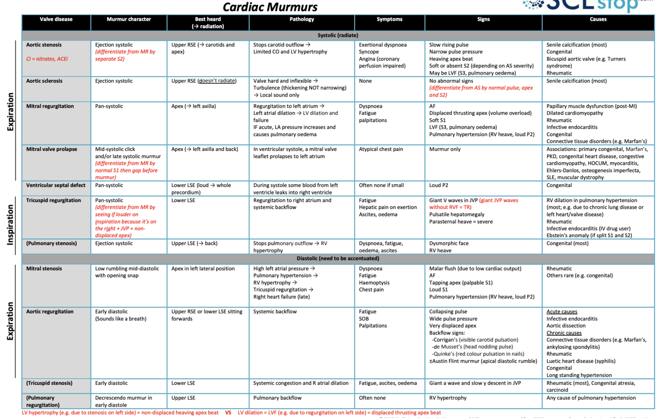
Key heart murmurs? Accentuation manoeuvres?

Causes? Left vs right heart valve abn epidemiology?

Mx? Valve types? Complications of prosthetic heart valves?

A

Key murmurs:

  • AS = ejection systolic + radiates to carotids, slow rising pulse, narrow pulse pressure, heaving apex beat
    • Sound: Wooooshhh
    • Severe AS - absent/soft 2nd heart sound, reversed splitting of 2nd HS, heaving apex beat
      • A longer murmur is worse (small space for blood to pass through = takes longer)
  • AR = early diastolic + sitting forward (LLSE), collapsing pulse, wide pulse pressure, displaced apex
    • Sound: de woooshhhh
    • Severe AR –> Austin-flint murmur = ‘Rumbling mid-diastolic murmur’
      • Best heard at apex, caused by blood flowing back through aortic valve and over mitral valve
      • Shorter murmur is worse (quicker to flow back through large hole)
  • MR = pan-_systolic_ + radiates to left axilla, AF, displaced thrusting apex, LVF/pul HTN
    • Sound: Woooooshhh (holosystolic)
  • MS = mid-diastolic + LLP, malar flush, AF, loud/palpable S1 “tapping” apex, pul HTN (loud P2 - pul thrill)
    • Sound: Wooosh de (loud S1) de (early diastolic snap)
  • NOTE: same pattern for pulmonary & tricuspid (pul stenosis & tricuspid regurgitation = systolic)
    • TR - pulsatile liver
    • PS - radiates to back, assoc w/ Noonan’s (AD, webbed neck, wide-spaced eyes etc.)

Accentuation manoeuvres:

  • R-sided murmurs (tricuspid + pulmonary) –> louder on INspiration = blood goes IN to right-side of heart
  • L-sided murmurs (aortic + mitral) –> louder on EXpiration = blood EXits left-side of heart
  • AS radiates to the carotids + louder on leaning forward + listen on right sternal edge
  • MS louder on turning to the left, MR radiates to axilla

Causes:

  • AS (stenosis/sclerosis):
    • Older - senile calcification
    • Younger - bicuspid valve
  • AR:
    • Acute (aortic root dilation, infective endocarditis)
    • Chronic (CTD, RHD, bicuspid aortic valve)
  • MR:
    • Acute: - IHD (papillary-muscle dysfunction post-MI), infective endocarditis, RHD
    • Chronic - myxomatous degeneration (can be assoc with Marfan’s/Ehlers-Danlos)
  • MS: rheumatic heart disease (RHD)

Left vs Right valve abn:

  • Left = more common as higher pressure system, more likely in damaged valves, commonly Strep Viridans
  • Right = more common in IV drug users –> tricuspid valve is first valve reached, commonly S. aureus

Ix:

  • ECHO (Dx & severity) - Transthoracic incl doppler (regurg jet) ± TOE
    • If the above is inconclusive:
      • 2nd - Cardiac MRI - analysis of cardiac function
      • 3rd - Cardiac catheterization (invasive) - pressure gradient
  • ECG (LVH), CXR (pul HTN)

Management:

  • AS:
    • C: Asymptomatic –> monitor (6 monthly ECHO)
    • S: Symptomatic –> aortic valve replacement:
      • Fit (req midline sternotomy & cardiopul bypass) = Surgical aortic valve replacement (SAVR)
      • Not fit = Transcatheter aortic valve implantation (TAVI)
    • M:
      • HF Sx (diuretics, ACEi)
      • RF optimisation (statins, HTN, DM)
  • AR:
    • If no left ventricular dysfunction –> medical Mx
      • ACEi, vasodilators e.g. nifedipine/hydralazine (if HTN)
    • If left ventricular dysfunction & low surgical risk/another indication for cardiac surgery –> aortic valve surgery (replacement > repair)
  • MR - primary (valve abn/damage):
    • Mitral valve surgery (repair > replacement)
    • Medical - ACEi, B-blocker & diuretics
      • Tx AF if present (incl. anticoagulate)
  • MS:
    • C: If non severe + asymptomatic –> monitor
    • S: Otherwise = Balloon valvuloplasty/mitral valve replacement
      • if C/Is (persistent left atrial thrombus/rigid calcified valve) –> need open surgery
      • NOTE: valvuloplasty = lateral thoracotomy scar
    • M: Tx AF if present (incl. anticoagulate)

Valve types:

  • Tissue valve - porcine xenograft
    • For older patients & females of childbearing-age - lasts 10-15yrs, less if active
    • No need for Warfarin
  • Mechnical valve - quiet clicking noise
    • Younger people (<60yrs) - no need for repeat surgery, last 30yrs+
    • Lifelong Warfarin

Complications of prosthetic heart valves: FIBAT

  • Failure
  • Infection
  • Bleeding - MAHA
  • Anaemia
  • Thromboembolic phenomena
29
Q

Atrial fibrillation (AF)

  • Def? Causes? Ix? Mx?
A

Def: rapid, chaotic, and ineffective atrial electrical conduction

  • ECG def: irregularly irregular narrow complex tachycardia with no p waves

Causes: idiopathic, cardio (IHD, valvular disease, cardiomyopathy), resp (PE, pneumonia), hyperthyroidism, alcohol

Ix: ECG (absence of p-waves, irregularly irreg rhythm)

Mx:

  • Haemodynamically unstable (≤90 BP, chest pain, acute HF) –> DC Cardioversion

OR

  • Rate control –> B-blocker (bisoprolol) OR rate-limiting CCB (verapamil - asthma)

OR

  • Rhythm control - ONLY if clear reversible cause
    • Sx onset <48hrs –> DC/chemical cardioversion (amiodarone/flecanide)
      • NOTE: IV heparin started prior to cardioversion
    • Sx onset >48hrs –> anticoagulate for 3wks –> elective cardioversion (also anticoag for 4wks after)

AND

  • Stroke risk - CHADS-Vasc Vs Orbit/HAS-BLED score –> DOAC (Apixaban)
    • If metallic heart valve –> warfarin INR 3-3.5
    • Otherwise DOAC
    • NOTE: if incidental non-symptomatic AF - normal rate, no other RFs, CHA2DS2-VASc 0 –> anticoagulation not recommended
    • CHF, HTN, Age ≥75rs (2), DM, Stroke (2), Vascular disease, Age 65-74, Sex - female
      • Score 1 - consider; ≥2 - DOAC/Warfarin needed
      • Lifetime risk = annual risk x estimated years of life left (up to 80 yrs e.g. if 60 then x annual risk by 20)
33
Q

Infective endocarditis - RFs? Ix? Dx criteria? Mx?

Acute vs subacute bacterial endocarditis - what hearts affected? who are commonly affected? What bacteria most likely?

A

Def: infection of heart valves (typically mitral/aortic or tricuspid in IVDU)

RFs: bacteraemia (long-term lines, IVDU, dental work), abn valves (prosthetic, RHD), prev endocarditis, VSD, piercings

Presentation: low-grade fevers, night sweats

  • Exam:
    • Splenomegaly
    • Splinter haemorrhages, osler’s nodes, Janeway lesions, petechiae, Roth spots (eyes)
    • Chronic = clubbing (rare, mostly acute now)

Ix:

  • Urine dip - haematuria
  • Serial BCs (x3 but start empirical abx), ESR
  • Transoesophageal Echo (TOE - vegetations)

Dx: DUKE’S CRITERIA (2 major OR 1 major + 3 minor OR 5 minor):

  • Major: +ve BC (typical organism), new regurg murmur/veg on echo
  • Minor: RF, fever (>38), embolic (vascular) phenomena, immune phenomena, +ve BC (another organism)
  • Mx: IV abx for 6wks – fluclox/vanc/gent

Acute in structurally normal heart – In IV drug user the first valve met is tricuspid valve, commonly S. aureus (also most common cause in prosthetic valve endocarditis)

Subacute in structurally abn heart – mitral & aortic valves more commonly affected as high pressure system, more likely damaged valves, commonly Strep Viridans (overall most common cause of endocarditis)

34
Q

3rd & 4th heart sounds - sounds & cause?

A

3rd = rapid ventricular filling = volume overload e.g. HF (reduced EF/systolic)

  • KEN…TU.CKY (deee. de.de)

4th = atrial contraction against stiff ventricles = pressure overload e.g. longstanding AS & other causes of left ventricular hypertrophy (HTN heart disease, HOCM, HF with preserved EF/diastolic)

  • TE.NE..SSEE (de.de.deee)
36
Q

2 days of chest pain following 4 days of generalised muscle aches

  • Worse on inspiration & lying flat
  • Low-grade fever
  • Exam: pericardial rub

Causes? Dx? Ix? Mx?

A

Pericarditis

Causes:

  • Viral (most common)
  • MI (can be Dressler’s syndrome)
  • TB (constrictive)
  • Uraemia (CKD where urea high –> pericarditis) = indication for haemodialysis (HUMP)
  • Hydralazine (AI pericarditis)
    • NOTE: also causes drug-induced lupus
  • SLE, RF, radiation

Presentation:

  • Pleuritic chest pain, worse lying flat
  • Exam: pericardial rub - “creaking/scratching”
    • Tip - put on all-fours, put stethoscope on sternal edge, hold inspiration

Ix:

  • ECG: ST elevation widespread
  • Only slightly raised/normal troponin

Mx: colchicine (3 months) + NSAIDs (ibuprofen, max 2wks)

37
Q

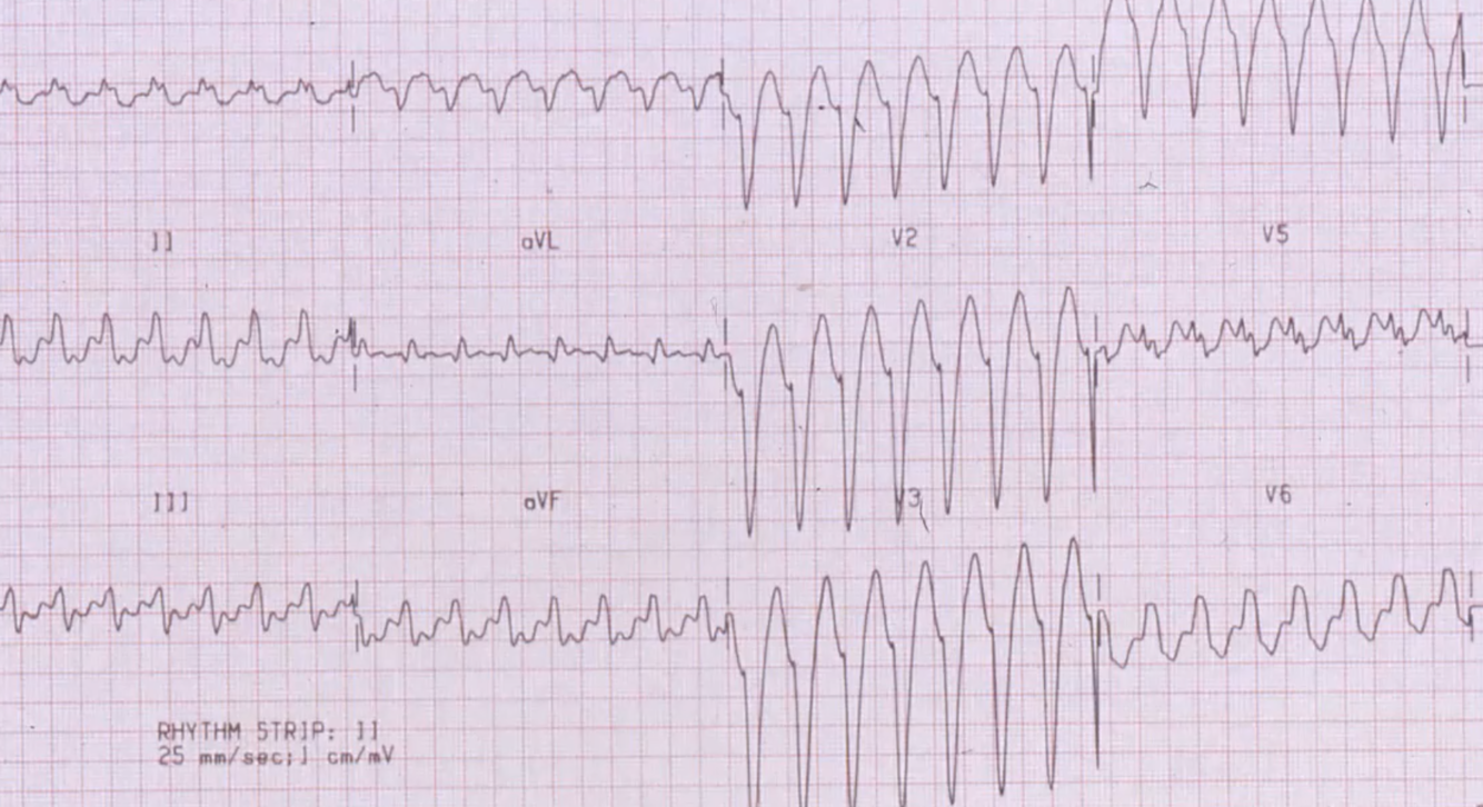
Ventricular tachycardia - Dx? Presentation? Ix - appearance on ECG? Mx?

A

VT or SVT w/ aberrancy

  • SVT >200bpm, also often irregular
  • VT more likely if LAD
  • Acutely treat any broad complex tachy as VT until proven otherwise

Presentation: palpitations, light-headed, chest pain, syncope, seizure

  • Tachycardia, LVF
  • ACS most common cause
  • NEVER IGNORE palpitations & light-headedness

Ix: ECG - regular broad complex tachycardia

  • U&E (Mg, Ca, K), TFTs, Troponins

Mx:

  • Unstable tachycardia (BP <90, chest pain, acute cardiac failure) = DC cardioversion
  • Stable:
    • IV amiodarone, b-blocker –> prepare for DC cardioversion
38
Q

Bradycardia arrhythmia with a palpable pulse (peri-arrest) - Mx?

A

Innitial: A-E

  • If unstable - 500mcg IV atropine (/5mins up to 3mg)
    • Also considered unstable if:
      • Recent asystole >3s/Mobitz T2 AV block/3rd degree heart block
    • Caution in acute MI, C/I if heart transplant
  • If persistent –> transcutaneous pacing + analgesia/sedation (very painful)
    • If can’t be achieved properly –> IV isoprenaline/adrenaline (specialist help)
  • Arrange transvenous pacing (temporary if recent asystole >3s/Mobitz T2 AV block/3rd degree heart block)
39
Q

Heart block causes? types? Ix? Mx? Complications?

A

Causes:

  • MI/IHD (MOST COMMON)
  • Inf (RHD, IE)
  • Drugs (digoxin)
  • Metabolic (hyperkalaemia)
  • Infiltration of conducting system (e.g. sarcoidosis)
  • Degeneration of conducting system

Types:

  • First Degree AV block - fixed prolonged PR interval (> 0.2 s) - ASYMPTOMATIC
  • Second degree AV block:
    • Mobitz TI (Wenckebach) - progressively prolonged PR interval –> P-wave NOT followed by a QRS complex = ‘going, going, gone’
      • Normally asymptomatic
    • Mobitz Type II - intermittently P wave NOT followed by a QRS
      • May be regular pattern of P waves not followed by QRS (e.g. 2:1 or 3:1)
      • Can cause:
        • Stokes-Adams Attacks (syncope caused by ventricular asystole)
        • Dizziness, palps, chest pain, HF
  • Complete AV heart block - no relationship between P waves and QRS complexes
    • Presentation as in Mobitz T2

Ix: ECG

  • Bloods: TFTs, Digoxin, cardiac enzymes (troponin, CK, BNP)
  • CXR (cardiac enlargement, pulmonary oedema)
  • Echo (wall motion abn, aortic valve disease, vegitations)

Mx:

  • Acute block - if clinical deterioration:
    • IV atropine
    • Consider temporary transcutaneous pacing
  • Chronic block:
    • 1st degree monitored
    • Permanent pacemaker in:
      • Symptomatic Mobitz T1
      • Advanced Mobitz T2
      • Complete heart block

Complications: asystole, cardiac arrest, HF, surgical complications of pacemaker insertion

40
Q

Types of pacemaker? When to use each type? Complications?

A

Types:

  • Implantable Cardioverter Defibrillator (ICD, has a thicker end)
  • Single-chamber pacemaker (right ventricle)
    • Used in permanent AF (no organised atrial contraction so atrial lead not required to sense contraction)
    • Rarely can have atrial lead only - if SA disease in young with good AV conduction
  • Dual-chamber pacemaker (right atrium & ventricle)
    • Can have ICD dual-chamber pacemaker
    • Used in paroxysmal AF/all other scenarios (there is sometimes organised atrial contraction - this is sensed by the atrial lead)
  • Cardiac Resynchronisation Therapy/Biventricular pacemaker (right ventricle, left ventricle ± right atrial lead)
    • Can have ICD biventricular pacemaker

When to use each type:

  • Atrial lead only → Sino-atrial disease in young people with good AV node conduction
  • RV lead only → Pacing whilst in permanent atrial fibrillation
  • Dual-lead → All other scenarios (paroxysmal AF, bradycardia)
  • CRT → LV dysfunction + broad QRS –> end-stage HF
  • Indications for ICD:
    • Primary prevention = @risk of serious ventricular arrhythmia
      • Familial cardiac conditions (hypertrophic cardiomyopathy, long QT)
      • Previous surgical repair of congenital HD
      • Previous MI + LVEF <35% + HF Sx
    • Secondary prevention = had previous serious ventricular arrhythmia wo/ treatable cause
      • Cardiac arrest from VT/VT
      • Spontaneous sustained VT AND:
        • Syncope/haemodynamic compromise OR
        • LVEF <35% + sign HF Sx (NYHA 3+)
      • NOTE: VT/VF from STEMI has treatable cause (open occluded vessel)

Complications:

  • Surgical complications - infection, bleeding, damage to underlying structures
  • Displacement (of lead)
  • Pacemaker syndrome (if ventricular lead with no atrial) –> AV node conducts in retrograde direction = mitral/tricuspid regurge + HF Sx
42
Q

How do you know this is the JVP and not the carotid pulse?

A
  • Not palpable
  • Double pulsation
  • Obliterated when pressure applied at base of neck
  • Rises with hepatojugular reflux
  • Height changes with respiration
43
Q

Causes of hepatic decompensation in CLD? Key features of decompensation?

Dx & Mx of decompensated chronic liver disease?

A

Cause of hepatic decompensation in CLD:

  • Hypokalaemia
  • Constipation (given lactulose in hospital)
  • Alcohol
  • GI bleed (lots of protein (Hb) enters the bowel –>liver can’t cope)
  • HCC

Decompensated CLD –> Ascites, jaundice & encephalopathy

  • Severely scarred liver (cirrhosis) in CLD –> back pressure on portal vein –> PORTAL HTN = splenomegaly, ascites, varices - caput medusae, oesophageal & rectal

Ix:

  • Serum Ascites Albumin Gradient (SAAG) - serum albumin conc vs ascites conc - 11.1g/L
    • <11.1g/L = exudative cause - peritonitis (infection), peritoneal malignancy OR n_ephrotic syndrome_ (pee out albumin so low serum albumin)
    • Otherwise = transudative cause - cirrhosis, renal failure, HF
  • >250 neutrophils = spontaneous bacterial peritonitis (SBP) –> Tazocin/3rd gen cephalosporin
    • If protein conc <15g/L give prophylactic oral ciprofloxacin

Mx:

  • Paracentesis (ascitic drain) –> post-paracentesis circulatory dysfunction (drops BP) SO if >5L drained give human albumin solution (HAS) 8g/L drained
  • Spironolactone (2nd line - Furosemide) - to prevent fluid accumulation
  • (Salt restrict)
  • Hepatic encephalopathy (liver not dealing with toxins) - give Lactulose + Rifaximin to prevent
  • Coagulopathy - OGD (check for varices) + vit K (needed for clotting)
44
Q

Chronic liver disease

  • Functions of liver? Outcome of failure?
  • Causes? Presentation? Ix?
  • Important complication?
  • Scoring?
A

Functions of the liver –> failure:

  • Albumin (plasma oncotic pressure) –> oedema
  • Bilirubin metabolism –> jaundice
  • Clotting factors –> coagulopathy
  • Detoxification –> encephalopathy

Causes:

  • Common - alcoholic liver disease, viral hepatitis, NASH (non-alcoholic steatohepatitis)
  • Less common - AI hepatitis, PSC/PBC, HF, alpha1-antitrypsin def, haemochromatosis, Wilson’s disease

Presentation:

  • Spider naevi (≥5, SVC distribution, flush inside to out), palmar erythema, gynecomastia, Dupuytren’s contracture (alcoholic liver disease), clubbing
  • Specific signs:
    • Needle marks/tattoos - hep C
    • Parotid swelling - alcohol-related liver disease
    • Bronzed complexion/insulin injection signs - haemochromatosis
    • Obesity/DM - non-alcoholic fatty liver disease
    • Xanthelasma - cholestatic disorder

Ix:

  • Alcohol history
  • Hep B/C serology
  • Ferritin, transferrin, A1AT, ceruloplasmin (Wilson’s)
  • Ig, auto-abs (ANA in AI hep, AMA in PBC)

Important complication = VARICES

  • Normal venous return: GI tract –hepatic portal vein –> liver –> hepatic vein –> systemic circulation
  • Physiological hepatosystemic anastomoses (connection of portal vein to systemic circulation) sites - oesophagus, spleen, umbilicus, rectum
    • MEMORY AID: BUTT, GUT, CAPUT
  • Pathological process:
    • In the case of cirrhosis - nodules impede flow of blood through the liver to the hepatic vein –> reducing blood flow to the systemic circulation
    • Backflow of blood to the hepatic portal vein = increased –> backflow to hepatosystemic anastomoses:
      • Oesophagus –> Oesophageal varices
      • Spleen –> Splenomegaly
      • Umbilicus –> Caput Medusae
        • ​Only from portal HTN if running from below umbilicus up
      • Rectum –> Rectal varices

Score for prognosis & need for liver transplant = Child-pugh score (A = 5-6; B = 7-9; C = 10-15 –> C is most severe)

45
Q

Chronic diarrhoea - ddx? Sx? Distribution? Histology? Ix? Mx?

A

IBD

  • Crohn’s disease
    • Sx: RIF pain (terminal ileum), failure to thrive (incl. between attacks)
    • Distribution: mouth to anus (esp. terminal ileum), skip lesions
    • Histology: transmural, non-caseating granuloma (also in sarcoidosis)
    • Ix:
      • OGD/colonoscopy/capsule endoscopy
      • Gastografin follow-through
    • Mx:
      • Induction:
        • Steroids (induce remission)
        • 5-ASA (mesalazine)
      • Maintenance:
        • Steroid-sparing agents (methotrexate, azathioprine, mercaptopurine)
        • Biologics e.g. Anti-TNF (infliximab)
    • Complications: strictures, obstruction, fistulae
  • Ulcerative colitis
    • Sx: gen. abdo pain, PR blood & mucus
    • Distribution: ONLY rectum & colon, continuous
    • Histology: mucosa & submucosa, mucosal ulcers, crypt abscesses
    • Ix:
      • Flexible sigmoidoscopy/colonoscopy
    • Mx:
      • Induction:
        • 5-ASA
        • Steroids
      • Maintenance:
        • 5-ASA (mesalazine)
        • Steroid-sparing agents (azathioprine)
        • Biologics (infliximab)
      • Surgery (proctocolectomy, IJ pouch)
    • Complications: toxic megacolon, colonic carcinoma, PSC
  • Extra-intestinal signs of IBD - A PIE SAC
    • Aphthous ulcers (crohn’s)
    • Pyoderma gangrenosum
    • Iritis, uveitis, episcleritis (Crohn’s)
    • Erythema nodosum
    • Sclerosing cholangitis (PSC in UC)
    • Arthritis
    • Clubbing fingers (CD > UC)
  • Truelove & Witts’ severity index = severity criteria for IBD

Coeliac disease

  • Def: inflammatory response to gluten (immune reaction to gliadin)
    • DQ2/8 (on HLA typing)
  • Sx: abdo Sx (steatorrhoea, diarrhoea, bloating), failure to thrive
    • Exam: blistering rash on both knees. aphthous mouth ulcers
    • Assoc: T1DM
  • Distribution: duodenum
  • Histology: subtotal villous atrophy with crypt hyperplasia
  • Ix: (maintain normal diet - eat gluten for 6wks before testing)
    • Haematinics - macrocytic anaemia, low Fe, B12/folate
    • Serological testing:
      • Screening: Anti-TTG & total IgA (check for selective IgA def in case this caused false ‘-ve’ anti-tTg abs)
      • If weakly +ve –> anti-endomysial abs (more sensitive & specific)
    • OGD & duodenal biopsy
      • Histology: sub-total villous atrophy with crypt hyperplasia
  • Mx: gluten avoidance

Irritable bowel syndrome - Dx of exclusion

  • Sx: bloating, related to stress, diarrhoea/constipation, relieved by defecation
    • “Pellet-like stools”
  • Ix: faecal calprotectin (an inflammatory marker of GI tract = less likely to be IBS, more likely IBD)
  • Mx: dietary changes, stress Mx, anti-spasmodics, probiotics, laxatives/loperamide, anti-depressants
50
Q

Mesenteric ischemia:

  • What is the blood supply to the GI tract?
  • What area of GI tract is vulnerable to ischaemia?
  • Breakdown of mesenteric ischemia? Sx? Ix? Mx?

What is the difference between Mesenteric ischemia & ischemic colitis? Sx? Ix?

A

Blood supply

  • Coeliac axis - oesophagus, stomach, start of small intestine
  • Superior mesenteric artery (SMA) - most of the small intestines up to 2/3 transverse colon
  • Inferior mesenteric artery (IMA) - from 2/3 along transverse colon down to rectym
  • NOTE: area 2/3 along transverse colon @splenic flexure = WATERSHED ZONE (limited supply from superior & inferior mesenteric arteries) –> vulnerable to ischaemia!!

_​_Mesenteric ischemia

  • Acute - occlusion of SMA by thrombus/embolus
    • Sx: severe abdo pain, normal abdo exam, shock
    • Ix:
      • AXR (gasless abdo)
      • VBG (lactic acidosis)
      • CT abdo with contrast (if suspect mesenteric ischaemia)/CT angiogram (if LA)
        • If not primary surgery –> followed by mesenteric angiography (Dx non-occlusive mesenteric ischaemia)
    • Mx: resus + CCOT + NBM
      • Empirical abx
      • Bypass ± bowel resection
      • Heparin post-op
  • Chronic - narrowing of SMA by atherosclerosis (stable angina of bowel)
    • Sx: colicky post-prandial abdo pain (more work for bowel to do –> pain, just like in stable angina & exercise)
    • Ix: as above
    • Mx: medical optimisation + bypass (angioplasty + stent if not suitable for surgery)

Ischemic colitis

  • Occlusion of inferior mesenteric artery (IMA) by thrombus/embolus
  • Sx: abdo pain, PR bleeding
  • Ix: sigmoidoscopy/colonoscopy
  • Mx: seek GI & surgical input (can develop chr ulcerating IC)
    • Mild - conservative (IV abx, fluids, bowel rest, DM control)
      • Prophylactic LMWH
    • Segmental resection + stoma
54
Q

How do you tell between spleen & kidney on examination?

A

Spleen (vs kidney):

  • Moves down with inspiration
  • You can’t get above it
  • Has a notch
  • Dull to percussion & not ballotable
56
Q

Upper GI bleed - scoring for need for intervention? Mx?

A

Blatchford score

Variceal bleed

  • Massive haemorrhage –> balloon tamponade
  • A-E assessment –> IV fluids, blood transfusion
    • F1 Essentials:
      • 2x large bore cannula
      • VBG
      • G&S/X-match
      • Bleep the bleed reg
  • Drugs with prognostic benefit:
    • IV Terlipressin (ADH analogue –> vasoconstriction)/Somatostatin (used for same reason)
    • Prophylactic abx - Ceftriaxone/Norfloxacin (abx)
  • Intervention (discuss with on-call bleed registrar) –> endoscopic band ligation
57
Q

Peptic ulcer disease - RFs? Types? What artery is most likely to bleed in duodenal ulcer?

Presentation? Ix? Mx? Scoring to evaluate risk of re-bleed/death?

A

RFs:

  • H. pylori exposure
  • Aspirin/NSAID use

Types:

  • Gastric - pain worsened by meal (pain 30m-1hr after meal), loose weight, vomiting, assoc w/ NSAIDs
  • Duodenal - MORE COMMON, pain relieved by meal (pain 2-3hrs after meal), assoc w/ H. pylori, worse by stress/at night –> radiates to back, put weight on, malaena
    • Gastroduodenal artery - runs posterior to 1st/2nd parts of duodenum –> likely cause of bleeding in PUD

Presentation:

  • Chr/recurrent upper abdo pain - related to eating & nocturnal
    • Can be severe/radiate to back in Duodenal ulcers if ulcers penetrates pancreas
  • Pointing sign on exam - show where pain is with 1-finger
  • NOTE: ‘Coffee grounds’ vomiting = upper GI bleed (mostly due to PUD > gastric erosions)

Ix:

  • OGD endoscopy = gold-standard
    • Immediately if dyspepsia + upper GI bleed
    • Within 2wks if ≥55yrs + weight loss + dyspepsia/reflux/upper abdo pain –> exclude malignancy
    • Repeat within 6-8wks to ensure ulcer healing/rule out malignancy
  • H. pylori stool antigen/breath test (2wk wash out period after PPI or 4wks after abx, retest 6-8wks after starting Tx)
  • Bloods - FBC

Mx: consider STOPPING NSAIDs

  • Active bleed:
    • Urgent evaluation (A-E + Blatchford score) + blood transfusion
    • OGD endoscopy (Dx & Tx) –> high-dose IV PPI afterwards (continue oral PPI for 6wks)
    • Rockall score (for risk of rebleed/death) –> if re-bleed –> repeat endoscopy & Tx endoscopically/emergency surgery
  • If H. pylori +ve:
    • H. pylori eradication - triple therapy for 7 days (PPI + 2 abx = Amox + clari/metro) –> retest 6-8wks after starting Tx (leave washout 2wks after PPI, 4wks after abx)
      • If pen allergic –> PPI + Clari + Metro
    • If long-term NSAID/aspirin use:
      • Consider stopping NSAIDs/aspirin
      • Ulcer healing Tx - full-dose PPI/H2 antagonist for 8wks –> H. pylori eradication afterwards
  • If H. pylori -ve: treat underlying cause + PPI (4-8wks, 2nd line = H2 antagonist)
  • If recurrent/refractory ulcers –> long-term PPI/H2 antagonist
58
Q
  • Joint pain in both knees, XR shows chondrocalcinosis
  • Wakes at night frequently to urinate, PMHx T2DM, low albumen on blood

Dx? Classification? Presentation? Ix? Mx?

A

Dx: haemochromatosis

  • Risk of HCC (hepatocellular carcinoma)

Classification:

  • Hereditary - AR, gene on chr6 (carried by 1:10 Europeans)
  • Secondary e.g. from frequent blood transfusions (SCD)

Presentation - from Fe-deposition in various tissues:

  • Arthritis (esp hands), bronze DM (tan), cirrhosis
    • Hypogonadism (accumulates in testis)
    • Dilated cardiomyopathy (accumulates in heart)
  • XR - chondrocalcinosis is assoc w/ pseudogout & haemochromatosis

Ix:

  • Blood iron profile:
    • TF saturation high (>55% men, >50% women)
    • Ferritin normal/slightly high (>500)
    • Low TIBC (as all transferrin saturated)

Mx:

  • Venesection (until transferrin saturation normal)
  • Desferrioxamine (iron chelator to prevent recurring)
  • Monitoring ; TF saturation <50% & serum ferritin <50ug/l
59
Q

Case:

  • 57yrs, burning pain after eating & lying flat, improved with cold drinks, high BMI, smokes, drinks alcohol, occasional bitter taste in back of the mouth
  • Difficulty swallowing in recent months

Dx? Presentation? Ix? Mx? Complication?

A

Dx: GORD

  • Decreased lower oesophageal sphincter (LOS) pressure:
    • Drugs - nitrates, CCBs (smooth muscle relaxation)
    • Alcohol, smoking
    • Hiatus hernia

Presentation: RFs (above), heartburn (worse lying down/at night), reflux incl bitter taste in mouth (after meals)

Ix:

  • 8wk PPI trial
  • If dyspepsia consider OGD for: anaemia, weight loss, dysphagia, >55yrs
  • Oesophageal pH study (<4 for >4% of time = GORD)

Mx:

  • Conservative: weight loss, smoking cessation, head of bed elevation/avoid eating late at night, reduce alcohol
  • Medical:
    • Standard-dose PPI (20mg omeprazole) - continued long-term (if Sx continue after stopping/erosive oesophagitis/Barrett’s oesophagus)
    • H2 antagonist (e.g. cimetidine)
  • Surgery (only if PPIs work but don’t want long-term medical Tx) e.g. laparoscopic fundoplication

Complication: Barrett’s oesophagus –> oesophageal adenocarcinoma

60
Q

Anal fissure - def? RFs? Presentation? Ix? Mx?

A

Def: tears of the squamous lining of the distal anal canal

  • <6 weeks = acute, ≥6wks = chronic
  • 90% @posterior midline

RFs: constipation, IBD, STIs (HIV, syphilis, herpes)

Presentation: painful, bright red, rectal bleeding

Ix: PR exam

Mx:

  • Acute <1wk:
    • Soften stool - high-fibre diet + high fluid intake, fybogel (2nd - lactulose)
    • Lubricants (for passing stool), topical anaesthetic, analgesia
  • Chronic: maintain above + topical GTN
    • After 8wks (if above not effective) –> GI referral for sphincterotomy (or botulinum toxin)
62
Q

Haemorrhoids - presentation, grading, Ix, Mx?

A

Presentation: rectal bleeding & perianal pain

Grading:

  • 1 - prominent BVs (no prolapse)
  • 2 - prolapse on bearing down + spontaneous reduction
  • 3 - manual reduction
  • 4 - can’t be manually reduced

Ix:

  • Bedside: Anoscopic exam, stool (occult haem)
  • Bloods: FBC
  • Imaging: Colonoscopy/flex sigmoidoscopy

Mx:

  • Conservative - fibre, fluids
  • Medical = G1 - topical CS
  • Surgical:
    • G2/3: band ligation
    • G4: surgical haemorrhoidectomy
63
Q

Diabetes: presentation? RFs? types? criteria for Dx? Mx? Complications?

A

Presentation: polyuria, polydipsia, dehydration

  • Ketosis - malaise, vomiting
  • FHx, other endo disorders
  • If known DM:
    • Previous DM control (hyp/hyper)
    • Micro/macrovascular complications
    • Diabetic eye disease (Dx & Tx)

RFs: overweight, FHx (DM), PMHx (GDM), PCOS, HTN, dyslipidemia, CVD

Criteria for Dx (repeat test needed for Dx):

  • Fasting plasma glucose of ≥7.0 (normal ≤6)
  • OGTT (BM 2hours after 75g glucose-load)/ Sx + random plasma glucose of ≥11.1mmol/l (normal <7.8)
  • HbA1c ≥48mmol/mol (≥6.5%) - not for young/T1DM, acutely ill, haem disease, preg, iatrogenic

T1DM Mx: exogenous insulin to avoid DKA & long-term complications

  • Diet - lower fat, higher carbs = counting carbs (adjust insulin around diet rather than limiting eating)
  • Diabetic specialist nurse - EDUCATE:
    • Self-adjust dose - DAFNE course for T1DM (D for DM)
    • Fingerprick glucose
    • Calorie intake & carb counting
    • Phone support
  • Don’t stop insulin during acute illness, maintain calorie intake
  • Insulin regimens:
    • 1st line - Basal-bolus regimen
      • Basal (background) - BD insulin detemir (or Levemir/Lantus/Tresiba) as basal insulin
      • Bolus (before meals) - analogue rapid-acting insulin e.g. insulin Lispro (Humalog)/Aspart (Novorapid)/Neutral (Actrapid)
    • Other:
      • BD biphasic (premixed insulin, hypos common) e.g. Novomix, Humulin M3, Humalog Mix
      • OD before bed long-acting (for T2DM)
      • NOTE: intermediate-acting insulin e.g. Humulin I, Insulatard

T2DM Mx:

  • 1st line - Lifestyle changes - DESMOND course for T2DM (D for DM), dietician input, self-BM monitoring (individual HbA1c target <6.5)
    • HbA1c targets:
      • No hypoglycaemics - 48mmol/mol
      • Hypoglycaemics - 53mmol/mol
      • Escalate Tx - 58mmol/mol
  • Medication:
    • 2nd - Metformin (SEs: diarrhoea, LA - avoid if eGFR <30)
    • 3rd - ADD Sulphonylurea e.g. Gliclazide (SEs: hypoglycaemia, weight gain)
    • 4th - ADD other DM med:
      • Pioglitazone (SEs: hypoglycaemia, weight gain, oedema, fractures in elderly)
        • C/I in HF, bladder cancer
      • SGLT-2 inhibitor e.g. Empagliflozin (SEs: Hypoglycaemia, weight loss, UTI)
        • Not recommended in impaired renal funct
      • DPP-4 inhibitor e.g. Linagliptin (APPROVED FOR USE IN CKD, weight neutral)
      • GLP-1 analogues e.g. Exenatide/Liraglutide (SE: weight loss - useful if BMI >35; vomiting)
        • Not recommended in impaired renal funct
    • 5th - If on triple therapy & not providing control –> commence insulin
  • CVD risk Mx - anti-HTN, anti-lipid, QRISK-3 score
  • Diabetic nephropathy Mx:
    • Monitor albumin-creatinine ratio (ACR)
    • Consider ACEi/ARB early
  • Diabetic neuropathy Mx:
    • Annual Sx review (erectile dysfunction, autonomic neuropathy - orthostatic hypotension, gastroparesis, bladder emptying difficulties)
    • Annual foot screen + specialist foot Mx, monitor for diabetic foot/ulcers ± amputation
  • Diabetic retinopathy: retinal screen annually (age ≥12yrs)
    • Background: need to tighten control
      • Venodilation, microaneurysms (dots), hard exudates (lipid deposits)
      • Tx: tighten glycaemic control, refer if near macula
    • Pre-proliferative (mild) - soft exudates (cotton wool spots e.g. infarcts)
    • Proliferative - neovascularization (+ floaters, reduced acuity)
      • Tx: pan-retinal photocoagulation
    • Diabetic maculopathy - hard exudates, oedema (+ blurred vision, reduced acuity)
      • Tx: intravitreal triamcinolone acetonide decreases macula oedema
    • NOTE: Pre-diabetic –> refer to diabetes prevention programme (DPP)

Diabetes complications:

  • Microvascular:
    • Eye - diabetic retinopathy (± cataracts, glaucoma)
    • Kidney - diabetic nephropathy
    • Neuropathy - damage to PNS –> diabetic neuropathy (peripheral neuropathy - glove & stockings distribution) –> diabetic ulcers/gangrene
  • Macrovascular:
    • Brain - stroke/TIA/cog impairment
    • Heart - coronary heart disease
    • Extremities - PVD, diabetic ulcers/gangrene
65
Q

Hyperosmolar Hyperglycaemic State

  • What does insulin do? Pathophysiology of HHS?
  • HHS criteria? HHS Mx? HHS Mx Targets?
A

Insulin:

  • High level of insulin –> reduces serum BM (pushes into surrounding tissues & hepatic glucose store)
  • Low level of insulin –> switches off ketone production

Pathophysiology:

  • HHS = complication of T2DM
  • In HHS have enough insulin to switch of ketone production but not enough to reduce BM lvls
  • High glucose - osmotically active –> polyuria –> dehydration

HHS criteria:

  • Hypovolaemia
  • Glucose >30mmol/L
  • NO ketonaemia
  • Serum osmolality >320mOsmol/kg

Mx: REHYDRATE = IV 0.9% NaCl (3-6L by 12hrs, deficit 110-220mL/kg)

  • Targets:
    • Reduce Na by less than 10mmol/L/day (otherwise risk osmotic demyelination syndrome)
    • Reduce BM by over 5mmol/L/hr
    • NOTE: if targets not met by 0.9% saline –> 0.45% instead
  • If fluid alone are not enough –> 0.05 units/kg/hr fixed-rate insulin infusion
66
Q

Diabetes insipidus - Sx? Types? Ix? Tx?

A

Sx: hypernatremia (lethargy, thirst, irritable, confusion, coma, fits), polyuria, urine plasma osmolality <2 (very dilute)

Types:

  • Central (lack of ADH prod in hypothalamus) – pituitary surgery, irradiation, trauma, infarction
  • Nephrogenic (resistance to ADH effects) – hypercalcaemia, hypokalaemia, meds (lithium)

Ix: rule out DDx (BM, review drugs, K/Ca lvls)

  • U&E, Ca, BM, serum & urine osmolarities
  • Dx: 2-step fluid deprivation test = give desmopressin (DDAVP):
    • Normal/primary polydipsia –> urine concentrates on fluid deprivation (>600)
    • Cranial DI - giving desmopressin allows ADH prod –> urine conc
    • Nephrogenic DI: urine never concentrates
    • NOTE: DI excluded if urine to plasma (U: P) osmolarity ratio >2:1
  • Cranial MRI - if suspect cranial DI

NOTE: Fluid deprivation test process:

  • Baseline urine osmolality
  • Fluid deprivation for 8hrs, take urine osmolality
  • Give desmopressin, wait 8hrs, take urine osmolality

Tx:

  • Nephrogenic DI –> fluids (+ monitor UO) + thiazide diuretics (hydrochlorothiazide - weird/does not make sense)
  • Central DI –> fluids + desmopressin
67
Q

Thyroid disease - types? presentations? causes? Mx? What is a thyroid storm?

A

Hypothyroidism:

  • Causes:
    • Primary:
      • Hashimoto’s thyroiditis - most common hypothyroidism
        • Goitre, AI disease (vitiligo, pernicious anaemia, T1DM, Addison’s)
        • Elderly females
        • May be initial ‘Hashitoxicosis’
        • +++ Autoantibody titres
          • Antithyroid peroxidase (TPO) & antithyroglobulin (TG)
      • Atrophic AI thyroiditis
        • No goitre, anti-TPO/TSH
      • Iodine def, meds (carbimazole, lithium), thyroid surgery
    • Secondary: pituitary gland fails to produce TSH
      • Tumour, vascular (Sheehan syndrome), radiation, inf
  • Ix:
    • Bedside: ECG (pericardial effusion, ischaemia)
    • Bloods: FBC, U&E, TFTs, Lipids, Prolactin, Anti-TPO/TG
    • Imaging: CXR (effusions, HF)
  • Mx: thyroid replacement therapy (levothyroxine)
    • Monitor with serum TSH, careful with dose in elderly (prone to IHD)
    • Myxoedema coma - A-E & active warming, T3 slow IV, hydrocortisone IV
  • Associated complications:
    • Cardiac - HF
    • Serous effusions (pleural, peritoneal, pericardial, joint)
    • Neuro - carpal tunnel, proximal myopathy, cerebellar dysfunction, myxedema coma
    • Systemic - dyslipidemia, anaemia

Hyperthyroidism:

  • Presentation:
    • Weight loss, increased appetite
    • Heat intolerance, tremor, sweating, palpitations
    • Mood (anxious, irritable)
    • Bowel habit (diarrhoea), menses (oligomenorrhoea)
    • Goitre
    • Eye Sx (watering, gritty, red)
    • Other AI disease (vitiligo)
    • Signs:
      • Lid lag (eyes)
      • Palmar erythema
      • Brisk reflexes
      • Sinus tachy/AF
  • Ix: TSH low, T3/4 high, TPO (may be raised), anti-TSHr ab (in Graves), technetium 99m uptake scan (diffuse/reduced)
  • Causes:
    • High-uptake:
      • Graves’ disease: MOST COMMON, autoantibodies ++ (anti-TSH-R ab),
        • Eye signs: proptosis –> exophthalmos, diplopia, visual loss
        • Peripheral signs: pretibial myxoedema, thyroid acropatchy, onycholysis, bruit over thyroid
      • Toxic multinodular goitre aka Plummer’s disease: hot nodules, painless
      • Toxic adenoma: 5%, single ‘hot nodule’ on isotope scan (1 area of uptake)
    • Low-uptake:
      • Subacute De Quervains (viral) thyroiditis: self-limiting post-viral painful goiter. Initially hyperthyroid, then hypothyroid
        • Tx: NSAIDs
      • Postpartum thyroiditis (like De Quervain’s but postpartum)
  • Mx:
    • Conservative: lifestyle changes, corneal lubricants
    • Medical:
      • Sx relief – B-blockers
      • Antithyroid meds - Carbimazole (or propylthiouracil)
        • SEs: rashes, agranulocytosis - monitor​
      • Radioiodine:
        • acts slowly, effective, best Tx for toxic multinodular goitre
        • Risk of permanent hypothyroidism (10%)
        • CI in preg/lactating women
    • Surgical: subtotal thyroidectomy (in large goitre/relapse)
    • Monitoring with TFTs annually
    • Thyroid storm: acute state of shock, pyrexia, confusion, vomiting
      • HDU/ITU support
      • Propylthiouracil 600mg –> 200mg QDS
      • Stable iodine (Lugol’s iodine) ≥1hr later
      • Propranolol, dexamethasone & IV fluids
68
Q

Adrenal anatomy? Adrenal disease breakdown? Causes? Presentations? Ix? Mx?

A

Adrenal anatomy (outside –> in):

  • Zona glomerulosa –> mineralocorticoids e.g. aldosterone
  • Zona fasciculata –> glucocorticoids e.g. cortisol
  • Zona reticularis –> androgens e.g. testosterone
  • Adrenal medulla (centre) –> catecholamines e.g. NA/adrenaline

Cushing’s #HighCortisol

  • Causes:
    • ACTH-dependent:
      • Pituitary tumour (85%) = “Cushing’s disease”
      • Ectopic ACTH-prod tumour (5%) - SCLC/carcinoid tumour
    • ACTH-independent:
      • Adrenal tumour (10%)
      • Iatrogenic steroid use
  • Presentation:
    • Thin skin, easy bruising, proximal myopathy, obesity (moon face, buffalo hump), other (hirsutism, acne)
    • Exam: vertical abdo striae
    • HTN, DM, osteoporosis (back/bone pain)
  • Ix:
    • Exclude pseudo-Cushing’s - depression, alcohol excess, chr inf (no muscle weakness)
    • Confirm Dx: low-dose dex suppression test - fail to suppress cortisol in all
      • Overnight dexamethasone suppression test (1g @11pm) OR
      • 48hr low-dose 0.5mg/6h PO dexamethasone supression test OR
      • 24-hr urinary free cortisol (test x3 as low sensitivity)
    • Plasma ACTH measured:
      • High - CRH test identifies if pituitary or ectopic
        • Could also do bilateral inferior petrosal sinus sampling –> if no mass –> CT contrast CAP for ectopic
      • Low - adrenal tumour –> CT/MRI adrenal glands –> if nothing –> adrenal vein sampling
  • Mx: treat underlying disease e.g. withhold steroids or surgical removal of the lesion
    • Cushing’s disease (pituitary tumour) –> trans-sphenoidal hypophysectomy (complications - DI, hypopituitarism)
      • 2nd - pituitary radiotherapy (also used if still high cortisol post-surgery) –> progressive anterior pituitary failure
      • Metyrapone & ketoconazole - sometimes before/after surgery
      • 3rd - laparoscopic bilateral adrenalectomy in refractory (but require lifelong glucocorticoid/mineralocorticoid replacement + risk of Nelson’s syndrome (increased skin pigment from high ACTH

Hypoadrenalism

  • Causes:
    • AI: Addison’s disease (>70%)
    • Malig: mets (lung, breast. kidney)
    • Inf: TB (>10%), HIV (CMV adrenalitis), Fungal
    • Infiltration: granulomatous disease, hemochromatosis, amyloidosis
    • Iatrogenic: ketoconazole, rifampicin, phenytoin, bilateral adrenalectomy
    • Congenital: congenital adrenal hyperplasia (late-onset)
  • Presentation:
    • Anorexia/weight loss, fatigue/weakness, postural dizziness, reduced libido, sweating
    • N&V, diarrhoea, salt craving
    • Signs: hyperpigmentation in skin creases/lip/mouth, vitiligo, sparse axillary/pubic hair
  • Ix:
    • Bedside - postural hypotension, IGRA (TB inf)
    • Bloods - FBC, U&E (↑ K+, ↓ Na+), low glucose, serum Ca, AI/auto-ab screen, plasma renin & aldosterone, short snACTHen 250mcg IM test (not Addison’s if 30min cortisol rises >550nmol/L
    • Imaging: CXR (past TB), adrenal CT (for TB/mets)
  • Mx:
    • Addisonina crisis: Tx underlying cause, ICU input & alert bracelet
      • Ppt factors: inf, trauma, surgery, missed steroid doses
      • Ix: Cortisol, ACTH, U&E
      • Mx: IV hydrocortisone + IV fluids ± fludrocortisone (if primary adrenal lesion)
        • Monitor BM ± IV dextrose
        • Bloods, urine, sputum culture ± abx
    • Chronic: hydrocortisone + fludrocortisone (Na homeostasis), education (compliance, increase steroid dose if ppt factors, follow-up

Hyperaldosteronism (incl. Conn’s)

  • Primary (HTN, ↑ Na+ ↓ K+):
    • ​Conn’s - aldosterone-secreting adrenal adenoma
    • Bilateral adrenal hyperplasia
  • Secondary - decreased renal perfusion (high renin –> Na reabsorption –> HTN) - RAS, HF, liver failure
  • Ix: aldosterone-renin ratio (ARR), CT/MRI adrenals, adrenal vein sampling
    • Conn’s is very high (high aldosterone, low renin) VS BAH has both high (high aldosterone, high renin)
  • Mx: spironolactone, unilateral adrenalectomy (for Conn’s)

PHaeochromocytoma #HighCatecholamines

  • Cause: adrenal medulla tumour –>↑ catecholamines (adrenaline/NA)
  • Presentation: episodic headache, sweating, tachycardia, hypertension, arrhythmias, death if untreated
  • 10% exta-adrenal, 10% bilateral, 10% malignant
  • Ix: ECG (LVF, increased ST seg, VT), 24hr urinary metanephrines, abdo CT/MRI
  • Mx: specialist endo centre - alpha blockade = PHenoxybenzamine then beta-blockade –> then surgery when BP controlled (after 4-6wks)
71
Q

DDx for tiredness & lethargy? Ix?

A

DDx:

  • Anaemia - PUD, altered bowel habit? occult blood loss?
  • Thyroid disease - thyroiditis?
  • DM - polyuria, polydipsia, weight loss
  • Adrenal insufficiency
  • Primary hyperparathyroidism - constipation, urinary stones, abdo pain, depression
  • Hypopituitarism
  • Anxiety & depression - sleep disturbances, poor appetite, recent stress

Ix:

  • FBC, U&E, LFTs, CRP, bone profile, HbA1c, TFTs, haematinics, BM, serology (anti-TSH/TPO), 9am cortisol lvls
  • CXR
74
Q

Diabetic Ketoacidosis (DKA)

  • Normal glucose transport & during starvation?
  • The problem in insulin deficiency (diabetes)? How does this relate to Sx of DKA?
  • Ix? Dx criteria? Mx? Monitoring Tx & hourly targets?
A

Normal glucose transport: diet –> blood –insulin–> hepatic glucose store –GH, Cortisol, Adrenaline, Glucagon–> blood

  • During starvating - GH, Cortisol, Adrenaline, Glucagon make sure there is enough glucose in the blood BUT liver also produces ketones
  • Ketones can cross BBB providing an alternative source of fuel for the brain when low glucose
  • Pancreatic beta cells –> insulin prod –> reduces glucose (high glucose has–> feedback on pancreatic beta cells –> produce more insulin)
  • High insulin (associated with high glucose) –> -ve feedback on ketone prod

In insulin deficiency - high glucose but unable to produce insulin + no -ve feedback on ketone prod –> high glucose, high ketones

  • High glucose - osmotically active (moves along concentration gradient into urine –> pulling more water with it) –> POLYURIA –> DEHYDRATION
  • High ketones - acidic (metabolic acidosis) –> enzyme dysfunction –> COMA & DEATH

DKA Ix:

  • Bedside - urine dip, ECG, continuous cardiac monitoring
  • Bloods - VBG, FBC, U&E, BC, BM
  • Imaging - possibly CXR

DKA Dx:

  • BM: ≥11mmol/L
  • Ketones: ≥3mmol/L (serum) OR ≥2+ (urine)
  • Acidaemia: pH <7.3 OR Bicarb <15mmol (ketoacidosis)

DKA Mx: A-E assessment

  • IV FLUIDS (rehydrate)
    • ​Bolus –> 1L over 1hr –> 2hr –> 3hr –> 4hr
    • Add 40mmol KCL to fluids after bolus
  • 0.1 U/kg/hr fixed-rate INSULIN infusion (reduce ketones)
    • If BM <14 –> start 125ml/hr 10% dextrose
    • Insulin infusion continues until ketones normalise (not BM) ​
  • NOTE: follow local trust guidelines for DKA Tx as varies slightly between trusts

Monitor - BM, ketones, VBG (K conc)

  • Hourly targets:
    • Fall in BM ≥3
    • Fall in ketones ≥0.5
    • Rise in HCO3 ≥3
  • Continue until: blood ketones <0.6, pH >7.3, HCO3 >18
75
Q

Acromegaly cause? presentation? Ix? Mx? Complications?

A

Cause: macroadenoma in anterior pituitary

Presentation: coarse facial features

  • Skin (acanthosis nigricans - thick & dark in creases), sweating, carpal tunnel syndrome (bilateral)
  • Arthropathy, sleep apnoea, reduced sexual functioning, visual field defects (bitemporal hemianopia)
  • Assoc: DM, HTN, MEN type 1 (15%)

Ix:

  • Bedside: CN VI palsy, urine dip (glucose), ECG (cardiomyopathy)
  • Bloods: elevated IGF-1, GH lvl during OGTT (not suppressed in acromegaly)
  • Imaging: MRI brain (macroadenoma), sleep studies, colonoscopy (screening), visual field testing (driving)

Mx:

  • Transsphenoidal hypophysectomy (curative)
  • Medical: somatostatin analogue (octreotide)
  • External pituitary radiotherapy (long-term 5-10yrs)

Complications:

  • General: HTN, DM, carpal tunnel syndrome, sleep apnoea, colorectal cancer, LVH/cardiomyopathy/IHD
  • Anterior pituitary hypofunction: hypogonadism
  • Local compressive: bitemporal hemianopia
81
Q

Hyperprolactinaemia - causes? presentation? Ix? Mx

A

Causes:

  • Prolactinoma (macroprolactinoma >6000)
  • Adenoma (non-funct pituitary adenoma 1000-5000, from compression & DA lvls)
  • DA antagonist (metoclopramide, anti-psychotics, methyldopa)
  • Hypothyroidism (increased TRH stimulates prolactin prod)
  • Physiological (pregnancy, breastfeeding)

Presentation: irreg periods, decreased libido, galactorrhoea

  • Bilateral hemianopia

Ix:

  • Bedside - pregnancy test, visual fields assessment
  • Bloods - serum prolactin, TFTs, U&E
  • Imaging - pituitary MRI

Mx:

  • Asymptomatic pre-menopausal/post-menopausal with microadenoma - observation
  • Symptomatic pre-menopausal/macroadenoma post-menopausal/male:
    • Dopamine agonist (Cabergoline/Bromocriptine), 2nd line - COCP (not if want pregnancy)
    • Trans-sphenoidal surgery (if visual Sx/pressure effects refractory to medical Tx)
    • Follow-up: monitor prolactin, check visual fields
83
Q

DDx for weight loss?

A

DDx:

  • Thyrotoxicosis - heat intolerance, palpitations, SoB, mood (irritable/anxious), bowel habit, menses, goitre, exophthalmos, recent pregnancy
  • GI-related conditions (IBD, upper GI pathology, coeliac disease, chr pancreatitis) - dysphagia, vomiting, regurgitation, abdo pain, altered bowel habit
  • Eating disorders (Anorexia nervosa) - reduced eating, compulsive exercise
  • Systemic conditions (malignancy) - FLAWS

Ix:

  • FBC, U&E, CRP, LFTs, fasting BM, bone profile, TFTs, anti-TTG, IgA levels, haematinics
  • CXR
84
Q

Compartment syndrome Key Signs & Sx? RFs? Ix? Mx?

A

Classic = 6Ps: pain, pallor, perishingly cold, pulseless, paraesthesia, paralysis (BUT these tend to be more associated with PVD than compartment syndrome)

Typical presentation: tightness in compartment + acute disproportionate pain (worse on stretching) after trauma:

  • Acute is following fracture, soft tissue/reperfusion/thermal injury
  • Chronic (pain on exercise) often in long-distance runners
  • Other early sign = paraesthesia
  • Late signs: pallor, pulseless, paralysis

RFs: Trauma, bleeding disorder (haemophilia), compartment support (tight cast/wrappings), burns, intense muscular activity (long distance runner)

Ix:

  • Compartment differential pressure measurement - within 20-30mmHg = fasciotomy needed
  • +/- serum CK, urine myoglobin (rhabdomyolysis)

Acute Mx: dressing release + morphine

  • 2nd line = complete fasciotomy (open all 4 compartments of lower leg)
  • 3rd line = amputation
  • If rhabdomyolysis —> hydration +/- Na bicarbonate (urine alkalisation) +/- haemodialysis (metabolic acidosis)

Chronic Mx: limit exercise + NSAIDs

  • 2nd line = fasciotomy
85
Q

Extra-articular manifestations of RA –> what would you also examine after hands?

A

NO HIVE:

  • Nodules - rheumatoid nodules (25%)
  • Osteoporosis (worse by steroids & immobilisation)
  • Heart & haem - pericarditis, anaemia, Felty’s syndrome (RA + splenomegaly + neutropenia)
  • Interstitial - pul fibrosis (& pleural effusions, Caplan’s syndrome - nodules from RA + pneumoconiosis), interstitial nephritis (nephrotic syndrome)
  • Vasculitis (high titre RF)
    • Peripheral neuropathy presenting as mono-neuritis multiplex
      • Other neuro: carpal tunnel, cervical myelopathy
    • Splinter haemorrhages, cutaneous ulcers, distal gangrene
  • Eye pathologies - secondary sjogren’s syndrome (dry eyes, mouth & cough), episcleritis
86
Q

RA - Def? Presentation? Ix? Findings on X-ray? Mx?

A

Def: AI process causing synovial joint inflammation (synovitis) with destruction of peri-articular structures

  • Genetics: HLA DR1/4

Presentation: joint pain @rest, swelling & stiffness in morning

  • Exam:
    • Symmetrical swelling w/ DIP sparing
    • Muscle wasting, ulnar deviation, volar subluxation @MCPs
    • Swan-neck & Boutonnieres’ & Z-thumb deformities
    • NOTE: if nail changes/extensor rash = psoriatic arthritis
  • Extra-articular manifestations of RA = NO HIVE:
    • Nodules - rheumatoid nodules (25%)
    • Osteoporosis (worse by steroids & immobilisation)
    • Heart & Haem - pericarditis, anaemia, Felty’s syndrome (RA + splenomegaly + neutropenia)
    • Interstitial:
      • Pul fibrosis (& pleural effusions, Caplan’s syndrome - nodules from RA + pneumoconiosis)
      • Interstitial nephritis (nephrotic syndrome)
    • Vasculitis (high titre RF)
      • Peripheral neuropathy presenting as mono-neuritis multiplex
      • Other neuro: carpal tunnel, cervical myelopathy
      • Splinter haemorrhages, cutaneous ulcers, distal gangrene
    • Eye pathologies - secondary sjogren’s syndrome (dry eyes, mouth & cough), episcleritis

Ix: anti-RF/CCP, ESR/CRP, X-rays

  • Findings on X-ray = LESS
    • Loss of joint space
    • Erosions
    • Soft tissue swelling
    • Soft bones (periarticular osteopenia)

Mx: MDT - physio & OT

  • Create an end target e.g., remission
  • NSAIDs for Sx relief
  • Bridging pred until Sx resolved
  • 1st - Monotherapy with conventional DMARD (methotrexate/sulfasalazine)
  • Target met?
    • Yes = keep drug regime + lifestyle changes (stay active, healthy diet, weight loss if appropriate, alcohol/smoking)
    • No = titrate cDMARD/trial alternative cDMARD ± dual therapy
  • Target met?
    • Yes = keep drug regime + lifestyle changes
    • No = biological agents if DAS28 score >5.1 (severe burden of disease)
87
Q

Osteoarthritis - presentation? Ix? Mx?

A

Pathogenesis - loss of cartilage + bone remodelling –> inflammation & osteophytes

Presentation: pain worse with activity, crepitus + reduced RoM

  • Hands - DIP & PIP swelling = Heberden’s (DIP) & Bouchard’s nodules (PIP)
    • Common site = base of the thumb (1st carpometacarpal joint)
  • Weight-bearing joints affected e.g. knees
  • NOTE: midline knee scar = total knee replacement –> fixed flexion deformity post-replacement

Ix: XR (2 views e.g. AP & lateral)

  • LOSS: Loss of joint space, Osteophytes, Subchondral sclerosis, Subchondral cysts

Mx:

  • Conservative – physio, exercise, weight loss
  • Medical:
    • Analgesia (paracetamol –> topical NSAID –> oral NSAID/opioid)
    • IA CS injection
  • Surgical – arthroplasty (e.g. TJR)
88
Q

Psoriatic arthritis - Signs? Types? Criteria name? Mx?

A

Signs:

  • Extensor plaques with scales (scalp + behind ears)
  • Nails - pitting, hyperkeratosis, onycholysis
  • Other forms: guttate (drop-like), Koebner phenomenon

Types:

  • Mono/oligoarthritis
  • Spondylitis (back)
  • Asymmetrical polyarthritis (incl DIP-specific)
  • Arthritis mutilans
  • Rheumatoid-like

Criteria name: CASPAR criteria - established inflame arthritis/enthesitis + ≥3 of options

Mx:

  • Conservative:
    • Psoriasis - precipitant avoidance (alcohol, smoking, stress, B-blockers)
    • Exercise & physio for arthritis
  • Medical:
    • Psoriasis:
      • Topical Tx:
        • Topical steroids (hydrocortisone 2.5%/betamethasone 0.05%)
        • Calcipotriol
      • PUVA = Psoralen + UVA phototherapy
  • Arthritis = Methotrexate, anti-TNFs
89
Q

Septic arthritis - presentation? Mx?

A

Presentation - hot swollen joint with reduced range of motion

Mx: needle aspiration before abx –> fluids for 3Cs & G (cells, culture, crystals, gram stain) –> empirical IV abx (based on aspiration)

  • S. aureus is most common
  • NOTE: if prosthetic joint - biofilm can be created - early (<3m post-surgery)/delayed (>3m) –> more complicated, coag neg staph more common
90
Q

Ankle fracture rules? Classification? Ix? Mx?

A

Rules = Ottawa rules - for what ankle fractures qualify for an X-ray

  • Ankle XR:
    • Pain over malleolar zone
    • Tenderness along distal 6cm of the posterior edge of tibia/fibula
    • Inability to weight bear
  • Foot XR:
    • Pain over midfoot zone
    • Tenderness at base of 5th metatarsal/navicular bone
    • Inability to weight bear

Classification = Weber classification - location of the fibula fracture in relation to the Syndesmosis (Distal Tibiofibula Syndesmosis - ligament between tibia & fibula)

  • Normal
  • A - fracture distal to the syndesmosis
  • B - fracture @level of the syndesmosis
  • C - Fracture above the syndesmosis
  • If also affecting the medial malleolus = bimalleolar fracture

Ix: AP, lateral & oblique ankle X-ray

  • CT (distal tibia & hindfoot) - comminuted fracture
  • MRI - ankle ligaments & tendons

Mx: depends on Weber classification & level of displacement

  • No to all Ottawa rules = conservative Mx: RICE – rest, immobilisation, compression, elevation
  • Unimalleolar:
    • No evidence of ligamentous injury on X-ray –> discharge w/ cast + fracture clinic follow-up X-rays
    • Evidence = UNSTABLE
  • Bimalleolar/unstable –> closed reduction (if joint dislocated on X-ray) –> refer for surgical fixation
95
Q

Neck of femur anatomy? Breakdown?

RFs? Presentation? Ix? (incl. fracture type & classification) Mx?

A

NoF anatomy - blood supply:

  • Deep femoral artery (the main branch of the femoral artery) - gives rise to:
    • Medial & lateral circumflex femoral arteries - gives rise to:
      • Retinacular arteries - progress superiorly up to the head of the femur
  • Artery of ligamentum teres - only a very small component of perfusion of the head/neck of the femur
  • NOTE: retrograde blood supply from distal to proximal

NOF anatomy - joint capsule (ligaments surrounding the head of the femur and acetabulum):

  • Intracapsular - fracture within the joint capsule (proximal to the trochanteric line) - high risk of blood supply compromise as retinacular vessels sheared –> Avascular Necrosis (AVN)
  • Extracapsular - fracture outside the joint capsule (distal to the trochanteric line) - less risk of blood supply compromise

RFs: elderly, freq falls, osteoporosis, high impact trauma, post-menopausal women

Presentation: externally rotated + shortened leg

  • Typically old, frail, osteoporosis/malacia

Ix:

  • Examine joints above & below, NV exam of lower limb
  • FBC, U&E, LFTs, CRP, G&S (if NoF - need surgery)
  • Hip XR –> CT/MRI (if normal X-ray but high suspicion)
    • NOTE: NoF fracture Xrays: Shenton line - if not smooth = likely fracture
    • NoF fracture types - based on location:
      • Sub-capital (slightly below the head)
      • Transcervical
      • Basicervical
      • Intertrochanteric
      • Subtrochanteric
    • Garden classification - level of displacement:
      • Stage 1 - incomplete (not all the way through the bone)
      • Stage 2 - complete (but fracture components intact)
      • Stage 3 - complete with some displacement
      • Stage 4 - complete with significant displacement

Mx:

  • Conservative - analgesia
  • Medical - bisphosphonates (bone protection) + Vit D + DEXA
  • Surgical:
    • Subtrochanteric - internal fixation = femoral nail
    • Intertrochanteric – internal fixation = dynamic hip screw (DHS)
    • Intracapsular:
      • Undisplaced (Garden I&II) – cannulated hip screw or DHS
      • Displaced (Garden III & IV) - higher risk of AVN:
        • <55yrs – reduction & fixation w/ screws
        • >65yrs – replace:
          • Fit = THR (HoF & acetabulum replaced)
            • Independent, mobile with ≤1 stick, no cognitive impairment
          • Not fit = hemiarthroplasty (just HoF replaced)
96
Q

Wrist fracture - types? Mx?

A

Types:

  • Colles’ fracture - common in A&E, fall on an outstretched hand –> fracture of distal radius, displaced dorsaly (dorsum of the wrist)
  • Smith fracture - fall on flexed wrist, displaced in a palmar direction (palm of the wrist)
  • NOTE: displaced in the direction that is facing upwards (e.g. in Colles’ the dorsal aspect is facing the sky; in Smith’s the palmar aspect is facing the sky)

Mx:

  • Check neurovascular intact
  • Imaging (visualise fracture)
  • Reduction under haematoma block > Hold (e.g. cast) > Rehabilitate
  • NOTE: Smith’s fracture is often more complicated –> surgical intervention
99
Q

How do you approach an MSK X-ray?

Describing a fracture on XR approach?

A

MSK X-RAY APPROACH:

  • NOTE: likely knee or hip x-ray in the exam

General:

  • Name, DoB ±PC
  • XR - date & time, views (AP/lateral), area of body, rotation, penetration (RI_P_E)
  • NOTE: always do ≥2 views, compare to previous X-rays, look at imaging for joints above and below

ABCS approach: Alignment & joint space, Bone texture, Cortices, Soft tissues

  • Alignment & joint space:
    • Changes suggest –> fracture, subluxation (still touch) or dislocation
    • Displacement - describe the position of fragment distal to fracture site described
    • Joint space:
      • Narrowing due to cartilage loss/calcification (chondrocalcinosis)/new bone (osteophytes)
      • Subchondral sclerosis is increased bone density along joint lines (OA)
  • Bone texture - altered density (subchondral cyst - increased density, OA)/disruption (blurry - osteomyelitis) in trabeculae (inside of bone)/cortex (outer coating)
  • Cortices - trace around outside of each bone
    • Step = possible pathology:
      • Fracture
      • Bony destruction - inf or tumour (primary/secondary)
    • Periosteal reaction (new bone in response to injury/stimuli, appears as pale bone on the outside) - can be only sign of stress/healing fracture, mild osteomyelitis, tumour)
  • Soft tissues - swelling, foreign bodies (lipohaemarthrosis caused by fracture), effusions

DESCRIBE A FRACTURE:

Where - what bone? location (proximal, middle, distal OR epiphysis, metaphysis, diaphysis)? Does it involve articular surface (intra/extra-articular)?

Types:

1. Simple vs Compound:

  • Simple: closed fracture i.e. only bone involved
  • Compound: open fracture i.e. bone exposed to the external environment - (↑risk of infection → ↑fracture non-union) Open fractures are emergencies and require urgent management with:
    • IV antibiotics
    • Tetanus prophylaxis
    • Wound debridement

2. Subtype:

  • Complete (all the way through the bone)
    • Transverse: perpendicular to long axis of bone
    • Oblique: tangential to long axis of bone
    • Spiral: oblique and rotating around the shaft
    • Comminuted: > 2 fragments –> CT to further assess
    • Impacted: broken ends of bone are jammed together by the force of injury, fracture line is indistinct
    • Linear: parallel to axis of the bone
    • Avulsion: bone attached to tendon/ligament is pulled away from main bone
  • Incomplete (not whole cortex, most common in children):
    • Greenstick: bone bends and cracks, occurs < 10yrs
    • Salter-Harris: growth plate involvement

Displacement - describe the position of distal fragment to body (anterior/posterior)

  • Angulation: change in bone axis (varus/valgus, dorsal/palmar, radial/ulna)
  • Translation: movement of fractured bones away from each other (% of bone width)

OVERALL: 1) Type (simple/compound > subtype) 2) Relevant region, side and name of bone 3) Displacement (& angulation, translation)

  • Example: Simple oblique fracture of the proximal right tibia with posterior displacement
103
Q

Gout - presentation? Ix? Mx?

A

Presentation: 1st MTP joint, monoarticular (can be poly)

Ix: Bloods (FBC, CRP, Urate), X-ray (erosive), joint aspiration (cells, culture, crystals, gram stain)

Mx:

  • Acute: NSAIDs/Colchicine/CS inj (monoarticular)/oral (polyarticular)
  • Chronic:
    • Dietary advice - low purine, weight loss
    • Xanthine oxidase inhibitor e.g. allopurinol/febuxostat
    • Consider - uricosuric agents e.g. sulphinpyrazone, probenicid, benzbromarone
    • (NOTE: Rasburicase - tumour lysis syndromes)
104
Q

Systemic sclerosis (scleroderma) - def? Subsets? Presentation? Ix? Raynaud’s Mx?

A

Def: multisystem disease - inflammation, vascular abn, fibrosis

Presentation: digital ulcers, tight skin around hands/mouth

Subsets:

  • Limited cutaneous SS (CREST) - skin changes limited to forearm/calf/distal/peri-oral area
  • Diffuse cutaneous SS - more extensive, upper arms, legs, trunk

Presentation:

  • Swelling of hands & feet, Reynaud’s phenomenon, skin thickening
  • Heartburn/reflux/dysphagia, bloating
  • Arthralgia/myalgia
  • Dyspnoea & dry crackles (pul fibrosis, pul HTN)
  • Scleroderma renal crisis (renal failure, marked HTN)

Ix:

  • Bedside: ECG, urine dip, pulmonary function tests (pul fibrosis)
  • Bloods:
    • FBC, U&E, LFTs, CRP/ESR,
    • Abs: ANA (also in SLE, AI hep), anti-centromere (limited cutaneous), SCL-70 aka topoisomerase (diffuse cutaneous)
  • Imaging: CXR, Echo, barium swallow

Mx:

  • Specialist MDT
  • Conservative: smoking cessation, emollients, avoid cold/gloves (Raynaud’s), physio, OT
  • Medical:
    • CCB (nifedipine) for Raynaud’s
    • Omeprazole, prokinetic (domperidone) for GI Sx
    • Analgesia (for joint pain)
    • ACEi (for HTN)
    • Steroids + IS (methotrexate) –> if diffuse disease/complications (pul fibrosis)
  • Scleroderma renal crisis - ACEi + renal dialysis/transplant
105
Q

GCA - Presentation? Ix? Mx?

A

GCA is most common primary vasculitis

Presentation: headache, scalp pain/tenderness, aching & stiffness, jaw/limb claudication

  • Loss of vision - can have amaurosis fugax, blindness if vision not treated within hrs
  • Commonly associated with polymyalgia rheumatica (PMR)

Ix:

  • ESR & CRP
  • USS temporal artery –> bilateral temporal artery biopsies - up to 2wks after high-dose steroids

Mx: prednisolone, if visual Sx consider IV methylprednisolone

  • If persistent active/relapsing - IL-6 receptor blocker (Tocilizumab)
110
Q

Polymyalgia Rheumatica (PMR) - def? presentation? Associated condition? Dx & Ix? Mx?

A

Def: inflammatory rheumatological condition

Presentation: muscle aches and joint stiffness (takes 2-3hrs to loosen up in the morning)

  • Profile: elderly woman (>70yrs)
  • Difficult to rise from seated/prone position
  • Shoulder/hip stiffness & bursitis
  • muscle tenderness & oligoarthritis

Associated with GCA (15-20% have GCA; 40-60% GCA have PMR)

  • GCA is most common primary vasculitis
  • Can have amaurosis fugax, blindness if vision not treated within hrs
  • Ix: bilateral temporal artery biopsies - up to 2wks after high-dose steroids

Dx: clinical + ESR, CRP, FBC (to rule out haem disorders)

Mx: prednisolone + osteoporosis prevention (Ca, Vit D, Bisphosphonates)

  • Other: methotrexate + folic acid
111
Q

Tenosynovitis of hand & wrist - def? RFs? Presentation of different types? Ix? Mx?

A

Tenosynovitis def:

  • Involves extrinsic tendons of hand & wrist & corresponding retinacular sheath
  • Characterised as tendon irritation manifesting as pain –> progress to catching and locking when tendon gliding fails.

RFs: insulin-dep DM, pregnancy/lactation, dom-hand involvement

Presentation: location over retinacular sheat, pain increases with motion,

  • Trigger finger:
    • Painful popping on finger flexion/extension (catching of flexor tendon)
    • Palpable nodule @level of metacarpal head in palm
  • De Quervain’s disease:
    • ​Pain, tenderness, swelling localised to radial wrist (1-2cm proximal to radial styloid)
    • Worse on thumb mov/ulnar deviation
    • Finkelstein test (thumb in hand + ulnar deviation –> pain)
  • Intersection syndrome:
    • ​Pain & swelling 4cm proximal to wrist joint ± redness/palpable crepitus
    • Much worse on resisted wrist extension
  • Extensor pollicis longus:
    • Thumb IP mov –> pain @Lister’s tubercle
  • Extensor carpi ulnaris:
    • Ulnar wrist pain
    • Worse with extension/ulnar deviation against resistance
  • Flexor carpi radialis:
    • Pain @palmar wrist crease over scaphoid tubercle + along tendon ± localised swelling/ganglion cyst
    • Worse with resisted wrist flexion, radial deviation

Ix: high-res USS

Mx:

  • NSAID ±splinting
  • CS injection (sheath/compartment): flexor tendon sheath (trigger finger), 1st dorsal compartment (De Quervain’s disease)
  • Surgery (surgical release of compartment)
119
Q

Fibromyalgia

A

Def: chronic pain syndrome diagnosed by the presence of widespread body pain ≥ 3 months

Presentation:

  • RFs: AI rheum condition (RA, SLE), female
  • Chronic widespread body pain
  • Diffuse tenderness on physical exam
  • Fatigue, memory problems, sleep & mood disturbance

Ix: clinical Dx

  • To exclude other causes: FBC, TFT, ESR/CRP, vit-D, Ab-testing (RF, anti-CCP, ANA)

Mx:

  • 1st - TCA e.g. amitryptiline
  • Non-pharm: education, exercise, CBT ± referral to psych
120
Q

Benign prostatic hyperplasia (BPH) - def? Sx? Ix? Mx?

A

Def: slowly progressive hyperplasia of periurethral (transition) zone of prostate gland –> urinary outflow obstruction

  • Common - occuring primarily in elderly men

Sx: lower urinary tract symptoms (FUND HIPS)

Ix: urinalysis (UTI), PSA (cancer) ± US/CT (abdo/pelvis)/cystoscopy

Mx:

  • Acute retention = CATHETERISE
  • Conservative - watchful waiting
  • Medical:
    • Alpha-blocker (relax sm muscle around prostatic urethra) e.g. tamsulosin
    • 5-alpha-reductase inhibitor (reduce conversion of testosterone –> dihydrotestosterone - reducing androgenic stimulation of prostate) e.g. finasteride
  • Surgical: TURP (transurethral resection of the prostate - shave extra prostate), open prostatectomy
    • NOTE: TURP not curative - will continue to grow
121
Q

Urinary tract calculi - def? presentation? Stone types? Ix? Mx? When to ADMIT/what to do if managed @home?

A

Def: crystal deposition within the urinary tract

Presentation: severe loin to groin pain (ureteric colic - starts around back and moves towards groin)

Stone types:

  • Ca oxalate (80%)
  • Mg Ammonium Phosphate (struvite) - likely staghorn calculi
    • Associated with proteus mirabilis inf
  • Urate
  • Cysteine

Ix:

  • Bedside - urine dip (microscopic haematuria)
  • Bloods - U&E (post-renal AKI)
  • Imaging - gold standard non-contrast CT KUB (kidneys, ureters, bladder)
    • Contrast is excreted by kidneys - looking for bright white light of Ca –> if you give contrast it will mask the finding

Mx:

  • Infected & obstructed = pyonephrosis –> URGENT decompression via NEPHROSTOMY (or retrograde w/ JJ stent)
    • Opening between kidney & skin w/ nephrostomy bag for external collection
    • Nephro (kidney) stomy (opening)
  • <5mm (on CT) - allow spontaneous passing, retain stone for analysis
  • >5mm (on CT):
    • Ureteroscopic lithotripsy - endoscope into ureter & using mechanical force/laser to destroy stone
      • Uretero (through ureter) scopic (camera) Litho (stone) tripsy (crush)
    • Extracorporeal shockwave lithotripsy - external USS to destroy stone
      • Extra (outside) corporeal (body) shockwave (USS) Lithtripsy (crushing stone)
    • Percutaneous nephrolithotomy - ONLY if stone in kidney (e.g. staghorn calculi), extract stone through skin in back
      • Percutaneous (through skin) Nephro (kidney) lith (stone) otomy (cutting)

When to ADMIT:

  • Pain not controlled
  • Significantly impaired renal function
  • Single kidney
  • Pyrexia/sepsis
  • Stone >5mm

If managed at home:

  • High fluid intake + return if pain worsening/develop temperature
  • OPA in 4 weeks with CT-KUB on arrival –> if stone still present need lithotripsy/nephrolithotomy
126
Q

AD Polycystic Kidney Disease (PCK)

Presentation? Associations? Screening? Ix? Mx?

A

Presentation: HTN, abdo discomfort, blood in urine, FHx of IC haemorrhage

  • Cysts in kidneys prod renin –> HTN
  • Space occupying masses in abdo (renal/liver cysts)
  • Can bleed into cysts –> blood in urine

Associations:

  • Hepatic cysts
  • Berry aneurysms –> risk of ICH
  • Mitral valve prolapse

Family screening - renal USS

Ix:

  • Urine MC&S, ECG
  • FBC, U&E, fasting lipid profile
  • Renal USS, CT/MRI AP, CT head
  • Genetic testing - PKD1/2

Mx:

  • Renoprotective lifestyle - weight, exercise, no smoking
  • Tolvaptan, anti-HTN
  • Inf - abx, cyst drainage, nephrectomy
  • End-stage renal disease - transplant, dialysis
128
Q

Intracranial bleeds - layers of the outer brain? types? Appearance on CT-head? Presentations?

A

Outer layers of the brain:

  • Skull > dura mater (adherent to inside of skull) > arachnoid mater > pia mater > brain parenchyma

Types:

  • Extradural haemorrhage- bleed between dura mater & skull
    • Most common = skull fracture of pterion (temple) –> trauma to middle meningeal artery
    • Hx: clear trauma –> transient LoC –> lucid interval –> ongoing headache –> reduced consciousness
    • Convex (EGG) appearance on CT-head as blood sealed in by dura ± soft-tissue oedema outside skull (trauma site)
    • Mx: A-E approach, refer to neurosurgery
      • Monitor GCS = deterioration
  • Subdural haemorrhage - bleed between dura & arachnoid mater
    • Tearing of bridging veins going from outer surface of brain to dura mater
    • Common in elderly + alcoholics (both have cerebral atrophy)
    • Types: both concave (SICKLE) appearance on CT-head
      • Acute - assoc w/ trauma + blood white (hyperdense) on CT-head
      • Chronic - little/no Hx of head trauma e.g. fall several weeks ago and then not been normal since (elderly) + dark (hypodense) blood on CT-head
      • Reduced consciousness, if severe = focal neurology (esp. if midline shift)
    • Mx: A-E, neurosurgery referral
      • Monitor GCS, reverse Warfarin
  • Subarachnoid haemorrhage - between arachnoid & pia matter
    • Caused by an aneurysm (berry) or trauma
    • Presentation: sudden-onset worst headache ever, photophobia, neck stiffness
    • LP 12hrs after Sx-onset (xanthochromia)
    • Mx: A-E approach
      • Monitor GCS and neuro obs
      • Discuss with neurosurgery
      • Other – fluids, monitor Na, nimodipine
    • Complications:
      • Vasospasm – presents like a stroke
      • Hyponatraemia – can be SIADH
      • Rebleed - coil ASAP if possible
129
Q

Headache - types? Ix? Mx?

A

Types:

  • Tension - band-like, front of forehead, causes: dehydration, stress
  • Cluster - intense pain behind/around eye (worse @night + lacrimation & ptosis during attack), daily clusters for 4-6wks then nothing for months, M>F
    • Mx: 100% O2 + nasal triptans
  • Migraine - intense/throbbing, focal, aura, photophobia
    • Triggers: alcohol, chocolate, cheese, oestrogens (OCP)
    • Mx:
      • Acute - simple analgesia, dark room + Triptans (take as soon as Sx start)
      • Prevent - trigger avoidance + propranolol/ topiramate (anticonvulsant, terratogenic) /amitriptytline (TCA)
  • Meningitis - generalised, moves down neck, neck stiffness + fever + rash (meningococcal)
    • Ix: CT-head, LP (type of meningitis), BC (causative organism)
    • Mx:
      • Suspected in primary care - IM/IV Benzypenicillin
      • Secondary care - IV Ceftriaxone ± Amoxicillin (infants/>50yrs)
      • If could be encephalitis (seizure/behavioural) - IV Aciclovir
  • SAH - sudden-onset, worst headache ever ± neck stiffness, can get preceding sentinel bleed (minor), assoc w/ PCKD (Berry aneurysm in Circle of Willis)/Marfan’s
    • Ix: non-contrast CT –> LP for xanthochromia (after 12hrs)
    • Mx: nimodipine (CCB, give every 4hrs, reduces vasospasm in cerebral arteries)
  • Giant cell (temporal) arteritis - vasculitis –> scalp tenderness, jaw claudication, vision loss, assoc w/ polymyalgia rheumatica (>50yrs)
    • Ix: ESR, temporal artery biopsy
    • Mx: high-dose Prednisolone (to prevent blindness, do not wait for biopsy)
  • ​Trigeminal neuralgia - electric shock-like pain on touching face (extremely tender)
    • Divisions of trigeminal nerve (V2&3)
    • Ix: MRI brain
    • Mx: Carbamazepine (for pain), neurology referral
  • Raised ICP - worse at night/on waking/coughing/straining ± N&V, visual disturbances
    • Mx: osmotic diuresis - mannitol/hypertonic saline
  • Cerebral Venous Sinus Thrombosis (VTE in the brain) - papilloedema, 6th nerve palsy, seizures, Cushing’s reflex (HTN, bradycardia, Cheyne-Stokes breathing)
    • Ix: MRI w/ MR venography
    • Mx: LMWH
  • Idiopathic intracranial HTN (IIH) - Obese, female, papilloedema, postural headache worse with straining, assoc w/ OCP
    • Ix: visual field testing, fundoscopy, MRI brain, LP (≥25 opening pressure)
    • Mx: acetazolamide (carbonic anhydrase inh), weight loss, LPs + VP/LP shunt, ophthalmology follow-up
130
Q

Cardiovascular accident (CVA) - Def? Presentation? DDx? Classification? Ix? Mx?

A

Def: better to use cardiovascular accident (CVA)

  • Stroke ≥24hrs, TIA ≤24hrs

Presentation:

  • Pyramidal weakness (flexors stronger than extensors in upper limb, opposite in lower limb), hypertonia, hyperreflexia
  • Hemiplegic/scissor gait –> circumduction
  • TIA: Amaurosis Fugax (black curtain)
    • If ≥2 in 1wk = high risk of stroke –> ADMIT
    • Aspirin 300mg + secondary prevention (as for stroke)

DDx:

  • Cerebral lesion - 80% ischaemic, 20% haemorrhagic (reduced GCS & raised ICP)
  • Neoplastic - SOL
  • Rare: syphilis, Todd’s paralysis (post-ictal), migraine, MS, cerebral abscess, rheumatological (arteritis)

Classification: Bamford aka Oxford

  • Total anterior circulation stroke: hemiplegia, homonymous hemianopia, higher cortical dysfunction
  • Partial anterior circulation stroke: 2/3
  • Lacunar circulation stroke: hemi-motor OR hemi-sensory stroke only
  • Posterior stroke: cranial nerve problems, eye-movement disorder, cerebella presentation
    • Weber’s & Wallenberg’s (lateral medullary syndrome = posterior cerebellar artery infarct)

Ix:

  • BEFORE Tx = CT-head - 80% strokes ischaemic, 20% haemorrhagic (wrong Tx will make it much worse) –> haemorrhage has an area of hyperdense (white) bleeding
  • After initial Mx –> identify cause:
    • ​​Structural heart defect - echocardiogram
    • AF - ambulatory ECG (24hr tape if no obv sign of AF as outpatient)
    • Carotid atherosclerosis - carotid doppler –> carotid artery stenosis (70-99%) = carotid endarterectomy

Mx: A-E approach + URGENTLY contact stroke team –> dedicated stroke unit, if ischaemic:

  • Immediately:
    • Aspirin 300mg OD + STOP anticoagulants (high risk of haemorrhagic transformation in first 2wks)
    • <4.5hrs since Sx onset: thrombolysis (Alteplase = tPA)
      • C/I: any sign of active bleeding, very high BP, recent surgery, raised ICP
    • >4.5hrs since Sx onset: conservative Mx:
      • BM - keep controlled <11 (sliding scale insulin)
      • NG tube (nutrition)
      • MDT - dietician, SALT, PT/OT
    • < 6hrs (sometimes up to 24hrs, depending on size, area, damage) –> thrombectomy (neurosurgery)
  • After 2wks:
    • STOP aspirin –> start clopidogrel 75mg OD + consider anticoagulation (e.g. if AF) - DOAC/Warfarin
    • Manage vascular RFs (DM, HTN, QRISK etc.)
131
Q

Parkinson’s disease definition? Cardinal Sx? DDx? Ix? Mx? Complications?

A

Def: a neurodegenerative disease of dopaminergic neurones of substantia nigra (part of basal ganglia in the midbrain)

Cardinal Sx (from extrapyramidal involuntary dysfunction): resting tremor, rigidity (cogwheeling due to superimposed tremor), bradykinesia, postural instability

+ insomnia, hypomimia (mask face), depression, autonomic dysfunction

DDx:

  • Benign essential tremor - bilateral, postural, FHx, alcohol improves it (in 50%)
  • Drug-induced (anti-psychotic, metoclopramide) - drug chart
  • MSA (multisystem atrophy) incl Shy-Drager (autonomic) - cerebellar signs
  • PSP (progressive supranuclear palsy) - loss of downward gaze
  • LBD (Lewy body dementia) - visual hallucinations
  • Normal-pressure hydrocephalus - gait disturbance, cognitive impairment, impaired bladder control
  • Other:
    • Corticobasal degeneration - single-arm signs, alien limb syndrome (myoclonus)
    • Wilson’s disease (copper) - motor

Ix: clinical Dx –> dopaminergic agent trial ± MRI scan/DAT scan

Mx: problem = lack of dopamine in substantia nigra –> aim of Tx is to increase dopamine here

  • Dopamine agonist (cross BBB):
    • Good for younger patients to reduce time on levodopa (as get very sensitised)
    • Types:
      • Non-ergot - ropinirole, pramipexole - avoid below SEs = 1st line but still get dopamine dysregulation syndrome (hypersexuality & gambling)
        • SC Apomorphine for advanced disease
      • Ergot - cabergoline, bromocriptine –> retroperitoneal/pulmonary fibrosis
  • L-DOPA (levodopa) AND peripheral DOPA-decarboxylase enzyme (Carbidopa)’ = Sinemet/Medapar
    • Relevant physiology:
      • Substantia nigra contains dopaminergic neurones with DOPA decarboxylase (converts L-DOPA –> dopamine) - not happening as normal in Parkinson’s
      • Peripheral DOPA-decarboxylase in body
      • Chemoreceptor trigger zone (CTZ) –> triggers nausea & vomiting
    • Drug explanation:
      • If give L-DOPA by itself it will be broken down by peripheral DOPA-decarboxylase into dopamine - this can’t cross BBB to reach substantia nigra but can reach CTZ –> nausea/vomiting
      • Instead, give L-DOPA with peripheral DOPA-decarboxylase inhibitor to allow more L-DOPA to cross BBB to reach dopaminergic neurones in substantial nigra to be converted to dopamine (some still reaches CTZ –> SE of nausea/vomiting)
    • Other SEs: dyskinesia, on-off phenomena, postural hypotension
      • Amantadine - for dyskinesia secondary to levodopa
      • COMT inhibitors e.g. entacapone for tailing off betw/ doses of levodopa

Complications:

  • Falls, cognitive impairment, depression
  • Drool (SALT/glycopyrronium)
  • Meds SEs (e.g. vomiting)
135
Q

Gait abnormalities vs normal

A

SUMMARY:

  • Normal gait cycle: 1) heel strike 2) foot flat 3) mid stance 4) heel off 5) toe-off
  • Antalgic gait - limping due to pain e.g. osteoarthritis, inflammatory joint disease, fracture, sciatica
  • Hemi/Diplegic gait - uni/bilateral circumduction of leg - CNS lesion (bilateral is called a ‘scissoring gait’)
  • Parkinsonian gait - shuffling gait, no arm swing - Parkinson’s/DLB/Antipsychotics
  • Ataxic gait - wide/broad-based stance & imbalance - Cerebellar/Vestibular/Sensory neuropathy
    • NOTE: can’t do tandem walk
  • Neuropathic gait - high-step gait (from foot drop) - motor neuropathy e.g. L5 radiculopathy (e.g. herniated disc), common peroneal neuropathy, MS, Charcot-Marie-Tooth disease
    • Can’t walk on heels
  • Myopathic gait - waddling/Trendelenburg gait (hip sways to one/both sides - hip abductor muscles weak) - muscular dystrophy/myopathy
  • Choreiform gait - involuntary movements - Basal ganglia disease e.g. Parkinson’s meds, Huntington’s, Wilson’s, Cerebral Palsy
  • Stomping gait - sensory neuropathy (loss of proprioception & vibration sense = dorsal column disease)
    • Vibration –> proprioception (Romberg’s +ve) –> light touch (in order lost)
    • Causes: SACD (subacute combined degen cord - B12), Friedrich’s ataxia, Tabes/taboparesis & tabes dorsalis (syphilis),
  • Marche à petit pas - small steps = normal pressure hydrocephalus

IN-DEPTH:

Normal gait cycle: 1) heel strike 2) foot flat 3) mid stance 4) heel off 5) toe off

Antalgic gait - limping due to pain

  • Causes: osteoarthritis, inflammatory joint disease, lower limb fracture, nerve entrapment (sciatica)

Hemiplegic gait - unilateral circumduction of leg to prevent dragging foot

  • CNS lesion –> unilateral weakness & spasticity (spastic flexion of upper limb vs extension of lower limb)
  • Causes - UMN lesion:
    • Unilateral cerebral lesion e.g. stroke, SOL, trauma, MS
    • Hemisection of spinal cord (trauma)

Diplegic gait - bilateral circumduction of legs (scissoring gait_)_

  • CNS lesion as above but bilateral clinical findings - spasticity worse in lower limbs vs upper limbs ± hyperreflexia, clonus, Babinski +ve, reduced power, sensory level if spinal cord pathology
  • Causes:
    • Spinal cord lesion e.g. prolapsed intervertebral disc, spinal spondylosis, spinal tumour/infarct, transverse myelitis
    • Bilateral brain lesion - cerebral palsy, MS, bilateral brain infarcts, midline tumour (paraspinal meningioma)
    • MND - if also LMN findings (wasting, fasciculations)

Parkinsonian gait - shuffling gait

  • Loss of dopaminergic neurones in substantia nigra of basal ganglia –> extrapyramidal dysregulation = rigidity, bradykinesia, resting tremor, postural instability
  • Features of parkinsonian gait:
    • Initiation - slow to start walking
    • Step length - shuffling gait (short steps) & festinant gait (progressively smaller steps)
    • Arm swing - reduced (early feature)
    • Posture - flexed trunk & neck = stooped
    • Turning - postural instability
  • Causes: Parkinson’s, Dementia w/ LBs, Parkinson’s plus syndromes (Multisystem atrophy & progressive supranuclear palsy), drug-induced (antipsychotics/emetics)

Ataxic gait - wide/broad-based stance (to maintain balance)

  • Assoc w/:
    • Midline cerebellar disease (alcoholism/B12 def, MS, cerebellar stroke) - DANISH (dysdiadokokinesia, ataxia, nystagmus, intention tremor, slurred speech, hypotonia)
    • Vestibular disease (labyrinthitis, Meniere’s, acoustic neuroma) - vertigo, N&V
    • Sensory ataxia (peripheral neuropathy e.g. DM) - +ve Romberg’s, impaired proprioception, impaired vibration sense, lack of other cerebellar signs
  • Features of ataxic gaint:
    • Stance - broad-based ataxic gait
    • Stability - staggering, slow, unsteady –> veer towards side of lesion
    • Turning - very difficult

Neuropathic gait - high-step gait (from foot drop)

  • Weakness in muscles of distal limb (foot dorsiflexors) - damage to peripheral nerves providing motor innervation –> foot drop & dragging toes –> knee & hip flex excessively to compensate
  • Other features: peripheral vascular disease, peripheral sensory impairment, reduced/absent reflexes
  • Causes: motor neuropathy
    • Isolated common peroneal nerve palsy (trauma/compression)
    • L5 radiculopathy (disc prolapse)
    • Generalised polyneuropathy involving multiple nerves (DM, MND, Charcot-Marie Tooth disease)

Myopathic gait - waddling/Trendelenburg gait (hip sways to one/both sides)

  • Weakness of hip abductors –> can’t stabilise pelvis –> tilts down towards unsupported side
  • Assoc features: hard to stand from seat/squat/lying without arms, +ve trendelenburg’s sign (stand on one leg - if the hip on the side of the raised leg drops = +ve)
  • Causes: muscular dystrophy, myopathy (e.g. in hyperthyroidism, Cushing’s, acromegaly, Polymyalgia rheumatica)

Choreiform gait - involuntary movements

  • Oro-facial dyskinesia (grimacing/lip-smacking) & choreic mov of upper/lower limbs = dance-like/writhing @rest/mov
  • Causes: Basal ganglia disease
    • Huntington’s, Sydenham’s chorea, Cerebal palsy (Choreiform type), WIlson’s disease, Dopaminergic meds (e.g. Parkinson’s)
136
Q

Cerebellar syndrome causes?

A

MAVIS:

  • MS - eye (RAPD, INO), spastic paraparesis, catheterised
  • Alcohol - peripheral neuropathy, liver signs
  • Vascular (thromboembolic/haemorrhagic)
  • Inherited (Friedrich’s ataxia)
  • SOL
  • Other: hypothyroidism & paraneoplastic syndrome
137
Q

MS - def? key features? DDx? Clinical courses? Ix? Mx? Poor prognostic features?

A

Def: evidence of damage to CNS that is separated in time & space

Key features:

  • Profile: young, female (catheter & walking aid/wheelchair), eye & cerebellar signs
  • Initial:
    • Ipsilateral visual disturbance - greying/blurring ± pain on eye mov/loss of red colours
    • Sensory phenomena - wetness/burning, uncomfortable band around the chest
      • Lhermitte’s sign - electric shock down neck and along spine & may radiate along limbs
      • Uthoff’s - neuro Sx worsen with increased temperature (hoff = hot)
  • UMN Sx: hyperreflexia, hypertonia, pyramidal pattern (arm flexors stronger, leg extensors stronger)
  • Dorsal column disease - reduced vibration, proprioception, Romberg’s +ve
  • Eyes:
    • Internuclear ophthalmoplegia
      • Adduction paralysis and abduction nystagmus
      • From lesion in medial longitudinal fasciculus
    • Optic nerve damage:
      • Colour & visual acuity loss, RAPD
      • Central scotoma (central blindspot), optic atrophy
    • CN palsy (most commonly 6th - as longest)
  • Cerebellar signs (imbalance, can’t tandem walk)

DDx:

  • Cervical spondylosis - wo/ UMN signs but no cerebellar signs
  • Subacute combined degeneration of the cord (SACD) - dorsal columns & cerebellar - if low B12
  • Neuromyelitis optica aka Devic’s disease (IgG against aquaporin-4 channel) - optic neuritis & transverse myelitis but no cerebellar Sx

Clinical courses/patterns:

  • Relapsing-remitting - steady decline + strong flair
  • Primary progressive - steady decline without attacks
  • Secondary progressive - initial relapsing-remitting becoming primary progressive
  • Marburg variant - very severe, rapidly progressive

Ix:

  • Bedside:
    • Fundoscopy (optic neuritis), full functional assessment (physio, OT)
    • Nerve conduction studies - visual evoked potentials (VEPs) - if single nerve lesion, delayed response
  • Bloods - B12 (SACD), TFTs
  • Imaging:
    • MRI (T2) head (lesions disseminated in time & space) - McDonald’s criteria
      • ​Periventricular white matter lesions
    • LP: IgG oligoclonal bands, high protein

Mx:

  • Acute relapse: high-dose methylprednisolone (shortens acute attack) e.g. optic neuritis (painful eye mov + reduced visual acuity)
  • Long-term - MDT approach (physio, OT, SALT)
    • Mobility - mobility aids, physio, OT
    • Spasticity –> Baclofen/Gabapentin (Dantrolene if CKD)
    • Bowel (laxatives) & bladder (oxybutynin, LT catheter)
    • Fatigue –> Amantadine
    • Oscillopsia (visual jumping when actually still) –> Gabapentin
    • Emotional lability –> Amitriptyline

Poor prognostic features:

  • Brainstem/cerebellar disease at onset
  • >40yrs at onset
  • Primary progressive MS (no resolution
139
Q

Neurology Ix depending on UMN/LMN signs?

A

ALL: involve MDT for full functional assessment (physio & OT)

UMN:

  • Imaging (brain ± spinal cord)
  • CSF (LP)
  • Brain biopsy

LMN:

  • Nerve conduction studies & electromyography (NCS & EMG)
  • Bloods (metabolic, abs)
  • Muscle/nerve biopsy
140
Q

What are the nerve innovations for deep tendon reflexes in upper and lower limb neuro examination?

A

Nerve innovations:

  • S1,2 - buckle my shoe - ankle jerk (Achilles tendon)
  • L3,4 - kick the door - knee jerk (patellar tendon)
  • C5,6 - pick up sticks - biceps, brachioradialis reflex
  • C7,8 - lay them straight - triceps reflex
141
Q

Raised ICP - what is the Monro-Kellie Doctrine? Causes? Signs? Mx?

A

Monro-Kellie Doctrine: skull is a closed box, the sum of volumes of brain, CSF & IC blood is constant - increase in one should cause a decrease in one/both of others

  • In brain there is little wiggle-room, the only weak point is the foramen magnum –> herniation = coning –> death

Causes: IC haemorrhage (IC blood), tumour, (brain vol), cerebral oedema (CSF)

Signs:

  • Acute (pressure on brainstem): CUSHING’S TRIAD (HTN, bradycardia, irregular breathing) + reduced GCS
  • Chronic: long-term headache

Mx:

  • Conservative: sit up, hyperventilated (if intubated –> reduce pCO2 –> reduce vasodilation in brain)
  • Medical: mannitol (osmotic diuretic), hypertonic saline –> both draw fluid out of brain –> reduce ICP
  • URGENT neurosurgical input –> Burr hole surgery
142
Q

Cauda equina - def? Sx? Causes? Key Ix? Mx?

A

Def: compression of Cauda Equina (nerve fibres below L1-2)

Sx: can you feel it while you urinate/when you tug on catheter?

  • Severe back pain
  • Saddle anaesthesia (numb around the anus)
  • Bladder/bowel dysfunction (urinary retention, faecal incontinence)
  • Lower limb weakness
  • Reduced anal tone (on PR exam)

Causes: large disc herniation, cancer, trauma, abscess, haematoma

  • NOTE: if likely mets e.g. background of prostate cancer –> 16mg Dexamethasone (reduces swelling)

Ix: urgent MRI scan (+ PR exam)

Mx:

  • In normal disc herniation (above cauda equina):
    • Get the patient to keep moving (or muscles will seize up)
    • Analgesia - PR Diclofenac (neuropathic pain) + Diazepam
  • In cauda equina –> urgent referral to neurosurgery for decompression/laminectomy
144
Q

Meningitis & encephalitis - presentation? causes? Ix? Mx?

A

Meningitis:

  • Presentation: headache, fever, photophobia, neck stiffness
  • Causes:
    • Bacterial – N. meningitides, S. pneumo (TB; neonates/elderly – GBS/Listeria/E.coli) – Listeria also more common in alcoholics
    • Viral – enterovirus (Coxsackie, echovirus), mumps, HSV2
    • Fungal – cryptococcus neoformans (chr)
  • Ix: A-E, BC, CT head, LP (CT first, if raised ICP –> LP would cause coning)
    • Kernig sign - flex knee & hip to 90 degrees –> extend knee (painful & limited extension)
    • Brudzinski sign - raise head while flat –> hip & knee flexion
  • Mx: bacterial – IV ceftriaxone ± amoxicillin (Listeria - neonate/elderly)
    • IM BenPen for possible meningococcal infection (rash) in GP setting before sending to the hospital
    • Viral – supportive (self-limiting)
    • Altered consciousness (encephalitis?) –> add IV acyclovir

Encephalitis:

  • Presentation: headache, fever, seizures, drowsiness, confusion (viral HSV1 affects temporal lobes –> affecting consciousness)
  • Ix: BC, CT head, LP
  • Mx: IV acyclovir ± anti-convulsants
146
Q

Epilepsy - def? types & Tx? Status epilepticus def & Mx?

A

Def: tendency to have recurrent, unprovoked seizures

  • Seizure = episodes of abnormal uncoordinated excessive brain activity
    • Provoked seizures = consequence of inf/drugs
    • Unprovoked = epilepsy

Types:

  • Generalised (whole brain):
    • Tonic-clonic, Tonic, Atonic, Myoclonic –> Tx: Na Valproate (AVOID in girls/women of childbearing age)
    • Absence –> Tx: Ethosuximide/Na Valproate
  • Focal - aware (conscious) OR impaired awareness (impaired consciousness)
    • Tx: Lamotrigine

Status epilepticus = >5mins/repeated seizures without full recovery in between

  • 1st - IV lorazepam 4mg –> repeat
  • 2nd - phenytoin infusion
  • 3rd - general anaesthesia
  • If no IV access –> rectal diazepam/buccal midazolam
150
Q

Wasting of hand muscles - distinguishing different nerves?

A

Hand muscle wasting - dorsal guttering = first to be affected in ulnar lesions

  • First dorsal interosseous (next to thumb) = ulnar nerve
  • Abductor policies brevis (thenar eminence) = median nerve
  • Weakness of both - suspect T1 radiculopathy
  • NOTE: ulnar nerve innervates most of the intrinsic muscles of hand except median nerve serves LOAF: 2 radial Lumbricals, Opponenes pollicis, Abductor pollicis brevis & Flexor pollicis brevis

Ulnar nerve palsy = claw hand @rest (4th & 5th fingers in extension at MCP, flexion at PIP/DIP)

  • Commonest site of lesion = elbow (arthritis @wrist & elbow)
  • Froment’s sign (weak adductor pollicis brevis –> thumb flexion) = +ve if thumb arches to hold paper = ulnar nerve palsy
  • Ulnar paradox - higher lesion causes lesser deformity as lower lesions spares flexor digitorum profundus (causes flexion at DIP)

Median nerve palsy = sign of benediction on asking to close hand (thumb, index finger can’t close, middle finger can close partially)

  • Look for sensation over thenar eminence –> if lost can’t be carpal tunnel syndrome (still median nerve palsy but arises proximally to carpal tunnel)
  • Bilateral carpal tunnel syndrome causes: Acromegaly, Amyloid (periorbital purpura after sneezing), DM, Hypothyroid, pregnancy
156
Q

Sensory ataxia vs Cerebellar ataxia based on Romberg’s test?

A

Postural imbalance/swaying:

  • When eyes open & closed = Cerebellar ataxia
  • When eyes are closed = sensory ataxia (somatosensory nerve affected)
    • Dorsal column loss: Tabes dorsalis (syphilis), SCDC (B12), MS
    • Sensory peripheral neuropathy
159
Q

Lumbar disc herniation vs lumbar spinal stenosis - presentation? Ix? Mx?

A

Presentation

  • Disc herniation - acute unilateral radiculopathy (pain & numb in specific dermatome/weakness in muscle group)
    • Straight leg raise +ve
    • Worse on flexion (bending over/sitting)
  • Spinal stenosis - insidious neurogenic claudication (intermittent back pain worse on walking/standing) ± pain radiating down leg/leg paraesthesia
    • RFs: rev back injury/surgery, manual labour
    • Worse on extension (standing/walking)

Ix:

  • Disc herniation - erect lumbar x-ray & MRI spine
  • Spinal stenosis - plane x-ray, MRI (T2-weighted)

Mx: surgical decompression

160
Q

Brain anatomy “rules” summary - cerebellar, motor nerves, dorsal columns, spinothalamic tract

Clues to localisation - cortical, basal ganglia, cerebellar, nerve root

A

Cerebellar signs ipsilateral

Motor nerves travel laterally in brainstem & cross @medulla

Dorsal columns are posterior & cross @medulla

Spinothalamic tract is anterior in spinal cord & cross in spinal cord

Localisation:

  • Cortical - UMN pyramidal signs (hypertonia, hyperreflexia, babinski)
  • Basal ganglia - rigidity, tremor, bradykinesia
  • Cerebellar - DANISH
  • Nerve root (dermatome/myotome)/single nerve - LMN signs (hypotonia, hyporeflexia, fasciculations, wasting, sensory loss)
164
Q

Mononeuritis multiplex causes?

A

Vasculitis - GPA, EGPA, microscopic polyangiitis, polyarteritis nodosa

Rheum - RA, SLE, Sjogren’s, Sarcoidosis

165
Q

Muscle weakness causes by location?

A

Brain: stroke, SOL, MS

Spinal cord: MS, trauma, disc herniation/spinal stenosis, spinal cord infarct, syringomyelia

  • MS - weakness & paraesthesia based on UMN demyelination, disseminated in time & space
    • Optic neuritis, periorbital pain, RAPD, INO (damage to medial longitudinal fasciculus)
    • Ataxia, vertigo, chr constipation, blaadder dysfunct
    • Lhermitte’s sign - electric shock down back on neck flexion
    • Unhthoff’s phenomenon - worsening of Sx with increased body temp e.g. exercise, hot shower
    • Ix: MRI (brain & spinal cord) + LP (oligoclonal bands), nerve conduction study (evoked potential)
    • Mx: methypred for flare, INF-b long-term
  • Trauma - extremity weakness wo/ cortical signs/facial weakness e.g. hemisection of spinal cord (Brown-Sequard syndrome) - below level of lesion:
    • Ipsilateral hemiparesis & loss of vibration/proprioception
    • Contralateral loss of pain & temperature
    • At level of lesion - loss of sensation + flaccid paralysis of muscles supplied by this spinal cord segment
  • Herniated disc/spinal stenosis - weakness at level of lesion & lower extremities + bladder/bowel dysfunction + sciatica
  • Spinal cord infarct - occlusion of anterior spinal artery (complication of aortic surgery)
    • Affects anterior 2/3 spinal cord in affected segment
      • Sudden-onset bilateral flaccid paralysis –> developing into spastic paralysis after several days
      • Loss of pain & temp
      • Light-touch, vibration & proprioception-sense spared (dorsal columns not involved)
  • Syringomyelia - developmental disorder
    • Syrinx (cavity) grows from centre of spinal cord & spreads outwards
    • Normally affects cervical cord but can extend into brainstem
    • Early - bilateral loss of pain & temp in cape-like distribution affecting neck, shoulders, upper arms
    • Late - as syrinx expands anterior horn cells affected –> bilateral flaccid paralysis

Anterior horn (motor neurone lesions): MND, poliomyelitis

  • Poliomyelitis - enterovirus infection attacking anterior horn cells
    • 1-2wks prodrome (fever, headache, N&V)
    • Asymmetrical weakness + can develop years after inf (post-polio syndrome) + can take place after taking live-attenuated polio vaccine
  • MND
    • Mixed UMN/LMN presentation
    • Ix (exclude other causes): EMG/NCS, MRI - whole spine
    • Mx: no cure - MDT approach (SALT (swallowing), physio & OT)
    • Sx-control:
      • Quinine - muscle cramp
      • Anticholinergics e.g. hyoscine patches - drooling
    • Prognostic:
      • Riluzole - glutamate antagonist (extend life expectancy by 3-5 months)
      • NIV - if respiratory muscles no longer functioning

Neuropathy: diabetic neuropathy

  • Polyneuropathy (multiple nerves): DANG My THeRAPIST
    • Diabetic neuropathy (most common, T1/2)
      • Sensory - glove & stocking loss of sensation/paraesthesia
      • Motor - same distribution as above + CN palsies (3rd - pupil-sparing)
      • Autonomic - orthostatic hypotension, constipation, erectile dysfunction
    • Alcoholic neuropathy (2nd most common)
    • Nutritional (B1/6/12 def)
    • GBS (AIDP - acute inflam demyelin polyneuropathy)
      • AI demyelination of peripheral nerves
      • 2-4wks post-inf e.g. diarrhoea with C. jejuni
      • Ascending weakness in distal lower extremities over hrs/days (worst after 4wks)
      • Flaccid paralysis with reduced/absent deep tendon reflexes
        • Can involve diaphragm –> resp failure
        • Bilateral facial nerve palsy
        • Difficulty swallowing –> aspiration
      • Autonomic dysfunction - sweating, orthostatic hypotension, urinary retention
      • NOTE: sensory Sx rare
      • Ix:
        • LP (high protein, normal WCC)
        • NCS & EMG (reduced conduction velocity)
        • Serum abs (anti-glycolipid abs)
      • Mx:
        • Monitor FVC <1L, NIF <20cm H2O –> intubate + mechanical ventilation
        • Monitor BP –> IV fluids for hypo, Labetalol for hyper
        • IVIG, plasma exchange
    • M(y)-edications (colchicine, cisplatin, isoniazid –> niacin def)
    • Toxins (lead)
    • Hereditary - HMSN (CMT)
      • Progressive hereditary (AD) motor & sensory neuropathy (HMSN)
      • Distal back & lower extremity weakness:
        • Foot drop (damage to common peroneal nerve)
        • High-arch foot (pes cavus) - does not flatten with weight-bearing
        • Scoliosis
      • ± pain & sensory loss (can lead to foot ulcers)
      • Ix: nerve conduction study + EMG, genetic testing (electrophoresis/FISH)
      • Mx: physio, walking aids, foot & heel-support
    • Renal failure (uraemic nephropathy)
    • Amyloidosis
    • Porphyrias
    • Inf (HIV, syphilis)
    • Systemic (hypothyroidism)
    • Tumours (multiple myeloma)
  • Mononeuropathy (one nerve):
    • Facial nerve palsy - not forehead-sparing, crying/sensitive to noise/abnormal taste (ant 2/3 tongue)
      • Idiopathic = Bell’s palsy
      • Secondary:
        • Lyme disease
        • Ramsay-Hunt syndrome - HZV reactivation in the geniculate ganglion, causes painful eruption in auditory canal
        • Tumours - acoustic neuroma, parotid
        • Bilateral palsy - Sarcoidosis, GBS
      • Tx: treat cause or if Bell’s palsy –> pred within 72hrs Sx onset + eye protection
    • Carpal tunnel syndrome - entrapment of median nerve in flexor retinaculum
      • Repetitive use of wrist –> numbness & tingling in lateral 3 fingers (& 1/2 4th finger) –> weakness of thenar muscles
      • Tinel’s sign - tap nerve at wrist
      • Phalen’s sign - reverse prayer sign for 60s
      • Ix: EMG, wrist USS, MRI wrist (imaging to detect SOL e.g. ganglion cyst)
      • Tx: immobiliser wrist (splint), CS injection, carpal tunnel release

Neuromuscular junction: MG/LEMS

  • Myasthenia gravis (MG) - AI condition where Abs attach to NMJ post-synaptic nicotinic AChR in skeletal muscle
    • FATIGUABILITY with use
    • Initial - oculobulbar weakness (diplopia, ptosis, dysphagia, nasal voice)
    • Chr:
      • Asymmetrical proximal limb muscle weakness
      • Resp muscle failure
    • Assoc w/ thymic hyperplasia/thymoma
    • NOTE: normal sensation, normal deep tendon & pupillary reflexes
    • Ix:
      • Serological testing: AChR, MUSK abs
      • Repetitive nerve stimulation (CMAPs decrease)
      • CT chest - thymoma
      • Monitoring with serial pul funct tests (FVC & negative inspiratory force) if suspect myasthenic crisis
    • Mx:
      • Acetylcholinesterase inhibitors (Pyridostigmine) + IS (azathioprine)
      • Surgery - thymectomy
    • Myasthenic crisis = acute respiratory failure - FVC <1L, negative inspiratory force (NIF) ≤20cm H2O
      • Often trigger - meds/inf
      • Accessory muscle use/weak cough = sign. exp muscle weakness
      • Mx: intubation + mechanical ventilation (& Mx above)
        • IVIG/Plasmapheresis
        • Stop pyridostigmine (increases secretions –> aspiration risk), stop trigger (meds)
    • Cholinergic crisis - same presentation as above (resp muscle weakness) BUT cause = excess pyridostigmine –> overstimulation of AChR –> eventually stop working
      • Pyridostigmine also binds to nicotinic receptors –> cholinergic Sx - SLUDGE (Salivation, Lacrimation, Urination, Diarrhoea, GI cramps, Emesis)
      • Mx: intubation + mechanical ventilation
        • Atropine (anti-muscarinic) for Sx but does not address resp weakness
        • Reduce dose of pyridostigmine
  • Lambert-Eaton myasthenic syndrome (LEMS) - AI condition where Abs target pre-synaptic voltage-gated Ca channels –> reduces release of ACh from presynaptic vesicles
    • Proximal muscle weakness - IMPROVES with use
    • Reflexes reduced/absent, ± autonomic Sx
    • Assoc w/ SCLC, Hodgkin’s lymphoma
    • Ix:
      • NCS, repetitive nerve stimulation (CMAPs increase)
      • Serological testing: VGCC abs
      • CT chest (SCLC)
    • Mx: Tx underlying cause, Amifampridine
      • IVIg/plasma exchange
      • Supportive care incl. intubation + mech ventilation (if resp compromise)

Myopathy: dermatomyositis

  • Inflammatory: proximal muscle weakness & pain
    • Dermatomyositis - skin rash (heliotropic rash & Gottron’s papules)
    • Polymyositis
    • Assoc: ILD - in 10% patients
    • Ix: CK, ab panel (anti-Jo-1, ANA)
      • EMG, muscle MRI + muscle biopsy
    • Mx:
      • Induction: CS (PO/IV) ± IVIg
      • Maintenance: IS (methotrexate/azathioprine) ± IVIg
  • Inherited:
    • X-linked - Duchenne’s & Becker’s muscular dystrophy
    • AD - Myotonic dystrophy
      • Myopathic facies (facial wasting) + frontal balding
      • Distal wasting & weakness + loss of ankle jerk
      • Assoc: cataracts, dysphagia, cardiac, DM, hypogonadism
      • Exam - failure of immediate relaxation after voluntary contraction
        • Percussion myotonia - thumb contracts tapping thenar eminence
        • Slow releasing grip on shaking hands
      • Ix: CK, genetic testing, EMG ‘dive-bomber’ potentials, muscle biopsy
      • Mx: MDT approach
        • Phenytoin for myotonia, weakness has no Tx
        • Genetic counselling - trinucleotide repeat -severity/earlier with each generation
        • Avoid statins (can cause myopathy)
  • Endocrine:
    • Hypothyroidism - proximal muscle weakness & pain, deep tendon reflexes decreased, myoedema (swelling on percussion)
    • Also possible in Addison’s Cushing’s, Vit D def
  • Meds - statins (proximal muscle weakness & pain), glucocorticoids (muscle weakness)
  • Ix: serum CK, TSH lvls, EMG
167
Q

Acute limb ischaemia - Def? Presentation? Ix? Mx?

A
  • Def: a sudden decrease in limb perfusion that threatens the viability of limb
    • AF = major RF for acute limb ischemia
  • Presentation - 6Ps:
    • Pale
    • Pulseless
    • Painful
    • Perishingly cold
    • NOTE: need immediate vascularisation (<6hrs) if:
      • Paralysis
      • Paraesthesia (esp worrying)
  • Ix (after initial Mx):
    • Bedside: ABPI (PAD), ECG (AF)
    • Bloods: FBC, U&E, clotting, HbA1c, lipid profile
    • Imaging: duplex USS, CT/MR angiography
  • Mx:
    • Initial:
      • A-E, IV access, analgesia
      • IV heparin infusion - reduces the chance of the clot getting worse
    • Limb viability:
      • Immediate - tender muscles, loss of power, loss of sensation
      • Urgent - pale, pulseless, painful, cold
      • Irreversible - fixed, mottled skin, woody, hard muscles
    • Refer to vascular surgery:
      • Thrombotic - local intra-arterial thrombolysis, angioplasty, bypass
      • Embolic - embolectomy/local intra-arterial thrombolysis/bypass
168
Q

Peripheral vascular disease localisation & type of bypass graft?

A
  • Both sides all reduced pulse = aorta-iliac –> aorto-bifemoral
  • One side reduced pulse from the popliteal down = femoro-popliteal
  • One side reduced pulse distally in anterior tibial = femoro-distal
  • One side reduced pulse distally in posterior tibial = femoro-distal
169
Q

AAA key Sx & ruptured Sx? Ix? Mx?

A

Sx:

  • Central abdo pain
  • Radiates to back
  • Bloating
  • Pulsatile mass on palpation (expansile - moves to sides)
  • NOTE: always consider if abdo pain + RFs (male, >65yrs, HTN, smoking etc.)

Ruptured AAA Sx:

  • Severe pain radiating to back
  • Visible pulsating abdo mass
  • Shock (circulatory compromise)

Ix:

  • Abdo duplex USS if part of national screening - male age 65yrs
  • CT angiography if stable but suspicious of rupture

Mx:

  • <5.5cm –> Conservative: monitor w/ USS + RF modification
    • <4.5cm –> yearly USS
    • 4.5≤x<5.5com –> 3 monthly USS
  • Medical: optimise BP control, statin, aspirin
  • Sx/>5.5cm/expanding >1cm/yr –> Surgical: endovascular (catheter into aorta to insert stent)/open repair
171
Q

Peripheral vascular (arterial) disease - Def? RFs? Spectrum? Ix? Special test?

A

Def: limb ischemia (chronic) from atherosclerosis in lower limb vasculature

RFs: male, older, smoker, HTN, DM

Spectrum:

  • Intermittent claudication (mild) - cramping leg pain after walking (& have to stop) + relieved by rest
    • NOTE: the equivalent of stable angina (worse on exertion)
    • Worse going uphill/upstairs
  • Critical limb ischemia (severe) - ulcers, gangrene, night pain & rest pain
    • ​NOTE: the equivalent of unstable angina (present at rest)

Ix:

  • Bedside:
    • Exam special test = Buerger’s angle - elevation pallor –> sudden drop feet down = sunset sign
    • exercise-treadmill ABPI (ankle-brachial pressure index) - <0.8 (<0.3 = CLI)
  • Bloods - FBC, U&E, LFTs, CRP, clotting
  • Imaging:
    • Arterial duplex USS
    • CT/MR angiography

Mx: dealt with by vascular surgeons –> optimise meds + surgery (bypass)

  • Conservative: smoking cessation
  • Medical: ACEi, clopidogrel, statin, DM control
  • Surgery: angioplasty/stent/bypass graft/amputation
172
Q

Gynaecomastia - causes? presentation? Dx? Mx?

A

Causes:

  • Failures:
    • Liver cirrhosis due to alcoholism (failure of liver to metabolise oestrogens)
    • Renal failure
  • Neoplasm:
    • Bronchial carcinoma
    • Pituitary tumours (hyperprolactinaemia)
    • Testicular tumours
  • Hyperthyroidism, Hypogonadism
  • Drug-related (spironolatone)

Presentation: elderly/puberty

Dx: bilateral USS

Mx:

  • Cause
  • Tamoxifen
  • Surgery
  • NOTE: do not stop meds just because they are causing gynecomastia
173
Q

Aortic dissection - def? Sx? Ix? Mx?

A

Def: tear in tunica intima (inner layer of BV) –> blood collection between tunica intima and tunica media –> false lumen (can occlude blood flows through aorta) –> AR, myocardial ischaemia, stroke

Sx: sudden onset, central tearing chest pain –> radiating to between shoulder blades

  • Hx of intermittent claudication
  • Haemodynamic instability (high HR, low BP)
  • Before left subclavian artery - left arm smaller than right arm
  • After left subclavian artery - lower body less developed than upper body

Ix:

  • BP in both arms - radio-radial delay
  • ECG, CXR (widened mediastinum)
  • Gold-standard: CT-aortogram w/ contrast

Mx:

  • Stanford A (ascending aorta) - more WORRYING (compromise blood to brain, cause aortic regurg):
    • BP control - B-blockers & CCB (aim 100-120mmHg)
    • Immediate referral for vascular surgery
  • Stanford B (descending aorta)
    • BP control - B-blockers & CCB (aim 100-120mmHg)
    • Urgent referral to vascular surgery (repair likely if complicated)
174
Q

Comparing different types of vascular ulcers:

  • Hx
  • Location
  • Characteristics - ulcer & surrounding skin
  • Tx
A

Venous:

  • Hx: varicose veins, previous DVT, obesity, preg, recurrent phlebitis
  • Location: lower calf-medial malleolus
  • Characteristics: mild pain
    • Ulcer - shallow/flat margins, exudate, sloughing @base, granulation tissue
    • Surrounding skin - haemosiderin staining, eczematous, oedematous, thickening skin, (normal CRT)
  • Tx: compression bandaging, leg elevation, surgical Mx

Arterial:

  • Hx: HTN, DM, smoking, prev vascular disease
  • Location: pressure points, toes/feet, lateral malleolus, tibia
  • Characteristics: painful
    • Ulcer - punched-out/deep, irreg shape, necrosis, no exudate (unless inf)
    • Surrounding skin: thin, shiny, reduced hair, 6Ps (pallor, pain, perishingly cold, pulselessness, paraesthesia, paralysis)
  • Tx: revascularization (e.g. bypass), anti-platelet, manage RFs

Neuropathic:

  • Hx: DM (peripheral neuropathy), trauma, prolonged pressure
  • Location: plantar foot, tip of toe, lateral-fifth metatarsal
  • Characteristics: no pain
    • Ulcer - deep, surrounded by callus, insensate (no feeling)
    • Surrounding skin - dry, cracked, callus, insensate
  • Tx: debridement, topical GF
175
Q

Chronic venous insufficiency & varicose veins - presentation? Ix? Mx? Complications of varicose veins?

A

Presentation:

  • Oedema, haemosiderin deposition, lipodermatosclerosis (inverted-champagne bottle), eczema, venous ulcers
  • Varicose veins - dilated tortuous, superficial veins
    • Pain, swelling, itching, restless legs, cramps
    • Feel for thrombosis (hard = thrombophlebitis)
    • Cough impulse at SFJ (for Saphena Varix - dilation of saphenous vein @junction w/ femoral vein)
    • Trendelenburg test
      • Lying flat, lift up leg & empty veins
      • Compression over SFJ –> stand up (maintain pressure) - if do not fill = competent valves below SFJ
      • If do fill = incompetent valves below SFJ (blood flow from deep to superficial vein via perforating veins)
      • Repeat with pressure lower down until filling stops
    • Perthe’s test - apply tourniquet to mid-thigh + walk for 5-mins –> compresses superficial vein
      • Less distended - normal deep veins as calf compression pushes blood into deep venous system
      • Remain distended - impaired deep veins
    • Doppler US for reflux
  • Warfarin - previous DVT
  • Abdo mass with compression

Ix: duplex USS (allow DVT to be ruled out)

Venous insufficiency Mx:

  • ABPI > 0.8 –> Compression bandaging
  • Varicose veins:
    • Conservative - weight loss, avoid standing for prolonged periods
    • Minimally invasive procedures - injection sclerotherapy, endovenous radiofrequency ablation
    • Surgical - vein ligation

Varicose Veins complications:

  • thrombophlebitis - Tx for superficial: NSAIDs
  • Eczema
  • Bleeding
  • Haemosiderin deposition
  • Lipodermatosclerosis (champagne bottle)
  • Ulceration
176
Q

Benign breast diseases - types? presentation? Dx? Mx?

A

Breast pain

  • Presentation:
    • Benign breast pain, cyclic/non-cyclic (fibrocystic breast)
    • Other causes - inf, trauma (fat necrosis), haematoma (on anti-coag)
  • Dx: nothing if no masses/concerning features on exam
  • Mx:
    • Reassurance, supportive bra
    • Ibuprofen/voltarol gel
    • Evening primrose oil

Breast Cyst

  • Presentation:
    • Sudden tender, small, smooth lump –> disappears after a period
    • Multiple w/ previous similar episodes
    • Younger
  • Dx:
    • USS only <35ys; USS + MMG ≥35yrs
    • USS - fluid-filled, well-circumscribed collection
      • Disappears after aspiration, biopsy if solid posterior residual lesion
  • Mx: USS-guided aspiration only if large + painful

Fibroadenoma - common benign breast tumours of both glandular and stromal tissue

  • Presentation:
    • Mobile, easily palpable, can be lobulated
    • Young women
  • Dx:
    • Age-dependent
    • Biopsy >25yrs/very large/concerns over phyllodes tumour
    • Re-scan small FAD again in 3-6 months to monitor for rapid enlargement
  • Mx:
    • Surgical excision - if rapid enlargement/>4cm
    • Reassurance otherwise

Mammary duct ectasia (periductal mastitis)

  • Presentation:
    • Milk ducts dilate and fill with fluid –> breast pain + green-white nipple discharge
    • Assoc w/ smoking, menopause
  • Dx: can mimic breast cancer –> need triple assessment
  • Mx: conservative ± abx ± surgery for complications

Breast abscess

  • Presentation:
    • breast-feeding, DM, post-op, smokers
    • Red, hot, tender fluctuant mass, systemic (febrile, unwell)
    • Acute Hx
  • Dx:
    • Too painful to tolerate MMG - USS only initially
    • MMG on resolution if ≥35yrs
  • Mx:
    • USS-guided aspiration + Abx
    • Clinical review in 48hrs –> possible repeat aspiration
    • Incision & drainage under GA if overlying necrotic skin
177
Q

Nipple discharge - causes? presentation? Dx? Mx?

A

Causes:

  • Physiological (90%)
  • Duct ectasia - benign, normal in post-menopausal, multi-duct
  • Intraductal papilloma - papilloma growth in single duct
  • Epithelial hyperplasia
  • Galactorrhoea - pregnancy/prolactinoma

Presentation:

  • Smokers - green multi-duct
  • Duct ectasia (dilated ducts) - yellow multi duct –> cheesy watery discharge ± bloodstained
  • Prolactinoma/pregnancy - b/l milky, multi duct
  • Underlying pathology (cancer, PCIS, papilloma) - bloody discharge from a single duct (can be watery/serous)
    • Epithelial hyperplasia has a similar but less severe presentation

Dx:

  • Age-related ± MRI
  • USS retro-areolar w/ biopsy of visible lesions

Mx:

  • Treat cancer incl. excision of NAC
  • Papilloma - single/total duct excision
178
Q

Breast carcinoma - RFs? types (incl. receptors, axilla involvement)? Presentation? Dx? Mx?

A

RFs:

  • Increase:
    • FHx (breast, ovarian, colon)
    • Genetic mutations (BRCA1/2 - sign increase risk of breast/ovarian cancer)
    • Post-menopausal, ≥40yrs, early menopause/late menarche
    • OCP (decreases risk of endometrial/ovarian/colorectal)
  • Decrease: pregnancy, breastfeeding

Types:

  • Invasive ductal carcinoma - 70%, firm/hard
  • Lobular carcinoma - 10%, bilateral in 20%, multicentric
  • Tubular - 10%, small stellate lesions, picked up on screening
  • Other: medullary (soft/fleshy, good prog), mucoid (rare, good prog), Paget’s (nipple excoriation, underly intraductal tumour, poor prog)

Receptor involvement: ER (oestrogen), PR (progesterone), HER2 (Herceptin)

  • ER/PR-receptive = good prognostic signs
  • HER2 = bad prognostic sign (but now Tx for it)

Presentation:

  • Post-menopausal
  • Firm mass ± skin tethering/dimpling
  • LNs in axilla

Ix:

  • Triple assessment:
    • ≥35yrs - MMG + USS + biopsy (FNA/core biopsy)
    • <35yrs - USS + biopsy –if proven cancer–> MMG + MRI
  • Axilla - USS + biopsy if any abnormal LNs
  • Staging CT & bone scan if >3cm/LNs involved - TNM
    • T1-4 (size, 4= invasive), N0/1 (no/yes), MO/1 (no/yes)
    • Mets - bone, lung, liver
    • Nottingham prognostic index - size & grade of tumour & LNs
  • MRI if lobular cancer

Mx:

  • ALWAYS - Surgery:
    • _​_Mastectomy/wide local excision - depends on tumour size vs breast size
  • ALWAYS - SLNB/ALNC
    • ​Sentinel LN biopsy vs axillary LN clearance = remove all
  • Chemo - if involve LNs/large cancer/young patient/HER2 +ve/Triple -ve disease
  • Radio - >4cm, LN involvement, skin/muscle involvement
  • Endo: ONLY if ER +ve
    • Tamoxifen pre-menopausal
    • Letrozole if post-menopausal
    • Herceptin (Trastuzumab) for HER2+
  • MDT approach (incl psychological support)
179
Q

Pre-cancerous breast masses

A

Ductal carcinoma in-situ

  • Def:
    • Limited to mammary ducts by basement membrane –> can’t metastasise
    • Can transform into invasive (ductal) malignancy
  • Presentation: often not palpable, picked up on screening, can have nipple discharge (more commonly is papilloma)
  • Mx: breast-conserving surgery - wide local excision

Lobular carcinoma in-situ

180
Q

Describing a lump?

A

Localise + describe lump: 3Ss, 3Cs, 3Ts

  • Site, Size, Shape
  • Consistency, Contours, Colour
  • Tenderness, Temperature, Tethering
181
Q

DVT - def? RFs? Presentation? Scoring & Ix? Mx?

A

Def: occlusion of deep vein in lower limb

RFs: SICC - Surgery, Immobility, Cancer, COCP

Presentation: pain, swelling (if extends proximally to iliacs –> bilateral swelling), pitting oedema, warmth, erythema

Scoring & Ix: Well’s score

  • 0-1= D-Dimer –sign raised–> as below
  • ≥2 = proximal leg vein USS + D-Dimer
  • Obtain baseline before starting anti-coag: FBC, U&E, LFTs, clotting screen

Mx:

  • Ongoing anticoagulation - DOAC/Warfarin
  • Provoked - 3 months (SICC)
  • Unprovoked - >6 months + thrombophilia testing
182
Q

Breast Diagnostics

A

Screening MMG

  • ≥50yrs - 3yrly
  • >40yrs - attending clinics

USS focused:

  • <40yrs USS only
  • ≥40yrs USS + MMG

Biopsy:

  • all breast lumps in women >25yrs
  • USS-guided
183
Q

Hernia (surgery) - def? types? Mx?

A

Def: protrusion of a viscus through its wall

Types:

  • Incisional hernia - iatrogenic following surgery
  • Groin hernia - ASIS & pubic tubercle palpated:
    • Femoral (W>M) - below and lateral to the pubic tubercle, requires more urgent repair than inguinal hernia - high risk of strangulation
    • Inguinal - above & medial to the pubic tubercle
      • Direct = weakness in posterior wall of inguinal canal, abdo contents emerge medial to deep ring and through superficial ring
      • Indirect (75%, most common hernias in M/W) = abdo contents passes through inguinal canal through deep ring and exits via superficial ring
      • Reduce hernia & compression on deep inguinal ring ask the patient to cough if it is direct it will reappear
    • Other groin lumps:
      • Lymphadenopathy - along inguinal ligament
      • Vascular - pulsatile varicose veins below inguinal ligament

Mx: surgical (mesh)

NOTE: hernias are safer the larger the defect as less likely to strangulate

184
Q

Colorectal cancer - Presentation? Ix? Mx? Screen?

A

Presentation:

  • Bowel habit change
  • Palpable mass
  • IDA, PR bleed (clots, fresh)
  • Acute (obstruction, perforation)

Ix: colonoscopy

Mx: based on TNM staging post-CT

  • Colon cancer
    • T4 (local advancement) –> neoadjuvant Tx
    • Otherwise –> colonic resection –> if T3+/nodal disease –> adjuvant chemo
  • Rectal cancer
    • T3-4 –> neoadjuvant Tx –> colonic resection –> if T3+/nodal disease –> adjuvant chemo
    • T1-2NOMO –> transanal excision

Screen: 60-74yrs FIT testing (or FOB)

188
Q

Appendicitis - presentation? DDx? Scoring? Mx?

A

Presentation:

  • Acute umbilical to RIF pain (McBurney’s point)
  • Tenderness + guarding (if rigid abdo - perforated apendix)
  • N&V –> anorexia (not wanting to eat anything from nausea), change in bowel habit
  • Rovsig’s sign - pain in RIF on LIF palpation
  • Psoas sign & Obturator sign

DDx: ectopic, ovarian torsion, IBS/D, bowel obstruction

Ix:

  • Urinalysis, bloods - FBC, U&E, CRP
  • USS abdo/pelvis –> consider contrast-enhanced CT-AP

Alvarado score - >4 = likely appendicitis

Mx:

  • A-E (incl. fluids)
  • Sepsis bundle - abx when Dx confirmed
  • Laparoscopic appendicectomy
  • Conservative only if uncomplicated appendicitis
190
Q

Bowel obstruction - immediate Mx, small vs large bowel obstruction - causes, features, Mx?

A

Immediate Mx:

  • NBM, Ryles tube (aspirate stomach contents)
  • IV fluids
  • AXR -> CT (identify transition point of obstruction)

Small bowel obstruction:

  • Causes: Adhesions (prev surgery?), Hernia, Malignancy (incl. non-GI e.g. ovarian)
  • Features: central dilated loops of bowel, >3cm, valvulae coniventes
  • Mx: surgery (but can be counterintuitive if surgery was cause), gastrografin (oral contrast medium, acts as an osmotic laxative)

Large bowel obstruction:

  • Causes: tumour, volvulus
  • Features: peripheral dilated loops of bowel, >6cm, haustra
  • Mx: surgery, flatus tube (in sigmoid volvulus)

NOTE: 3/6/9 rule

191
Q

Right & left hemicolectomy - indications, process & what are you left with?

Panproctocolectomy - indications, process & what are you left with?

Total vs subtotal colectomy - process & what are you left with?

Hartmann procedure - indications, process & what are you left with?

Anterior vs AP resection - indications, process & what are you left with?

Urostomy (ileal conduit) - indications & process?

A

Right hemicolectomy - right colon

  • Indication: problems affecting ascending colon e.g. malignancy
  • Process: removing from ileocaecal valve to 1/3 the way along the transverse colon
  • What are you left with:
    • Primary anastomoses (ileum attached to the remaining colon)
    • Sometimes left with a stoma

Left hemicolectomy - left colon

  • Indication: problems affecting the descending colon e.g. tumour
  • Process: removing from 2/3 the way along the transverse colon to the sigmoid area
  • What are you left with: primary anastomoses from the transverse colon to the sigmoid colon

Panproctocolectomy vs total colectomy vs subtotal colectomy

  • Panproctocolectomy - total colon & rectum e.g. UC
    • Indication: a diffuse disease affecting the full colon e.g. FAP (familial adenomatous polyposis) or UC
    • Process: remove full colon & rectum (from ileocaecal valve to the anus)
    • What are you left with: end ileostomy (loose end of ileum brought to the skin)
  • Total colectomy:
    • Process: remove full colon but not the rectum
    • What are you left with: ileal-pouch anal anastomosis (loose end of ileum used to reform a rectum, don’t need a stoma)
  • Subtotal colectomy:
    • Process: remove colon except for the rectum and part of the sigmoid colon
    • What are you left with: end-ileostomy & rectal/sigmoid stump

Hartmann procedure - emergency sigmoid colon

  • Indication: emergency circumstances for diseases affecting sigmoid colon e.g. malignant obstruction, sigmoid volvulus, diverticular complication (e.g. perforation - healing suboptimal so don’t want to create anastomoses as unlikely to succeed)
  • Process: sigmoidectomy
  • What are you left with: end-colostomy and rectal stump –> at a future date can be reversed but many just stay with end-colostomy

Anterior vs AP resection - for rectal tumours (A higher; P lower)

  • Anterior:
    • Indication: higher rectal tumours
    • Process: removing sigmoid colon + top part of the rectum
    • What are you left with: end-colostomy/primary anastomoses ± defunctioning loop ileostomy
      • DLI - loop of ileum brought to surface, split so 2 lumens on surface –> divert faecal stream (into stoma bag) from distal anastomosis = gives best chance of healing
    • NOTE: sometimes low anterior resection is performed where the distal margin of resection is brought down (very little rectum remains)
  • AP (abdominoperineal):
    • Indication: low-lying rectal tumours
    • What are you left with: end-colostomy
    • Process: remove everything up to the top of the sigmoid colon

Urostomy (ileal conduit)

  • Indications:
    • Bladder cancer (had cystectomy)
    • Neurogenic bladder
      Radiation injury to the bladder
    • Chronic pelvic pain
  • Process: removing some ileum, forming pouch & bringing to surface so forms stoma - ureters are connected to this pouch
192
Q

Stoma differentiation & examination

A

Colostomy - RIF, flushed, normal faecal matter

Ileostomy - LIF, spouted (as contents irritant to skin), runny & green faecal matter

Urostomy (via ileal conduit) - if urinary flow not possible via bladder/urethra e.g. post-cystectomy with bladder cancer

  • NOTE: nephrostomy is an opening created between the kidney & skin if urine flow from the kidney to the ureter is blocked e.g. in pyonephrosis (renal stones + inf)

Stoma examination:

  • Hands around stoma and ask to cough –> check for hernia + check for skin changes
  • Offer stoma rectal examination (finger into stoma)
194
Q

Acute abdomen - def? immediate Tx & Ix?

A

Def: sudden, severe abdo pain that requires urgent medical/surgical attention

Immediate Tx & Ix:

  • Immediate Tx - NBM, IV fluids, analgesia
  • Bedside - urine dip
  • Bloods - basics, VBG (lactate), amylase, G&S, clotting
  • Imaging - errect CXR, AXR (bowel obstruction):
    • USS/MRCP/ERCP - gallstones
    • CT-KUB - renal stones
    • CT-AP - perforation/obstruction
    • CT-angiogram - ischaemic/bleed/AAA
200
Q

Acute pancreatitis - causes? Patterns of injury? Pathophysiology? Ix? Mx? Complications? Severity score?

A
  • Causes: alcohol, gallstones (GET SMASHED)
    • Duct obstruction: gallstones (50%), trauma, tumours
    • Metabolic/toxic: alcohol (33%), drugs, hypercalcaemia/hyperlipidaemia
    • Ischaemia: shock
    • Infection/inflammation: viruses (mumps), AI
  • Patterns of injury:
    • Periductal – cause: duct obstruction
      • Necrosis of acinar cells near ducts
    • Perilobular – cause: ischaemia (shock)
      • Necrosis @edges of lobule – blood supply comes w/ ducts = periphery most affected
    • Panlobular – develops as ½ progress
  • Pathophysiology: vicious cycle - activated enzymes –> acinar necrosis –> enzyme release
    • Lipase release –> fat necrosis (Ca ions bind to free fatty acids forming soaps = yellow/white deposits)
  • Ix:
    • Bloods – FBC, U&E, LFTs, CRP, Amylase
    • Imaging – USS (looking for biliary problem, sentinel loop – early sign of ileus), CXR
    • NOTE: Epigastric pain –> remember to do rectal exam (looking for melaena)
  • Mx - supportive (IV fluids + analgesia first)
    • Resus patient (IV fluids, analgesia, antiemetic, abx) –> ITU if glasgow score >3
    • VTE prophylaxis
  • Complications:
    • Pancreatic pseudocyst – cystic space wo/ epithelial lining, lined with necrotic & granulation tissue –> can be infected –> abscess
    • Systemic: shock, hypoglycaemia, hypocalcaemia

Severity criteria = GLASGOW score (PANCREAS):

  • PaO2
  • Age
  • Neutrophils (WBC)
  • Ca
  • Renal funct (urea)
  • Enzymes (LDH)
  • Albumin
  • Sugar (glucose)
201
Q

Diverticulosis vs Diverticular disease vs Diverticulitis? Acute diverticulitis classification?

Presentation? Ix? Mx?

A

Diverticulosis: colonic outpouching in mucosa & submucosa of colon - happen @weak points in colonic wall subject to increased pressure (common in elderly, low fibre diet)

Diverticular disease: above + Sx

Diverticulitis: infection & inflammation of diverticulum

  • Hinchey classification for acute diverticulitis:
    1. a) Pericolic phlegmon & inflammation, no fluid collection b) pericolic abscess <4cm
    2. Pelvic/interloop abscess/abscess >4cm
    3. Purulent peritonitis
    4. Faeculent peritonitis

Presentation:

  • RFs: >50yrs, low dietary fibre
  • LIF pain, raised WCC, fever, painless rectal bleed

Ix:

  • FBC, U&E, CRP, (BC, ABG & lactate if septic)
  • Contrast CT abdo (if suspected acute diverticulitis)
  • Other imaging:
    • CXR (rule out pneumoperitoneum)
    • Colonoscopy/sigmoidoscopy (if Dx unclear, could be cancer/ischaemia)
    • CT angiogram (if excess bleeding)

Mx:

  • Asymptomatic diverticulosis:
    • Increased fibre & fluids
    • Excercise, weight loss, stop smoking
    • Fybogel - if constipated
  • Symptomatic diverticular disease:
    • Above + paracetamol
    • PO Dicycloverine = anti-spasmodic (for abdo cramping)
  • Acute diverticulitis:
    • Uncomplicated: above + oral abx (amoxicillin)
    • Complicated:
      • Excess bleed - resus + endoscopic band ligation/surgery
      • Abscess/perf/fistulae/obstruct - radiological drainage/surgery + IV abx
    • Reccurent = open/laparoscopic resection
202
Q

RFs? Types of stone? RUQ pain Ix?

Biliary colic - Def? Presentation?

Cholecystitis - Def? Presentation? Ix?

Ascending cholangitis - Def? Presentation? Acute Mx?

A

RFs (5Fs): Fat, Female, Fertile, Forty, Fair

Types of gallstone: mixed (80%), cholesterol (10%, bigger & smaller in #), pigment (10%, smaller & more numerous)

RUQ Pain Ix:

  • Abdo USS
  • Stone in CBD (common bile duct) –> ERCP
  • Deranged LFTs/dilated ducts –> MRCP
  • None of above –> cholecystectomy

Biliary colic:

  • Def: pain due to blockage of bile flow due to a gallstone
  • Presentation: RUQ pain, worse after fatty meals (stimulates gallbladder contraction –> bile release for fat emulsification)

Cholecystitis:

  • Def: inflammation of the gallbladder (typically in context of gallstones)
  • Presentation: RUQ pain (+ tenderness - Murphy’s sign), FEVER
    • ​Murphy’s only +ve if not present on the left as well
  • Ix:
    • Urine dip – infection, bHCG
    • Bloods – FBC, U&E, LFTs, CRP, amylase, clotting screen
    • Imaging – USS (want to be fasted – so gallbladder larger), erect CXR (pneumoperitoneum – perforation of duodenum)

Ascending cholangitis:

  • Def: inf ascending the biliary tree, usually in the context of gallstone
  • Presentation (due to obstruction of Common Bile Duct - bile stagnates and becomes infected):
    • Charcot’s triad - RUQ pain, fever, JAUNDICE
    • Reynold’s pentad (severe) - above + shock + confusion
  • ​Surgical EMERGENCY - Mx via sepsis 6 protocol (abx = Tazocin) + drain obstruction:
    • Endoscopic retrograde cholangiopancreatography (ERCP) - Dx & Tx (dye can be used to enhance the obstruction) - endoscope passed up to Ampulla of Vater with wire passed into the biliary system
      • Complications: pancreatitis, bleeding (from dilation of AoV), perforation
    • Percutaneous transhepatic cholangiogram (PTC) - interventional radiology - passing a wire through the liver into a hepatic duct and then into the top end of the biliary tree –> dislodge stone/alternative route for bile to flow - leave in cholecystostomy
    • Cholecystostomy - interventional radiology - insert wire directly into the gallbladder and allow it to be drained - leave in cholecystostomy
    • Patients will likely require a later cholecystectomy (after dealing with the acute infection)
203
Q

Liver transplant - indications? Unsuitable group? Surgery scar? Post-transplant care?

A

Indications:

  • Acute liver failure - acute viral hep, paracetamol overdose
  • Chronic liver failure

Unsuitable for liver transplant:

  • Sign. co-morbidity (renal/heart failure)
  • Excess weight loss/malnutrition
  • Active hep B/C (other inf), end-stage HIV
  • Active alcohol - 6 months abstinence needed

Surgery: Rooftop/Mercedes Benz scar

Post-transplant care:

  • IS - steroids, azathioprine, tacrolimus (careful drug monitoring)
  • Conservative - no alcohol/smoking, monitor for disease recurrence, cancer, transplant rejection (fatigue, fever, jaundice, abn LFTs)
  • Tx opportunistic inf
204
Q

Ptosis causes?

A

Bilateral:

  • MG
  • Myotonic dystrophy
  • Tabes dorsalis (syphilis) = Argyll-Robertson pupil

Unilateral:

  • 3rd nerve palsy (down & out, dilated, ptosis)
  • Horner’s syndrome (ptosis, meiosis, anhydrosis)
205
Q

Proximal muscle weakness causes?

A

CONGENITAL MIND

  • CONGENITAL - mitochondrial
  • Metabolic - Cushing’s, hypothyroidism
  • Inflammatory - dermato/poly/inclusion body myositis
  • Neuromuscular - MG/LEMS
  • Dystrophy - Becker