Parotid, temporal, infratemporal, and pterygopalatine regions week 2 Flashcards
(32 cards)
What does the zygomatic arch do for the infratemporal fossa?
What passes through the petrotypanic fissure?
Zygomatic arch - protects the infratemporal fossa
Petrotympanic fissure - site of exit for the Chorda tympani nerve from the skull and into the infratemporal fossa (not shown in attached pic)

Identify the parts of this bone. What bone is it?


Identify the parts of the sphenoid bone. State what passes through each foramen.


Identify the parts of the mandible.


Identify the parts of the mandible.


What is the function of the following parts of the mandible:
condylar process
ramus
coronoid process
mandibular foramen
lingula
mylohoid line
- Condylar process - articulates with the temporal bone forming the temporomandibular joint.
- Ramus – a broad smooth surface acting as a strong attachment site for the masseter muscle.
- Coronoid process for attachment of the temporalis muscle.
- Mandibular foramen – entrance of the inferior alveolar nerve (a branch of V3).
- Lingula – attachment site of the sphenomandibular ligament.
- Mylohyoid line: where mylohyoid muscle attaches
What kind of joint is the temporomandibular joint (TMJ)? What kind of cartilage lines this joint?
What does the articular disk do?
What ligaments add further reinforcement to this joint?
The temporomandibular joint (TMJ) is a synovial joint and lined with fibrocartilage instead of hyaline cartilage. An articular disk divides the joint into upper and lower parts. The joint capsule is lined with a synovial membrane and reinforced with ligaments.
Three extracapsular ligaments add further reinforcement:
- Lateral ligament (part of the joint capsule
- Sphenomandibular ligament (attaches spine of the sphenoid to the lingula)
- Stylomandibular ligament (attaches styloid process to the angle of the mandible)

What are the movements of the TMJ? What muscles function in each movement?

4 movements:
- Protrusion (jaw juts forward)
- Retraction (jaw is pulled back)
- Elevation (mouth is closed)
- Depression (mouth is open)
Movements at the TMJ are most often a combination of these 4 motions.

What structures pass through the parotid gland?
The external carotid artery , superficial temporal and maxillary veins and facial nerve pass through the parotid gland.
Key concept: There are numerous structures that travel through the parotid gland. Swelling in the gland can compromise the gland as well as any structures passing through the gland. Compression/interruption of the facial nerve and its branches will produce paralysis of the muscles of facial expression on the side of the injury.
The parotid gland is positioned lateral to the temporomandibular joint and ramus of the mandible and extends from the zygomatic arch to the angle of the mandible. The parotid duct extends from the gland anteriorly to pierce the buccinator muscle and enter the oral cavity.

What is the most prominent structure in the temporal fascia?
The temporalis muscle is the most prominent structure in the temporal fossa. Place your fingers on the side of your head, make a chewing motion and you can feel the temporalis muscle.
What encloses the temporalis muscle?
What 2 things does the temporalis connect?
What is the prinicple function of the temporalis muscle?
The temporalis muscle is enclosed by the temporal fascia on its outer surface. The muscle attaches the temporal fossa with the coronoid process of the mandible. The principle function of this muscle is to close the jaw (elevate the mandible.)

Identify the arterial and nervous components of the temporal fossa.

What are the general contents of the infratemporal fossa?
The infratemporal fossa is filled with muscles and the nerves, arteries and veins that supply them.

Identify the muscles of the infratemporal fossa.


What does the medial pterygoid muscle do? What is its relationship to the mandibular nerve?
What is the action of the lateral pterygoid muscle?
The medial pterygoid muscle functions in elevation and protrusion of the mandible at the TMJ. It sits medial to the mandibular nerve (V3).
The lateral pterygoid functions in protrusion of the mandible at the TMJ (pull condylar process forward).

The ___ ___ is the major source of blood for nasal cavity, lateral wall and roof of oral cavity, teeth and dura mater. It passes through the infratemporal fossa and enters the pterygopalatine fossa by passing through the ____ ____.
- maxillary artery
- pterygomaxillary fissure
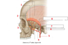
Identify the numbered arteries.


What foramen does the inferior alveolar aa enter? What does it supply?
What is the relationnship of the deep temporal artery to the temporalis muscle?
Maxillary Artery Branches:
- Middle meningeal – enter middle cranial cavity through foramen spinosum .
- Inferior alveolar – enters the inferior alveolar foramen to supply the teeth.
- Deep temporal - supplies the temporalis muscle
- Masseteric – supplies the masseter muscle
- Buccal – supplies the buccinator muscle

What vein(s) drain the region supplied by the branches of the maxillary artery? What is significant about this/these vein(s)?
The pterygoid plexus of veins drains the region supplied by the branches of the maxillary artery; these veins have no valves, thus pressure can force a backward/reverse flow of blood; this is a potential route for infection in the head and neck.

What kind of innervation does each branch of the trigeminal nerve provide? To what structures?
The mandibular division of the trigeminal nerve is the only division of the trigeminal nerve that is both sensory and motor.
V1 - Ophthalmic - sensory to skin of orbital region and nose
V2 - Maxillary - sensory to skin of maxillary region
V3 - Mandibular - sensory to skin of mandibular region, anterior 2/3 of tongue and adjacent mucous. Motor to muscles of mastication

What are branches of the anterior division of the mandibular nerve (V3)? What do they innervate?
What are branches of the posterior division of the mandibular nerve (V3)? What do they innervate?
What is the relationship of the branches of the anterior and posterior divisions to the lateral pterygoid muscle?
Anterior Division - all branches pierce the lateral pterygoid muscle.
- Muscle branches - to muscles of mastication
- Meningeal - recurs thru foramen spinosum to dura
- Buccal nerve - sensory to mucous membranes of oral cavity and skin of cheek.
Posterior Division - Lies deep to the Lateral Pterygoid Muscle
- Auriculotemporal - splits around middle meningeal artery innervate to temporomandibular joint, parotid, superficial temporal regions. Provides secretomotor fibers to the parotid gland via postganglionic paraympathetic fibers from the otic ganglion. Also provides GSA innervation to the anterosuperior ear and part of external auditory meatus as well as the TMJ.
- Inferior Alveolar -dives into mandibular foramen - (sensory to teeth) ends as mental nerve (sensory to chin).
- Lingual - anterior to inferior alveolar nerve. GSA innervation from the anterior 2/3 of the tongue and floor of the mouth. Taste to anterior 2/3 tongue and parasympathetic to submandibular and sublingual glands (via branches of chorda tympani)
see slides 33-34 of notes!!!
What is the pterygopalatine fossa? What is the general purpose of it?
Where is it located?
What are the boundaries of the pterygopalatine fossa?
The pterygopalatine fossa can be best understood as a distribution center for innervation to orbital, nasal and other facial structures.
The Pterygopalatine Fossa is:
- in the middle of the head – just behind the face.
- shaped like an “upside down tear drop”.
- communicates with numerous regions
The walls of the pterygopalatine fossa are formed by parts of the palatine, maxilla, and sphenoid bones:
- anterior wall - posterior surface of the maxilla;
- medial wall - lateral surface of the palatine bone;
- posterior wall - parts of the sphenoid bone.

What is located within the pterygopalatine fossa?
What structures pass through the pterygopalatine fossa?
The pterygopalatine fossa is:
- a major site of distribution for the maxillary nerve [V2].
- a site of the terminal part of the maxillary artery.
- a location for parasympathetic fibers from the CN VII and sympathetic fibers that join branches of V2 in the pterygopalatine fossa.
- contains the pterygopalatine ganglion.
attached is slide 36 of notes. must know this slide!

The ____ ____ and its branches are the major arterial supply for the infratemporal fossa and center portions of the face.

The maxillary artery and its branches are the major arterial supply for the infratemporal fossa and center portions of the face.


