Pediatric Pictures Flashcards
(94 cards)
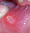
What is this?

Dermoid cyst
- saclike growths present at birth
- are like teratomas (can contain hair & teeth)
- often associated with tufts or sinuses
- grow slowly
- can get infected
- should be REMOVED
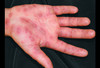
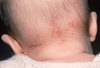
What type of rash is this?

Psoriasis
- Auspitz sign - punctate bleeding when scales removed
- can go into inguinal folds
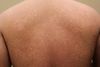
What type of rash is this?

Nummular Eczema
- on extensor surfaces of extremities
- lesions are uniform
- may ooze, crust or have a scaling pattern
- treat with steroids
What is this?

Pediculosis Pubis
Pubic Lice or Crabs
- infection in the groin
- red, crusted suprapubic macules and possibly bluish-gray dots.
- STRONG ASSOCIATION with sexual abuse in children.
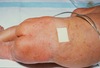
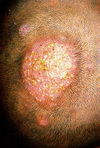
Whats is this?

Erythema Chronicum Migrans
- caused by BORRELIA BURGDORFERI, that causes LYME DISEASE.
- large, flat lesion (> 5 cm); a “bulls eye” lesion. Shows up 1–2 weeks after the bite.
- transmitted via the Ixodes deer tick.
- Lyme antibody titers. If these are positive, confirm with a Western blot.
- Treat: ORAL medication (doxycycline if >8 years old, or penicillin or amoxicillin if < 8 years old). If the patient has CARDITIS, neuritis (encephalitis/meningitis), or RECURRENT arthritis, treat with IV medication (PCN or ceftriaxone)

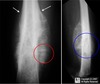
What syndrome is this?

Klippel-Feil Syndrome

- fused cervical vertebrae
- torticollis-like appearance
- short, webbed neck, limited range of motion at neck
- associated with Sprengel’s anomaly (elevated & medial rotation of scapula)
What type of rash is this?

Harlequin Ichthyosis
- covering is hard (“armor-like”) and horny
- movement is restricted
- poor prognosis
What is this caused by?

Scabies

- linear, papular, erythematous, pruritic, vesicular, and crusting lesions
- often seen in areas with CREASES (wrist, groin, webbing of fingers). You may see burrows.
- Treat: permethrin overnight from head to toe for the entire family.
- Re-treat if the patient is still having symptoms after 14 days and LIVE MITES are found
- persisting pruritis can be from residual inflammation.
What syndrome is this seen in?

Klippel-Trenaunay Syndrome
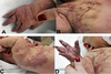
- associated with AV fistula, causing skeletal or limb overgrowth (HEMIHYPERTROPHY)
- port wine stain
- Look for unilateral limb overgrowth and CHF
What type of rash is this?

Lamellar Ichthyosis
Collodion Baby
- noted at the time of birth
- thin transparent film
- eyelashes are missing
- eyelids seem everted (ectropion)
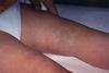
What is this?

Livedo Reticularis
AKA Cutis Marmorata
- a mottled, reticulate patterned rash and may be described as a lacy rash.
- benign
- resolves by 1 month.
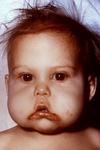
What syndrome is this?

McCune Albright Syndrome
Polyostotic Fibrous Dysplasia
- irregular cafe-au-lait macules (> 3 cm or multiple)
- precocious puberty
- bone problems (long bone fractures & bowing of arms)
- endocrine issues (hyperthyroidism)
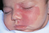
What causes this type of rash?

Zinc Deficiency
- SCALY and EXTREMELY ERYTHEMATOUSdermatitis in the perioral and perianal area (around the natural orifices) that can DESQUAMATE.
- The rash is sometimes described as erosive and eczematous.
- It can also be associated with ALOPECIA and poor taste.
- Breastfeeding helps with zinc absorption.
What is this seen in?

Incontinentia Pigmenti
- severe X-linked DOMINANT; only seen in FEMALES
- DEATH IN ALL MALES
- 4 stages of the rash: inflammatory vesicular phase; followed by a verrous phase; hyperpigmented phase along the lines of Blaschko; atrophy or hypopigmentation
- delayed dentition, mental retardation, paralysis, peg teeth, seizures
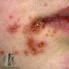
What is this?

Impetigo
- Bullous impetigo
- honey colored crusting lesions + bullae
- staph auresu
- Non-bullous impetigo
- honey colored crusting lesions w/o bullae (more crusting/oozing)
- staph or strep
What type of rash is this?

Lichen Sclerosus
- found in genital area
- no thickening or sclerosis
- no symptoms or some pruritis
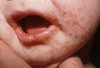
What is this rash?

Neonatal Acne
AKA Neonatal Cephalic Pustulosis
- occurs within the first month of life
- resolves by 4 months of age.
- inflammatory pustules on the cheeks and forehead without comedones.
- a benign rash that requires no treatment.
What is this?

Hutchinson Teeth
- found in late CONGENITAL SYPHILIS
- have nothes on the biting surface

What is this?

Alopecia Areata

- round/well-circumscribed area(s) of alopecia.
- can be on the scalp or in other areas.
- Hairs at the periphery of the areas are short, pluckable, and may resemble an exclamation point!
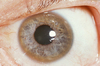
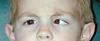
What Syndrome is this associated with?

PHACES SYNDROME
- Postrior fossa malformation (Dandy Walker)
- Hemangioma (large segmental hemangioma on the face) - associated with strokes
- Arterial cerebrovascular anomaly
- Cardiac anomalies (Coarctation of aorta)
- Eye anomalies (micropthalmia, strabismus)
- Sternal defect
What is this hair condition?

Alopecia Totalis
- the loss of all hair on the HEAD.
- Alopecia universalis is the loss of all hair on the entire BODY. There is usually a SYSTEMIC etiology such as hypothyroidism, a nutritional deficiency, or even lupus (SLE).
What is this type of hair loss called?

Telogen Effluvium
- a form of acute hair shedding that occurs diffusely.
- “thinning” of the hair.
- The hair that is shed can be recognized by a small bulb of keratin on the root end.
- often related to a psychological or medical stressor.
- Treat with REASSURANCE because the hair will grow back.
What is this?

Erythema Marginatum
- a transient, erythematous, macular and light colored.
- “SERPENTiginous” (snakelike) and the MARGINs are noted to progress as the center clears.
- It is part of the Jones criteria for Rheumatic Fever.
What is this?

Miliria Rubra
- very superficial vesicles that are easily ruptured
- occurs due to obstruction of sweat glands
- also called “prickly heat rash.”