peds Flashcards
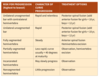
Legg-Calvé-Perthes Disease (Coxa Plana)
prognosis
goal is sphericity of femoral head
asperical - early DJD
poor prognosis w/ >6 years onset, female, lateral column C (regardless of age), adcreased abduction
Septic arthritis - aspiration results
>50K WBC, glucose 50 lower than serum level
Septic arthritis - who gets it from osteomyelitis?
neonates, in whom transphyseal vessels allow proximal spread into the joint in joints with an intraarticular metaphysis (hip, elbow, shoulder, ankle
Sprengel Deformity -associated diseases
Klippel-Feil syndrome (fused cervical vertebra w/ short neck; one third have Sprengel deformity) Kidney disease Scoliosis Diastematomyelia (split spinal cord)
Slipped Capital Femoral Epiphysis
technique
The pin should be started anteriorly on the femoral neck, ending in the central portion of the femoral head

Developmental Dysplasia of the Hip - associated problems and natural hx
other problems w/ positioning - torticollis (20%) and metatarsus adductus (10%) hip contracts and acetab becomes dysplastic and filled w/ pulvinar (fibrofatty tissue)
Developmental Dysplasia of the Hip - radiographic studies
dynamic u/s before ossification of femoral head at 4-6 months
Slipped Capital Femoral Epiphysis
xrays and grading
AP and frog-leg pelvic views
If the slippage is unstable, a cross-table lateral view is required
Grade I: 0% to 33% slippage
Grade II: 34% to 50% slippage
Grade III: more than 50% slippage
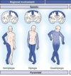
Brachial Plexus Palsy - what happens with significant IR contracture
progressive glenoid hypoplasia
Septic arthritis - treatment
aspiration, I&D
Legg-Calvé-Perthes Disease (Coxa Plana)
presentation
boys 4-8 years
pain (often knee), effusion, limp, decreased hip ROM (lack abd/IR)
Developmental Dysplasia of the Hip
-dynamic u/s angles
coronal view, the normal α angle is greater than 60 degrees, and the femoral head is bisected by the line drawn down the ilium.

Developmental Dysplasia of the Hip - risk factors in order
Breech>family hx>female >firstborn *left hip (67%) and girls (85%)
Osteomyelitis - why more common in kids?
rich metaphyseal blood supply and thick periosteum
Proximal Femoral Focal Deficiency- classification
A: femoral head present with normal acetabulum; B: femoral head present with dysplastic acetabulum; C: femoral head absent with markedly dysplastic acetabulum; D: both femoral head and acetabulum absent
Septic arthritis - joints with intraarticular metaphsysis prone to septic arthritis from osteomyelitis?

Proximal Femoral Focal Deficiency - associations
coxa vara, fibular hemimelia, ACL deficiency, knee contracture
Septic arthritis vs transient synovitis
Kocker criteria: 3/4 = >90% 1) WBC>12K 2) ESR>40 3) inability to bear weight 4) fever > 101.5/38.6
Rotational Problems of the Lower Extremities Femoral anteversion -features -treatment
3-6 years old, kids sit w/ legs in W position corrects by age 10 usually, no shoes/PT/braces are effective older children with less than 10 degrees of external rotation, femoral derotational osteotomy (intertrochanteric is best) may be considered for cosmesis, although this is not a functional problem
Osteomyelitis kids.- imaging findings
xray findings only after 5-7 days, MRI is key
Leg Length Discrepancy - when does bone growth stop?
age 16 in boys and age 14 in girls
open reduction for DDH
age?
approach and reason?
procedure steps?
6-18 months with failed closed reduction OR 18 months-3 years
Anterior approach (less risk to medial femoral circumflex) but may use medial if <12 months b/c less blood loss and direct access to obstacles for reduction, increased osteonecrosis
capsulorrhaphy, adductor tenotomy, femoral shortening + acetab procedure if dysplastic
Septic arthritis - when is an LP needed?
if H. influenzae because of association with meningitis