Pelvic region Flashcards

(61 cards)

1
Q

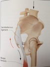
Sacrotuberous ligament - one of the strongest collagenous structures in the ms system (diagram)

A

wide as the thumb, found on palpation with px prone, between ischial tuberosity and inferolateral angle of sacrum

2
Q

Diagram showing how long head of the biceps femoris blends with the sacrotuberous ligament

A

Action of Biceps femoris : flex the knee joint and laterally rotate

3
Q

Impingement Test

Test to assess integrity of the acetabular labrum.

A

Patient is supine.

Practitioner initiates internal rotation.

Practitioner then passively flexes symptomatic hip to 90 degrees and adducts

Reproduction of symptoms in mid superior thigh inferior to the inguinal ligament indicates suspected labral tear or FAI

Symptoms may include pain and an audible click or just pain.

4
Q

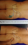
projection of the piriformis muscles (diagram)

A

a rounded structure that is firmer that its direct surroundings

5
Q

what happens when the arms are elevated whilst px is lying prone?

A

it tenses the thoracolumbar fascia and impedes palpate of lumbosacral structures

6
Q

Fitzgerald Test - Posterior Labrum

Test to assess integrity of the acetabular labrum

A

Patient supine

Practitioner passively fully flexes, adducts and internally rotates patient’s hip.

Practitioner then extends the hip whilst abducting and externally rotating in one continual movement.

Pain with or without an audible click is considered a positive test.

7
Q

FABER Test:

General screening test for hip joint pathology.

A

Patient supine, practitioner instructs patient to cross symptomatic leg so that the ipsilateral ankle rests

above the contralateral knee.

Practitioner then stabilizes the contralateral ASIS and supports the ipsilateral knee until end of range

Practitioner then applies gentle downward pressure to the medial aspect of the patients knee

Reproduction of pain or inability to perform test due to pain is considered a positive test.

8
Q

FABER (Patrick) test

- for hip joint pain

A

Flexion, abduction & external rotation of the hip joint passively with your patient supine.

This test can be used for the pelvis as part of a test cluster to differentiate hip joint pain from SIj pain.

It is assumed once the hip joint is at end of range this test will stress the anterior ligaments of the SIJ and the pubic symphysis

o Location of pain is an important factor in determining origin.

9
Q

MET - improve external rotation of hip joint

A

Piriformis MET – Px. Prone

Knee flexed,

px. Resists movement towards the midline.

Practitioner holds piriformis muscle.

Exhale and move leg outwards.

10
Q

What are the pelvic ligaments? (5)

A

interosseous sacroiliac ligaments

anterior sacroiliac ligaments

posterior sacroiliac ligaments

sacrotuberous ligaments

sacrospinous ligaments

11
Q

Which nerve gets affected in the dysfunctional abductor muscle of Trendelenburg gait?

A

Superior Gluteal nerve

12
Q

position of nerves in the gluteal region (diagram)

A
13
Q

Diagram showing the superolateral border of the gluteus maximus (approx)

A
14
Q

Femoroacetabular impingement image - Cam

A
15
Q

What type of joint is the SI joint?

A

Arthroidal.

Hyaline cartilage on the sacral side, fibro-cartilage on the ilial side.

16
Q

sciatica nerve exiting piriformis (diagram)

A

further down the sciatic nerve can be palpated laterally, between the ischial tuberosity and the superior aspect of g.t. (as line is drawn). It runs directly lateral to tuberosity.

17
Q

Seated Flexion Test

ROM test

A

Both thumbs on inferior slope of each posterior superior iliac spine.

Patient bends forwards with arms between the knees, as far as possible.

The PSIS that moves furthest in a superior or anterior is +ve, indicative of restricted mobility on that side.

Observe lumbar and thoracic vertebrae.

This test takes out lower extremities as an influencing factor.
If iliac crest is unlevel when seated initially then innominates are unequal size.

18
Q

Trendelenburg test
Test for weakness/atrophy of hip abductor muscles. (often occurs with a hip pathology)

A

Patient standing with feet hip width apart.

Practitioner instructs patient to stand on one leg and observes levels of iliac crests

Test is considered positive if the iliac crest is high on the stance leg and lower on the lifted leg

May indicate underlying hip joint pathology and weakness of gluteus medius

19
Q

How can you palpate the G.T. in obese patients?

A

in prone position, flex the knee and rotate laterally and medially to move the G.T.

20
Q

what structure might you find when palpating superiorly to the ischial tuberosity?

A

insertion of the sacrotuberous ligament

21
Q

What tends to be affected in a degenerative hip joint?

A

Medial rotation

22
Q

If long posterior sacroiliac joint is painful inferior to PSIS, what does this indicate?

A

SI joint pathology

23
Q

where are the PSIS in relation to the lumbar dimples?

A

approx 2cm lateral and 2cm inferior

24
Q

POSH (posterior shear) test

for the Sacro-iliac Joint

A

Patient supine

Practitioner passively flexes thigh at the hip and optionally places hand at the superior pole of the SIJ to palpate ROM, quality of movement and tissue texture.

Practitioner supports thigh at the knee with a broad secure contact using arm and axilla

Practitioner exerts a gradual even posterior compressive force into the SIJ taking care to note any signs of hip joint pain

Reproduction of SIJ symptoms is a positive test.

25
what do the interosseous sacroiliac ligaments do, and where are they? NB. these are very short and richly supplied with nociceptors
act to **maintain contact** between the surfaces of the SI joint located **directly posterior to SI joints**
26
Diagram of Iliotibial band What's the distal insertion How can the proximal aspect be palpated?
Gerdy tubercle (lateral condyle of the tibia) palpate laterally from the gluteus medius using transverse friction, a band 2-3 fingerwidths maybe felt betwen the iliac crest and the G.T.
27
**Ober’s test** Test for assessment of Hip joint pathology, trochanteric bursitis and ITB contracture.
Patient sidelying with LEX to be tested uppermost, lower LEX slightly flexed at knee and hip. Practitioner stands behind patient, **stabilizes pelvis and passively flexes upper knee to 90 degrees** whilst supporting knee and ankle **Practitioner then abducts and extends hip and lets supported LEX drop** to assess ITB contracture. **Pain over the greater trochanter is considered positive for trochanteric bursitis** Practitioner internally rotates hip to test for hip joint pathology.
28
what is the distance between the top of the iliac crest and the lowest rib?
two fingerwidths
29
**Sacroiliac compression test**
**Patient supine, using both hands, practitioner applies bilateral and medial compression to the ilium to compress the SIjs.** Reproduction of SIj. symptoms is a positive test. Test stress ligaments (mainly posterior) of the SIJ
30
MET - hip extension
MET for psoas. Px side-lying
31
what is ligament dynamization?
the extension of muscles into capsular-ligamentous structures
32
**Standing Flexion Test** **ROM test** **(test for SI evaluation)**
**Place thumbs on inferior slope of the posterior superior iliac spine** Patient bends forwards to try and touch the floor. Test +ve if one posterior superior iliac spine moves further than the other \* also observe thoracic and lumbar vertebrae.
33
* *_Thomas test_** * *Test for hip flexion contracture typically due to shortened Rectus Femoris or Iliopsoas.**
Patient supine and **passively flexes hip and knee to bring knee to chest,** whilst keeping other leg extended on the couch. **Practitioner observes for any elevation of the thigh or hip flexion in the extended leg.** Elevation of the straight leg indicates a positive test. Significant increase in lumbar lordosis is also considered a positive test.
34
How do you active the glut maximus? and.. what is its other main action?
prone. Px tries **active extension (raises leg off table)**. Can be done against resistance. Px can turn **heel inwards** to ext. rot. hip before extension. Operator can apply **additional pressure to inside of thigh** to stimulate adduction strong external rotator of the hip
35
which ligaments are in the first kinematic sacral-vertebral column chain?
iliolumbar ligaments
36
what can be felt between the sacrum and the PSIS, on the posterior surface of the sacrum?
the multifidus muscles If px increases lumbar lordosis then muscle contracts and tissue becomes firmer
37
**Stork Test** **ROM test** (test for SI joints) **Sensitive for SI restriction.**
**Place thumbs over left posterior superior iliac spine and the right thumb overlying the sacrum at the same level.** Patient flexes left hip and knee to minimum 90% flexion. **A negative test finds the left thumb on the PSIS move downwards in relation to the right thumb on sacrum.**
38
how do you palpate for the ischial tuberosity in prone position?
palpate medially along the gluteal fold with the thumb
39
**Gillets (Stork) Test**
as described previously. Can be used as a test. Positive if painful. ## Footnote **Normal motion would see PSIS move slightly inferiorly to S2 as the hip is flexed and the SIJ engaged.** With abnormal SIj motion the PSIS may appear to hitch up and move superiorly.
40
Pathologies of the hip (3)
41
what can be located at the level of the PSIS?
S2 SP
42
Diagram of the long posterior sacroiliac ligament
size - 3/4 cm long and 1-2 cm wide
43
Log Roll Test
Patient supine **Practitioner passively internally and externally rotates femur of asymptomatic side using hand on the mid thigh to test range and quality of movement.** Practitioner then tests symptomatic side in the same way **Reproduction of symptoms and increased external rotation of the hip is a positive test.**
44
what is a common cause od coccyx pain after falling?
overstretched ligamentous conections
45
where is the main trochanteric bursae located?
between the g.t. and the gluteus maximus or iliotibial tract
46
Diagram showing **positive** and **compensated** Trendelenburg sign
47
How can you palpate the posterior inferior iliac spine?
draw line from PSIS to sacral horn and half it. Firm deep pressure needed midway along line
48
MET - hip flexion
**Px. Supine.** **Hold flexed hip under armpit**, and leaning over px, hold contralateral leg to prevent flexion.
49
what ligament merges with the sacrotuberous ligament and is the ony ligament that restricts countermutation?
**long posterior sacroiliac ligament** connects PSIS with edge of sacrum, **about 3cm long.** felt as a grounded structure with transverse palpation
50
51
**Sacroiliac distraction test**
Patient supine. **Practitioner places both thenar eminence on medial aspect of ilium at the ASIS with arms crossed.** **Practitioner applies a lateral and slightly posterior compression force.** Reproduction of symptoms is a positive test This test stresses the anterior ligaments of the SIJ
52
**Fitzgerald Tests** - Anterior Test to assess integrity of the acetabular labrum.
Patient supine **Practitioner passively flexes, externally rotates and abducts hip.** **Practitioner then extends, internally rotates and adducts hip from previous position in one continual movement** Pain with or without an audible click is considered a positive test.
53
**SI Joint -** Active ROM testing Done in this order...
Standing flexion test Stork test Seated flexion test
54
which one ligament can help limit counternutation?
long posterior sacroiliac ligament
55
Image of Femoroacetabular impingement - Pincer
56
What is the job of the biceps femori tendon as it attaches into the sacrotuberous ligament?
It prevents the sacrum from nutating, and it stabilizes the SI joint directly before the landing phase.
57
58
Diagram showing sacroiliac joint projection
approx 2 cm lateral and parallel to the line connecting PSIS and sacral horn
59
Location of trochanteric bursa
60
**Special tests** for **SI joint/ lumbar-pelvic region (5)**
**POSH (posterior shear) test** for the Sacro-iliac Joint ## Footnote **Sacroiliac compression tests** **Sacroiliac distraction test** **FABER (Patrick) test** **Gillets (Stork) Test**
61
when palpating the PSIS, it could be painful on some px. Why?
**long posterior sacroiliac ligament** being sensitive to pressue due to possible SI pathology